. 2019 Jun 5;220(1):163-173.
doi: 10.1093/infdis/jiz074.

Interleukin 2 is an Upstream Regulator of CD4+ T Cells From Visceral Leishmaniasis Patients With Therapeutic Potential

Affiliations

Interleukin 2 is an Upstream Regulator of CD4+ T Cells From Visceral Leishmaniasis Patients With Therapeutic Potential

Shashi Bhushan Chauhan et al. J Infect Dis. .

Abstract

Control of visceral leishmaniasis (VL) caused by Leishmania donovani requires interferon-γ production by CD4+ T cells. In VL patients, antiparasitic CD4+ T-cell responses are ineffective for unknown reasons. In this study, we measured the expression of genes associated with various immune functions in these cells from VL patients and compared them to CD4+ T cells from the same patients after drug treatment and from endemic controls. We found reduced GATA3, RORC, and FOXP3 gene expression in CD4+ T cells of VL patients, associated with reduced Th2, Th17, and FOXP3+CD4+ T regulatory cell frequencies in VL patient blood. Interleukin 2 (IL-2) was an important upstream regulator of CD4+ T cells from VL patients, and functional studies demonstrated the therapeutic potential of IL-2 for improving antiparasitic immunity. Together, these results provide new insights into the characteristics of CD4+ T cells from VL patients that can be used to improve antiparasitic immune responses.

Keywords: Leishmania donovani; CD4+ T cells; IL-2; visceral leishmaniasis.


Figures

Figure 1.
Figure 1.
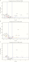
Nanostring analysis of immune-related genes in CD4+ T cells reveals more up- than down-regulated genes. The volcano plots show the distribution of immune-related genes in comparisons between visceral leishmaniasis (VL; n = 27) patients during infection (D0) and VL patients after treatment (DIS) (A), D0 and endemic controls (ECs; n = 15) (B), and DIS and ECs (C). Genes were determined to be up-regulated or down-regulated in the condition listed first in the title above each panel. Vertical lines in panels indicate absolute log2 fold-change (FC) = 0.5, and the horizontal lines indicate -log10 (P value false discovery rate [FDR]) = 1.3 (ie, FDR < 0.05). Genes of interest that were found to be differentially expressed (-log10 [FDR > 1.3], ie, FDR < 0.05) are indicated.
Figure 2.
Figure 2.
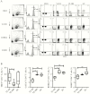
Changes in peripheral blood CD4+ T-cell subsets. The fluorescence-activated cell sorting plots show the gating strategy used to identify TBET+, GATA3+, RORCγ+, and FOXP3+ CD4+ T cells (A), and the frequency of these subsets among CD4+ T cells between visceral leishmaniasis (VL) patients during infection (D0; n = 7), VL patients after treatment (n = 7), and endemic controls (n = 6), median + minimum and maximum (B). *P < .05, **P < .01, Wilcoxon matched-pairs signed-rank test. Abbreviations: DIS, visceral leishmaniasis patients after treatment; D0, day zero; EC, endemic control; FMO, fluorescence minus one; FSC, forward scatter; SSC-A, side scatter-A; VL, visceral leishmaniasis.
Figure 3.
Figure 3.
Changes in immune molecule expression by CD4+ T cells. The fluorescence-activated cell sorting plots show the gating strategy used to identify CD38+, CTLA4+, CCR6+, CCR4+, CD40L+, IFN-γR1+, and CD96+ CD4+ T cells (A), and the frequency of these subsets among CD4+ T cells between visceral leishmaniasis (VL) patients during infection (D0; n = 6), VL patients after treatment (n = 6), and endemic controls (n = 6), median + minimum and maximum (B). *P < .05, **P < .01, Wilcoxon matched-pairs signed-rank test. Abbreviations: DIS, visceral leishmaniasis patients after treatment; D0, day zero; EC, endemic control; FMO, fluorescence minus one; FSC, forward scatter; IFN, interferon; SSC-A, side scatter-A; VL, visceral leishmaniasis.
Figure 4.
Figure 4.
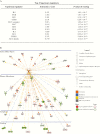
Interleukin 2 (IL-2) is the top upstream regulator of immune-related genes in visceral leishmaniasis patients. The table lists the top 12 upstream regulators (sorted by P value of overlap) that were identified by ingenuity pathway analysis (A). The network shows all up-regulated (red) and down-regulated (green) genes within the dataset that were predicted to be regulated by IL-2, and illustrates the predicted relationship between IL-2 and these genes (B).
Figure 5.
Figure 5.
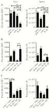
Mouse interleukin 2 (IL-2)/antibody complexes stimulate CD4+ T-cell–dependent antiparasitic immunity in mice infected with Leishmania donovani. C57BL/6 mice were infected with parasites for 14 days and then administered saline, anti–IL-2 monoclonal antibody (mAb) (JES6.1A12), recombinant mouse IL-2 (rmIL-2), or anti–IL-2 mAb complexed with rmIL-2 (IL-2Jc) on days 14 and 21 postinfection (p.i.), as indicated, and parasite burdens in the liver and spleen were measured 14 days later (day 28 p.i.) (A). Leishmania donovani–infected mice receiving saline or IL-2Jc were treated with control mAb (MAC5) or anti-CD4 mAb every 3 days, as indicated, over the same time period, and parasite burdens (expressed in Leishman–Donovan units [LDU]) in the liver and spleen were measured at day 28 p.i. (B). Leishmania donovani–infected B6.Foxp3.DTR mice receiving saline or IL-2Jc were treated with saline or diphtheria toxin (DT) every 3 days, as indicated, over the same time period, and parasite burdens in the liver and spleen were measured at day 28 p.i. (C). All values are mean ± standard error of the mean of at least 2 independent experiments, n = 5 mice per group. *P < .05, **P < .01, ***P < .001; significance assessed by 1-way analysis of variance.
Figure 6.
Figure 6.
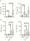
Human interleukin 2 (IL-2) stimulates antigen-specific interferon gamma (IFN-γ), but not interleukin 10 (IL-10) production by whole blood cells from patients with visceral leishmaniasis (VL). Antigen-specific IFN-γ and IL-10 production was measured in whole blood cells from VL patients on admission to clinic (n = 8–26) (A) and from endemic controls (n = 10–24) (B) cultured for 24 hours with media alone (NIL), soluble leishmania antigen (SLA), recombinant human IL-2, or SLA + IL-2, as indicated. Median + minimum and maximum, *P < .05, **P < .01, ***P < .001; significance assessed by Wilcoxon matched-pairs signed-rank test.

Similar articles

Cited by

References

    1. Zhu J, Yamane H, Paul WE. Differentiation of effector CD4 T cell populations (*). Annu Rev Immunol 2010; 28:445–89. - PMC - PubMed
    1. Rosas LE, Snider HM, Barbi J, et al. . Cutting edge: STAT1 and T-bet play distinct roles in determining outcome of visceral leishmaniasis caused by Leishmania donovani. J Immunol 2006; 177:22–5. - PubMed
    1. Janeway CA Jr, Medzhitov R. Innate immune recognition. Annu Rev Immunol 2002; 20:197–216. - PubMed
    1. Tubo NJ, Jenkins MK. CD4+ T cells: guardians of the phagosome. Clin Microbiol Rev 2014; 27:200–13. - PMC - PubMed
    1. Sakaguchi S, Yamaguchi T, Nomura T, Ono M. Regulatory T cells and immune tolerance. Cell 2008; 133:775–87. - PubMed

Publication types