

Connecting muscle development, birth defects, and evolution: An essential role for muscle connective tissue
- PMID: 30797508
- PMCID: PMC6449175
- DOI: 10.1016/bs.ctdb.2018.12.004
Connecting muscle development, birth defects, and evolution: An essential role for muscle connective tissue
Abstract
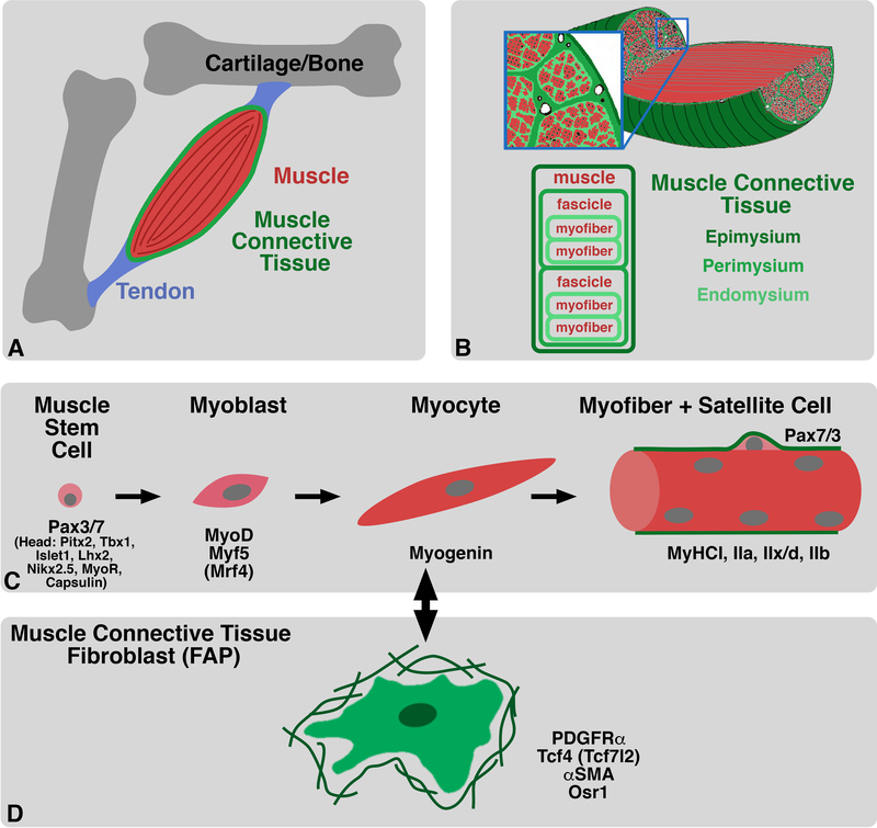
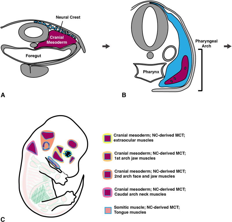
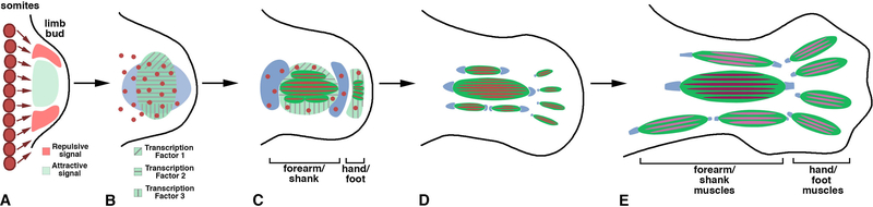
Skeletal muscle powers all movement of the vertebrate body and is distributed in multiple regions that have evolved distinct functions. Axial muscles are ancestral muscles essential for support and locomotion of the whole body. The evolution of the head was accompanied by development of cranial muscles essential for eye movement, feeding, vocalization, and facial expression. With the evolution of paired fins and limbs and their associated muscles, vertebrates gained increased locomotor agility, populated the land, and acquired fine motor skills. Finally, unique muscles with specialized functions have evolved in some groups, and the diaphragm which solely evolved in mammals to increase respiratory capacity is one such example. The function of all these muscles requires their integration with the other components of the musculoskeletal system: muscle connective tissue (MCT), tendons, bones as well as nerves and vasculature. MCT is muscle's closest anatomical and functional partner. Not only is MCT critical in the adult for muscle structure and function, but recently MCT in the embryo has been found to be crucial for muscle development. In this review, we examine the important role of the MCT in axial, head, limb, and diaphragm muscles for regulating normal muscle development, discuss how defects in MCT-muscle interactions during development underlie the etiology of a range of birth defects, and explore how changes in MCT development or communication with muscle may have led to the modification and acquisition of new muscles during vertebrate evolution.
Keywords: Axial muscle; Birth defects; Development; Diaphragm; Evolution; FAPs; Head; Limb; Muscle; Muscle connective tissue; Myogenesis.
© 2019 Elsevier Inc. All rights reserved.
Figures


Similar articles
-
Hox11 genes are required for regional patterning and integration of muscle, tendon and bone.Development. 2013 Nov;140(22):4574-82. doi: 10.1242/dev.096693. Epub 2013 Oct 23. Development. 2013. PMID: 24154528 Free PMC article.
-
Head muscle development.Results Probl Cell Differ. 2015;56:123-42. doi: 10.1007/978-3-662-44608-9_6. Results Probl Cell Differ. 2015. PMID: 25344669 Review.
-
Tbx4 and tbx5 acting in connective tissue are required for limb muscle and tendon patterning.Dev Cell. 2010 Jan 19;18(1):148-56. doi: 10.1016/j.devcel.2009.11.013. Dev Cell. 2010. PMID: 20152185 Free PMC article.
-
Developmental origin and morphogenesis of the diaphragm, an essential mammalian muscle.Dev Biol. 2018 Aug 15;440(2):64-73. doi: 10.1016/j.ydbio.2018.04.010. Epub 2018 Apr 19. Dev Biol. 2018. PMID: 29679560 Free PMC article.
-
To roll the eyes and snap a bite - function, development and evolution of craniofacial muscles.Semin Cell Dev Biol. 2019 Jul;91:31-44. doi: 10.1016/j.semcdb.2017.12.013. Epub 2018 Jan 10. Semin Cell Dev Biol. 2019. PMID: 29331210 Review.
Cited by
-
Distinct Embryonic Origin and Injury Response of Resident Stem Cells in Craniofacial Muscles.Front Physiol. 2021 Jul 1;12:690248. doi: 10.3389/fphys.2021.690248. eCollection 2021. Front Physiol. 2021. PMID: 34276411 Free PMC article. Review.
-
Hoxa5 Activity Across the Lateral Somitic Frontier Regulates Development of the Mouse Sternum.Front Cell Dev Biol. 2022 Apr 26;10:806545. doi: 10.3389/fcell.2022.806545. eCollection 2022. Front Cell Dev Biol. 2022. PMID: 35557949 Free PMC article.
-
Developmental fates of shark head cavities reveal mesodermal contributions to tendon progenitor cells in extraocular muscles.Zoological Lett. 2021 Feb 15;7(1):3. doi: 10.1186/s40851-021-00170-2. Zoological Lett. 2021. PMID: 33588955 Free PMC article.
-
Local retinoic acid signaling directs emergence of the extraocular muscle functional unit.PLoS Biol. 2020 Nov 17;18(11):e3000902. doi: 10.1371/journal.pbio.3000902. eCollection 2020 Nov. PLoS Biol. 2020. PMID: 33201874 Free PMC article.
-
Individual Limb Muscle Bundles Are Formed through Progressive Steps Orchestrated by Adjacent Connective Tissue Cells during Primary Myogenesis.Cell Rep. 2020 Mar 10;30(10):3552-3565.e6. doi: 10.1016/j.celrep.2020.02.037. Cell Rep. 2020. PMID: 32160556 Free PMC article.
References
-
- Ahmed MU, Maurya AK, Cheng L, Jorge EC, Schubert FR, Maire P, Basson MA, Ingham PW, Dietrich S, 2017. Engrailed controls epaxial-hypaxial muscle innervation and the establishment of vertebrate three-dimensional mobility. Dev Biol 430, 90–104. - PubMed
-
- Andrade W, Brandan E, 1991. Isolation and characterization of rat skeletal muscle proteoglycan decorin and comparison with the human fibroblast decorin. Comp Biochem Physiol B 100, 565–570. - PubMed
-
- Babiuk RP, Zhang W, Clugston R, Allan DW, Greer JJ, 2003. Embryological origins and development of the rat diaphragm. J Comp Neurol 455, 477–487. - PubMed
Publication types
MeSH terms
Grants and funding
LinkOut - more resources
Full Text Sources
