Review
. 2019:132:31-65.
doi: 10.1016/bs.ctdb.2018.12.001. Epub 2019 Jan 2.

Signals and forces shaping organogenesis of the small intestine

Affiliations
Review

Signals and forces shaping organogenesis of the small intestine

Sha Wang et al. Curr Top Dev Biol. 2019.

Abstract

The adult gastrointestinal tract (GI) is a series of connected organs (esophagus, stomach, small intestine, colon) that develop via progressive regional specification of a continuous tubular embryonic organ anlage. This chapter focuses on organogenesis of the small intestine. The intestine arises by folding of a flat sheet of endodermal cells into a tube of highly proliferative pseudostratified cells. Dramatic elongation of this tube is driven by rapid epithelial proliferation. Then, epithelial-mesenchymal crosstalk and physical forces drive a stepwise cascade that results in convolution of the tubular surface into finger-like projections called villi. Concomitant with villus formation, a sharp epithelial transcriptional boundary is defined between stomach and intestine. Finally, flask-like depressions called crypts are established to house the intestinal stem cells needed throughout life for epithelial renewal. New insights into these events are being provided by in vitro organoid systems, which hold promise for future regenerative engineering of the small intestine.

Keywords: Boundary formation; Crypt development; Epithelial-mesenchymal crosstalk; Intestinal lengthening; Intestinal organoids; Intestinal regionalization; Mesenchymal clusters; Signaling cascades; Tensile forces driving morphogenesis; Villus development.


Figures

Fig. 1
Fig. 1
Expression domains of CDX2, SOX2, and PDX1 during gut tube closure and formation of the anterior and caudal intestinal portals. At E8.0, SOX2 (green) and CDX2 (red) are expressed in the anterior- and posterior-most endoderm, respectively. At E8.25, the anterior intestinal portal is observed in the SOX2 domain (*) and at E8.5, the caudal intestinal portal (**) begins to form within the CDX2 domain. Between E8 and E8.75, the two domains extend toward each other as the endoderm wraps into a tube. The two domains meet at E8.5 and cells at the boundary co-express SOX2 and CDX2 (yellow). By E9.25, the boundary appears sharper in whole mount tissue (but see text) and PDX1 expression is seen in cells in the future pyloric region, extending from both sides of this boundary (blue). Images were modified with permission from Sherwood, R. I., Chen, T. Y., & Melton, D. A. (2009). Transcriptional dynamics of endodermal organ formation. Developmental Dynamics, 238, 2942.
Fig. 2
Fig. 2
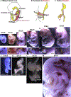
Intestinal hernia, rotation, looping, and retraction during development. (A-C) Schematic illustration of intestinal hairpin loop formation (A), herniation (B) and rotation (B, C) in amniotes. As the midgut elongates, it protrudes into the umbilical cord and forms an intestinal hernia. As it protrudes, the midgut loop makes a counterclockwise rotation. Outside of the body, the gut elongates dramatically; looping/coiling occurs as the result of differential growth of the intestine and the attached mesentery. As the gut returns to the body cavity, it makes a further rotation. (D-I) Mouse embryos at E10.5–16.5. (D′-I′) Higher magnifications of the white boxed regions in (D-I), respectively (herniation and retraction). Yellow dashed line outlines the intestine located outside of the body cavity. (J-L) Dissected mouse GI tracts at E10.5 (hairpin loop), E11.5 (cecum bulge formation), E13.5 (looping). (M) Convoluted GI tract in an opened abdominal cavity of an E16.5 mouse embryo, showing intestinal loops. Scalebars are 1 mm.
Fig. 3
Fig. 3
Small intestinal elongation and its epithelial configuration in Phase I and Phase II. Schematics of intestinal lengthening (drawn to scale) are shown at top. Accompanying circular diagrams represent intestinal cross sections at the point of the horizontal line in top figures. Bottom diagrams show detail of cell shape in the epithelium. In Phase I, the epithelium is pseudostratified with a small flat apical surface (red line) and nearly all cells are actively cycling (blue nuclei, cell cycle phases are noted). In Phase II, beginning at E14.5, mesenchymal clusters form beneath the epithelium (orange) and villi begin to emerge. Cells on villi stop cycling (black nuclei) and change shape, becoming columnar. Cells in the intervillus regions remain proliferative (blue nuclei). Around P7, crypts (purple) form and house proliferating cells.
Fig. 4
Fig. 4
The choreography of IKNM-associated cell division in Phase I small intestinal epithelium. As the nucleus migrates apically for mitosis, the cell maintains a basal connection via a thin filament, termed the basal process. This process splits into two, but only one of the two remains intact and is inherited by one daughter cell, leaving the other daughter disconnected basally. During G1, daughter pairs return their nuclei to the basal side in two distinct modes. In Mode I, the daughter with the basal process uses it as a “conduit” for a quick nuclear return; return of the other daughter’s nucleus is slower as this cell must generate a filopodium to build a new pathway to return its nucleus—the “pathfinding” strategy. In Mode II, both daughters utilize “pathfinding” and return their nuclei basally at a similar pace. Pathfinding sometimes fails in the absence of the mesenchymal cue, WNT5A. The two daughters remain apically connected throughout mitosis. Finally, two daughters separate from each other (they are not side-by-side in the epithelium) and prepare for the next round of division. Adapted from Fig. 7L from Wang, S., Cebrian, C., Schnell, S., & Gumucio, D. L. (2018). Radial WNT5A-guided post-mitotic filopodial pathfinding is critical for midgut tube elongation. Developmental Cell, 46, 173188.e3.
Fig. 5
Fig. 5
Villus formation. (A) At E13.5, the intestinal epithelium (yellow) is a tube with a flat lumenal surface that uniformly secretes Hh and PDGF signals (yellow) to subepithelial mesenchymal cells (orange). (A′) The flat apical surface is highlighted by EZRIN staining (green) and the even distribution of PDGF responsive cells is noted by PDGFRa staining (red). (A″) SEM image showing the flat lumenal surface at E13.5. (B) By E14.5 in the anterior duodenum, Hh/PDGF responsive mesenchymal cells begin to aggregate to form clusters (orange dots) in a regularly spaced field; spacing is determined by a Bmp-dependent Turing field, in which formation of one cluster prevents the initiation of another cluster in the immediate area. (B′) Mesenchymal clusters result in uneven compression in the epithelium, leading to membrane invaginations that demarcate villi, here shown as T-shaped extensions of the apical surface (arrows). Apical demarcations (arrows) form between mesenchymal clusters. (B″) SEM reveals demarcations of initial villi at the apical surface (arrows). (C and C′) Villi emerge at the site of each cluster; clusters remain associated with tips of emerging villi. (C″) Villus domes are obvious in SEM. (D) By E16.5, villi are longer and additional rounds of cluster formation occur in intervillus regions; villi are present throughout the small intestine. Panels (A′-C″): Reproduced from Freddo, A. M., Shoffner, S. K., Shao, Y., Taniguchi, K., Grosse, A. S., Guysinger, M. N., et al. (2016). Coordination of signaling and tissue mechanics during morphogenesis of murine intestinal villi: A role for mitotic cell rounding. Integrative Biology: Quantitative Biosciences from Nano to Macro, 8, 918928 with permission from The Royal Society of Chemistry.
Fig. 6
Fig. 6
Crypt development. (A-D) Whole mount staining of CD44v6 (magenta) in epithelium of ZO1-GFP labeled mouse intestines at P0, 4, 7 and 10. (E-G) Schematic summary of crypt morphogenesis. (E) The CD44v6 + compartment starts within the flat sheet of intervillus cells at P0. Myosin II-drives apical constriction of CD44v6 positive cells. (F) Forming crypts invaginate into the underlying mesenchyme. (G) Cells at the bend between the crypt and villus adopt a wedge-like shape—the “hinge” region—which morphologically separates crypts from villi. Panels (A-D): Reproduced with permission from Sumigray, K. D., Terwilliger, M., & Lechler, T. (2018). Morphogenesis and compartmentalization of the intestinal crypt. Developmental Cell, 45, 183197.e185 (Fig. B). Panels (E-G): Adapted with permission from the graphical abstract in Sumigray, K. D., Terwilliger, M., & Lechler, T. (2018). Morphogenesis and compartmentalization of the intestinal crypt. Developmental Cell, 45, 183197.e185.

Similar articles

Cited by

References

    1. Aoki R, Shoshkes-Carmel M, Gao N, Shin S, May CL, Golson ML, et al. (2016). Foxl1-expressing mesenchymal cells constitute the intestinal stem cell niche. Cellular and Molecular Gastroenterology and Hepatology, 2, 175–188. - PMC - PubMed
    1. Applegate KE, Anderson JM, & Klatte EC (2006). Intestinal malrotation in children: A problem-solving approach to the upper gastrointestinal series. Radiographics, 26, 1485–1500. - PubMed
    1. Arnaout R, & Stainier DYR (2011). Developmental biology: Physics adds a twist to gut looping. Current Biology, 21, R854–R857. - PubMed
    1. Barker N, Huch M, Kujala P, van de Wetering M, Snippert HJ, van Es JH, et al. (2010). Lgr5(+ve) stem cells drive self-renewal in the stomach and build long-lived gastric units in vitro. Cell Stem Cell, 6, 25–36. - PubMed
    1. Battle MA, Bondow BJ, Iverson MA, Adams SJ, Jandacek RJ, Tso P, et al. (2008). GATA4 is essential for jejunal function in mice. Gastroenterology, 135, 1676–1686.e1. - PMC - PubMed

LinkOut - more resources