Review
. 2019 Nov-Dec;23(6):467-475.
doi: 10.1016/j.bjpt.2019.01.011. Epub 2019 Feb 3.

Kinesiologic considerations for targeting activation of scapulothoracic muscles - part 2: trapezius

Affiliations
Review

Kinesiologic considerations for targeting activation of scapulothoracic muscles - part 2: trapezius

Paula R Camargo et al. Braz J Phys Ther. 2019 Nov-Dec.

Abstract

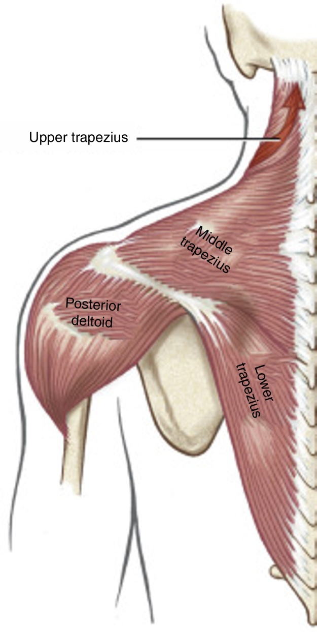
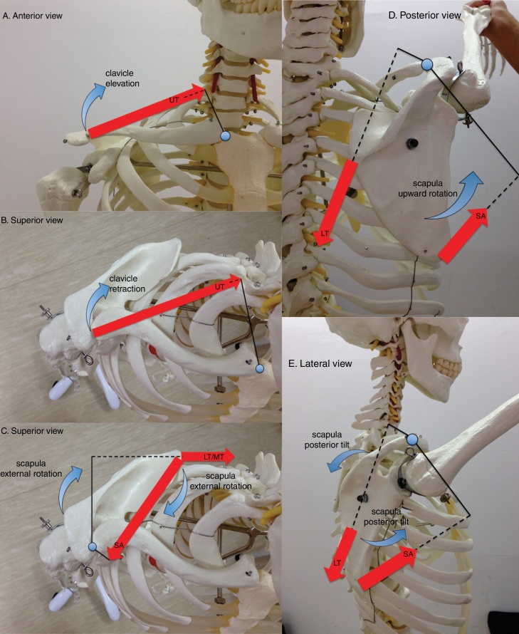
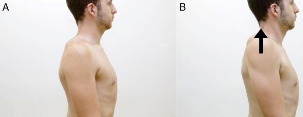
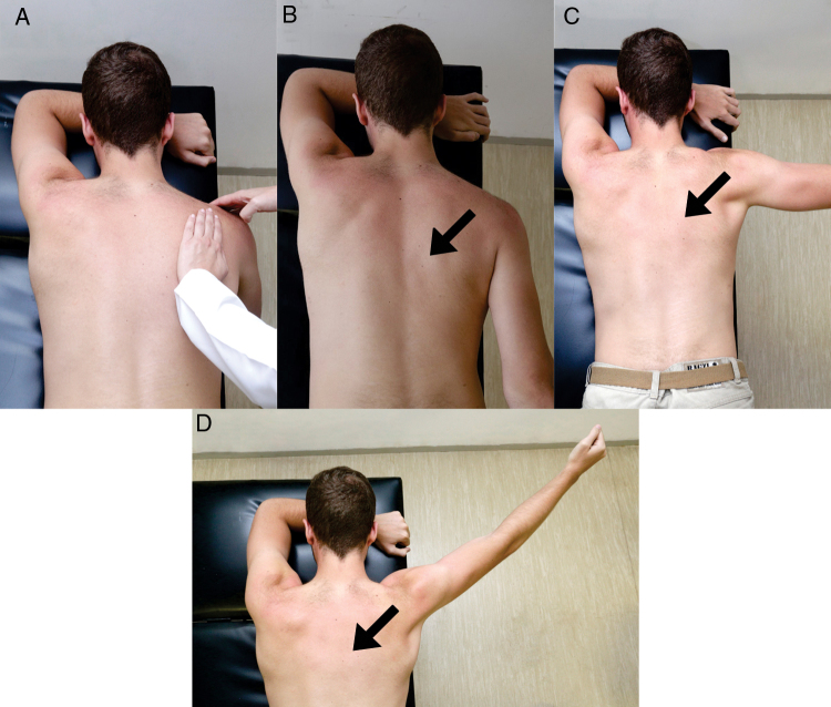
Background: The trapezius is an extensive muscle subdivided into upper, middle, and lower parts. This muscle is a dominant stabilizer of the scapula, normally operating synergistically with other scapular muscles, most notably the serratus anterior. Altered activation, poor control, or reduced strength of the different parts of the trapezius have been linked with abnormal scapular movements, often associated with pain. Several exercises have been designed and studied that specifically target the different parts of the trapezius, with the goal of developing exercises that optimize scapular position and scapulohumeral rhythm that reduce pain and increase function.

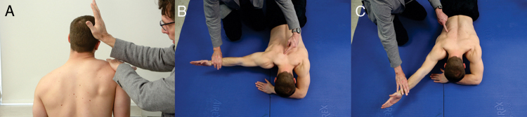
Methods: This paper describes the anatomy, kinesiology, and pathokinesiology of the trapezius as well as exercises that selectively target the activation of the different parts of this complex muscle.

Conclusions: This review provides the anatomy and kinesiology of the trapezius muscle with the underlying intention of understanding how this muscle contributes to the normal mechanics of the scapula as well as the entire shoulder region. This paper can guide the clinician with planning exercises that specifically target the different parts of the trapezius. It is recommended that this paper be read as a companion to another paper: Kinesiologic considerations for targeting activation of scapulothoracic muscles - part 1: serratus anterior.

Keywords: Physical therapy; Scapular dyskinesis; Scapulothoracic joint; Shoulder rehabilitation; Trapezius exercises.


Figures

Figure 1
Figure 1
The 3 parts of the trapezius muscle. The posterior deltoid is also illustrated. Note that the red arrow shows the line of action of the upper trapezius muscle.
Figure 2
Figure 2
Lines of action of selected scapulothoracic muscles are depicted by red large straight arrows (UT: upper trapezius, MT: middle trapezius, LT: lower trapezius, SA: serratus anterior). The muscles are shown contributing to clavicular elevation (A), clavicular retraction (B), scapular external rotation (C), scapular upward rotation (D), and scapular posterior tilt (E) during shoulder flexion. Internal moment arms for muscles are shown as a solid line from the axis of rotation to the line of action of each muscle. Dashed lines indicate a right-angle intersection between muscle's line of action and its moment arm. Note two axes of rotation: the sternoclavicular axis, located near the manubrium, and the acromioclavicular axis, located near the acromion.
Figure 3
Figure 3
Manual muscle test of strength of the upper trapezius (A), middle trapezius (B) and lower trapezius (C).
Figure 4
Figure 4
Shrug exercises and retraction. Exercise starts with the individual with the arms at the side of the trunk and shoulders in retraction (A). Perform the shrug movement (B) and return to the starting position.
Figure 5
Figure 5
Overhead shrug. Exercise starts with the individual standing, placing the arms in overhead position against the wall (A) and performing a shrug movement (B) and returning to the starting position.
Figure 6
Figure 6
Prone horizontal abduction. Exercise starts with the individual in prone and arm abducted to 90° with external rotation. Perform horizontal abduction.
Figure 7
Figure 7
Retraction overhead. Exercise starts with the individual standing, placing the arms in overhead position against the wall (A) and lifting both arms (black arrows) while performing retraction (blue arrows) of the shoulders (B), then return to the starting position.
Figure 8
Figure 8
Prone scapular setting. Exercise starts with the individual lying in prone with the arm in overhead position resting on the treatment table (A). Position the scapula in relative retraction and depression (black arrow). While maintaining this scapular position, lift the arm slightly off the treatment table (B). Avoid shrugging the shoulder and activity of the upper trapezius.
Figure 9
Figure 9
Elevation with external rotation. Exercise starts with the individual standing, elbows flexed to 90° and an elastic band held in hands. The elastic band is brought to tension with 30° of arms external rotation (A). Elevate both arms to 90° in the scapular plane while holding/maintaining the tension in the band (B).
Figure 10
Figure 10
Prone extension. Exercise starts with the individual in prone with the arm at 90° of forward flexion (A). Perform extension to neutral position with the shoulder in neutral rotation (B).
Figure 11
Figure 11
Side-lying external rotation. Exercise starts with the individual in the side-lying position with the shoulder in neutral position and elbow flexed to 90° (A). Perform external rotation of the shoulder with a towel between the elbow and trunk (B). Avoid compensatory movements.
Figure 12
Figure 12
Side-lying forward flexion. Exercise starts with the individual in the side-lying position and the arm parallel with the body (A). Perform forward flexion (B).
Figure 13
Figure 13
Prone: Making I, T, Y letters. Exercise starts with the individual lying in prone with arm hanging off the edge of the treatment table (A). Position the scapula in retraction and depression. The therapist can provide feedback with hands. Maintain this position while extending the arm posteriorly in line with the trunk (letter “I”, B), horizontally abducting the arm (letter “T”, C) or elevating the arm to about 120° (letter “Y”, D). Avoid activity of the upper trapezius. The black arrows indicate the importance of maintaining relative scapular retraction and depression during the exercise.

References

    1. Neumann D.A., Camargo P.R. Kinesiologic considerations for targeting activation of scapulothoracic muscles: part 1: serratus anterior. Braz J Phys Ther. 2019;23(6):459–466. - PMC - PubMed
    1. De Vita A., Kibler W.B., Pouliart N., Sciascia A. Scapulothoracic joint. In: Di Giacomo G., Pouliart N., Constantini A., DeVita A., editors. Atlas of Functional Shoulder Anatomy. 1st ed. Springer; Milan: 2008.
    1. Wiater J.M., Bigliani L.U. Spinal accessory nerve injury. Clin Orthop Relat Res. 1999;(368):5–16. - PubMed
    1. Chan P.K., Hems T.E. Clinical signs of accessory nerve palsy. J Trauma. 2006;60(5):1142–1144. - PubMed
    1. Neumann D.A. 3rd ed. Elsevier; St Louis: 2017. Kinesiology of the Musculoskeletal System: Foundations for Rehabilitation.