Review
. 2019 May 15:204:140-150.
doi: 10.1016/j.physbeh.2019.02.025. Epub 2019 Feb 21.

Hindbrain astrocytes and glucose counter-regulation

Affiliations
Review

Hindbrain astrocytes and glucose counter-regulation

Richard C Rogers et al. Physiol Behav. .

Abstract

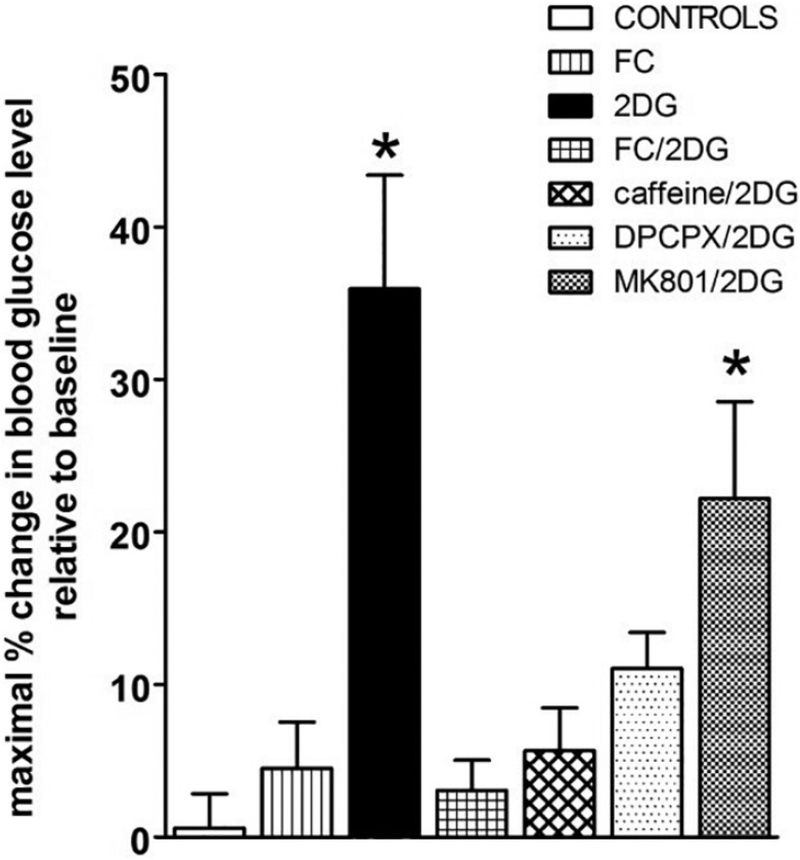
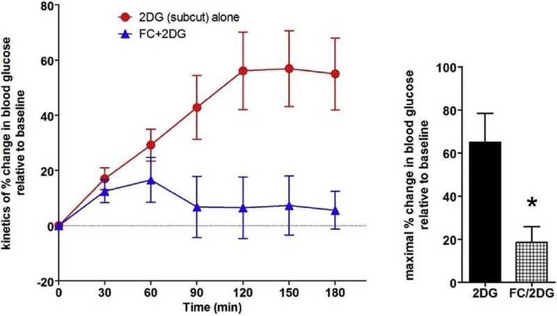
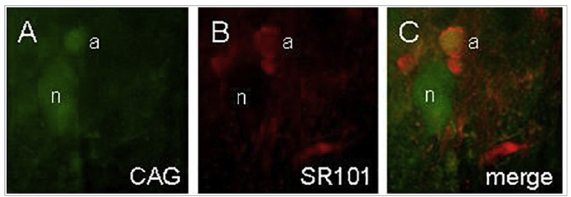
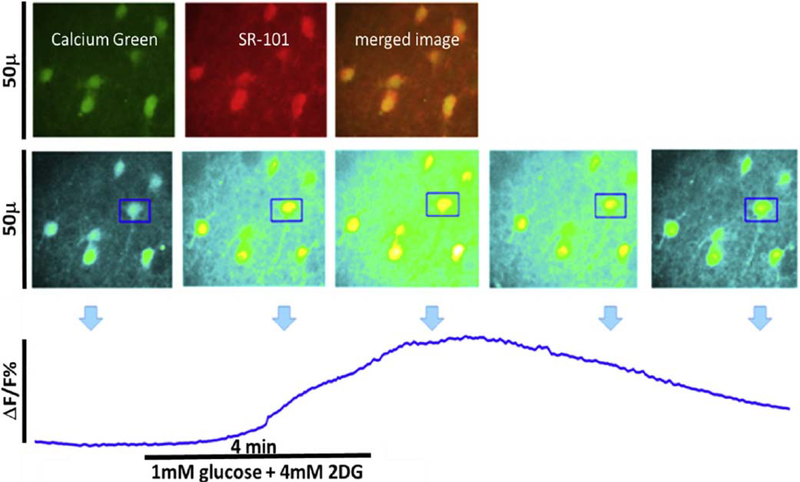
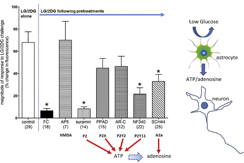
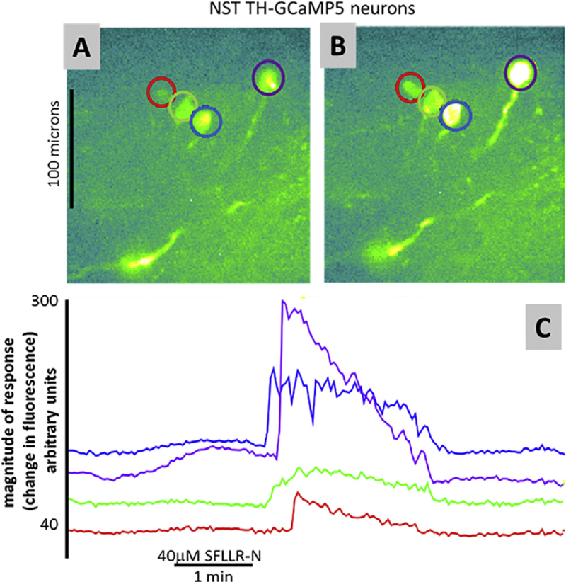
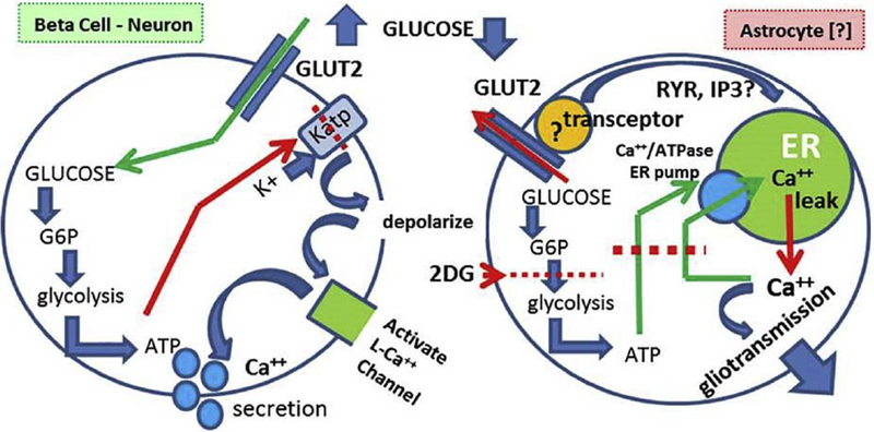
Hindbrain astrocytes are emerging as critical components in the regulation of homeostatic functions by either modulating synaptic activity or serving as primary detectors of physiological parameters. Recent studies have suggested that the glucose counter-regulation response (CRR), a critical defense against hypoglycemic emergencies, is dependent on glucoprivation-sensitive astrocytes in the hindbrain. This subpopulation of astrocytes produces a robust calcium signal in response to glucopenic stimuli. Both ex vivo and in vivo evidence suggest that low-glucose sensitive astrocytes utilize purinergic gliotransmission to activate catecholamine neurons in the hindbrain that are critical to the generation of the integrated CRR. Lastly, reports in the clinical literature suggest that an uncontrolled activation of CRR may as part of the pathology of severe traumatic injury. Work in our laboratory also suggests that this pathological hyperglycemia resulting from traumatic injury may be caused by the action of thrombin (generated by tissue trauma or bleeding) on hindbrain astrocytes. Similar to their glucopenia-sensitive neighbors, these hindbrain astrocytes may trigger hyperglycemic responses by their interactions with catecholaminergic neurons.


Figures

Fig. 1.
Fig. 1.
Gastric emptying rate in an awake, freely moving animal can be monitored by determining the rate of appearance of 13C in respired CO2 that had been ingested as 13C-tagged sodium octanoate-doped meal. 13C in respired CO2 is indicative of the transit of the carbohydrate meal from the stomach to the duodenum. This 13C method captures the entire time course of the transit event (samples taken at 15 min intervals) and is displayed in the right-hand graphs. Specific parameters of this transit can be extracted for comparisons: Tlag corresponds with the time at which rate of excretion of 13CO2 is maximal, T1/2 is the gastric half-emptying time (time when half of the label that is to be excreted has been excreted) and the GEC (a global index of rate of emptying). Each animal served as its own control. That is, the first session the animal receives the control injection (saline either i.p. or 4 V, as appropriate); the next session the animal receives the test injection. Therefore, paired t-test comparisons could be made between the saline control and the agonist condition for each animal. Examples of such paired gastric transit experiments of individual animals are seen in the right-hand graphs. In the left-hand column are group averages for the gastric transit parameters under each condition; statistical comparisons were only made within each condition group. Microinjection of the PAR1-selective agonist SFLLRN (10 nmol) into the fourth ventricle caused a significant reduction in gastric transit as measured by Tlag and T1/2 as well as a slowed overall GEC compared with their respective saline controls. (Adapted from Hermann et al., J Neurosci, 2009, 29 [29]: 9292–9300).
Fig. 2.
Fig. 2.
Proposed circuitry explaining how thrombin effects on astrocytes can suppress gastric motility in response to traumatic injury.
Fig. 3.
Fig. 3.
Preliminary studies indicate that fourth ventricular application of thrombin in the thiobutabarbital anesthetized rat provokes a significant increase in blood glucose levels compared to control saline applications [45]. This thrombin effect on glycemia is blocked by pretreatment with either the PAR antagonist, SCH79797 or the astrocyte calcium signaling inhibitor, fluorocitrate (FC). Thus, the presence of thrombin in the hindbrain can evoke hyperglycemia and this effect is dependent on functioning astrocytes.
Fig. 4.
Fig. 4.
Proposed circuitry responsible for hypoglycemic effects on astrocytes to elicit an increase in gastric motility.
Fig. 5.
Fig. 5.
Fourth ventricle 2DG evokes a counter-regulatory hyperglycemia that is suppressed by FC and adenosine antagonists. Maximal percent change in blood glucose levels relative to baseline levels were averaged within each group. Maximal peak responses for the 2DG (alone) group averaged 36.0 ± 7.4%change. While 4 V application of vehicle controls (saline or 1:4 DMSO/saline) or FC (alone) or caffeine (alone; data not shown here) had no effect on glucose levels, pretreatment of the 4 V with FC blocked the effect of subsequent 2DG to increase blood glucose (FC + 2DG: 3.1 ± 2.0%). Similarly, pretreatment with caffeine or DPCPX suppressed the glycemic effects of 2DG (caffeine+2DG = 5.7 ± 2.8%; DPCPX+2DG = 11.1 ± 2.4%). In contrast, the NMDA antagonist, MK801, did not block the 2DG effect to increase blood glucose (MK801 + 2DG = 22.2 ± 6.4%). One way ANOVA F7,38 = 9.78, p < .0001; Dunnett’s post-hoc test * p < .05. These results are consistent with a counter-regulatory hyperglycemia triggered by astrocytes utilizing adenosine as a gliotransmitter. (Adapted from Rogers et al., 2016, Am J Physiol Regul Integr Comp Physiol 310: R1102–R1108).
Fig. 6.
Fig. 6.
Subcutaneous 2DG (100 mg/kg/ml) in the thiobutabarbital-anesthetized rat produces a counter-regulatory increase in blood glucose. Pretreatment with 4 V FC (5nanomol) 30 min prior to subcutaneous delivery of 2DG significantly reduced the hyperglycemic effect of systemic 2DG (t = 3.074; p = .02). These results suggest that the principal mechanism connecting systemic glucopenia with counter-regulatory hyperglycemia involves hindbrain astrocytes. (Adapted from Rogers et al., 2016, Am J Physiol Regul Integr Comp Physiol 310: R1102–R1108).
Fig. 7.
Fig. 7.
Identification of astrocytes and neurons during live cell imaging. A) Calcium Green (CAG), the Ca++ reporter dye, is taken up by both astrocytes (a) and neuronal cells (n). B) SR101 astrocyte-specific vital dye staining. C) Same field as both A and B; demonstration of ex vivo glial and neuronal identification. (Adapted from Hermann et al., J Neurosci, 2009, 29 [29]: 9292–9300).(For interpretation of the references to colour in this figure legend, the reader is referred to the web version of this article.)
Fig. 8.
Fig. 8.
Live cell calcium imaging of NST astrocytes in response to glucoprivic challenge. Top panel: In vivo co-injection of the calcium indicator dye, Calcium Green (green) and the glial vital stain sulforhodamine 101 (SR101; red) allows for simple discrimination of astrocytes and neurons in the in situ brain stem slice preparation. Following a dual exposure utilizing the 488 nm and 561 nm laser lines, a composite image is obtained in which astrocytes are clearly labeled yellow, i.e. positive for both CAG and SR101, while neurons remain green. Middle panel: screen shots of field of astrocytes responding to glucoprivic challenge over time (time-lapse imaging with only the 488 nm laser line). Changes in intracellular calcium concentrations are indicated by a proportional change in the intensity of the fluorescence of the calcium green. One region of interest [ROI] was drawn around an individual astrocyte (blue box) for analysis of its time-lapse re-sponse to the challenge. Lower panel: plot of change in magnitude of fluorescence of individual astrocyte before, during, and after exposure to glucoprivic challenge. (For interpretation of the references to colour in this figure legend, the reader is referred to the web version of this article.)
Fig. 9.
Fig. 9.
Magnitude of changes in fluorescence due to intracellular calcium fluxes in TH-GCaMP5 NST neurons in response to glucoprivic challenge after specific pretreatment conditions (Number of neurons studied per each group is noted in parentheses). Exposure of TH-GCaMP5 neurons in hindbrain slices to the various pretreatment conditions produced significant differences in response to subsequent glucoprivic challenge (ANOVA F(10,175) = 7.39; p < .0001). Similar to the responses seen in the general population of NST neurons, TH-GCaMP5 NST neurons were robustly activated by glucoprivation and that effect is essentially blocked by pre-treatment with FC (Dunnett’s post hoc test q = 6.415; *p < .05). The NMDA antagonist (AP5) had no effect to inhibit the TH-GCaMP5 NST neuron response to glucoprivation. However, the non-selective P2 antagonist (suramin) also blocked the re-sponses to low glucose/2DG (Dunnett’s post hoc test q = 5.715; *p < .05). Lastly, both NF 340 (P2Y11 antagonist) and SCH442416 (A2a antagonist) sup-pressed TH-GCaMP5 NST neuronal responses to glucoprivic conditions (Dunnett’s post hoc test q = 5.160 and 4.047, respectively; *p < .05). These data suggest that the catecholamine neurons critical to activating counter-regulation in response to central glucopenia depend on purinergic gliotransmission. (Adapted from Rogers et al., 2018, Am J Physiol Regul Integr Comp Physiol 315: R153–R164.)
Fig. 10.
Fig. 10.
Preliminary ex vivo imaging studies using the TH-GCaMP5 transgenic preparation show that these identified catecholaminergic NST neurons are also responsive to the PAR1 agonist, SFLLRN. A) Identified catecholaminergic (TH-GCaMP5) cells at rest. B) Same cells as in (A) stimulated by perfusion of SFLLR-N. C) Individual response profiles of cells identified in (A) and (B). Of the 28 TH-GCaMP5 neurons in the NST that were responsive to glutamate/ATP challenge (i.e., viability test), 15 (i.e., ~54%) were also responsive to SFLLR-N. These data suggest that hindbrain catecholaminergic neurons are also involved in the hyperglycemia induced by thrombin.
Fig. 11.
Fig. 11.
Fundamental differences in how the “classic model” (i.e., beta cell) of glucodetection must differ from the astrocyte mechanism: 1. Activating levels of glucose are opposite. That is, glucopenia activates astrocytes; while elevated glucose activates beta cells and glucose-sensitive neurons. 2. 2. The mechanism of glucose activation of beta cells is well known; ATP generated by glycolysis inhibits a dominant K+ conductance, causing depolarization. Depolarization opens voltage-gated cation channels triggering action potential generation and/or secretion. 3. Mechanisms for activating calcium entry/release after low glucose exposure are not known but may involve ATP “starvation” induced shut-down of ER calcium storage followed by calcium leakage from the ER. The role of “transceptors” attached to GLUT2 possibly mediating ER calcium release (via inhibition or activation) are common in yeast and may exist in vertebrates, but this is not yet clear. (Adapted from Rogers et al., 2017, Hindbrain Astrocyte Glucodetectors and Counterregulation. In: Appetite and Food Intake: Central Control, edited by Harris RBS, p. 205–228.

Similar articles

Cited by

References

    1. Accorsi-Mendonca D, Bonagamba LGH, Machado BH, ATP released by glia increases the excitatory neurotransmission onto NTS neurons related to the peripheral chemoreflex, Soc. Neurosci 824 (809) (2012).
    1. Agulhon C, Boyt KM, Xie AX, Friocourt F, Roth BL, McCarthy KD, Modulation of the autonomic nervous system and behaviour by acute glial cell Gq protein-coupled receptor activation in vivo, J. Physiol. 591 (2013) 5599–5609. - PMC - PubMed
    1. Agulhon C, Fiacco TA, McCarthy KD, Hippocampal short- and long-term plas- ticity are not modulated by astrocyte Ca2+ signaling, Science 327 (2010) 1250–1254. - PubMed
    1. Andrew SF, Dinh TT, Ritter S, Localized glucoprivation of hindbrain sites elicits corticosterone and glucagon secretion, Am. J. Phys. Regul. Integr. Comp. Phys. 292 (2007) R1792–1798. - PubMed
    1. Angulo MC, Kozlov AS, Charpak S, Audinat E, Glutamate released from glial cells synchronizes neuronal activity in the hippocampus, J. Neurosci. 24 (2004) 6920–6927. - PMC - PubMed

Publication types