

Electrostatic Steering Enables Flow-Activated Von Willebrand Factor to Bind Platelet Glycoprotein, Revealed by Single-Molecule Stretching and Imaging
- PMID: 30797858
- PMCID: PMC6936110
- DOI: 10.1016/j.jmb.2019.02.014
Electrostatic Steering Enables Flow-Activated Von Willebrand Factor to Bind Platelet Glycoprotein, Revealed by Single-Molecule Stretching and Imaging
Abstract
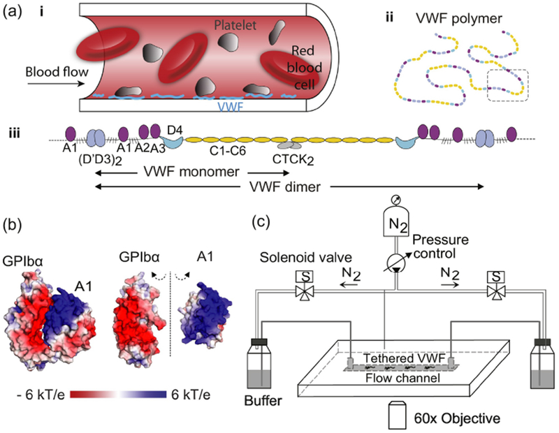
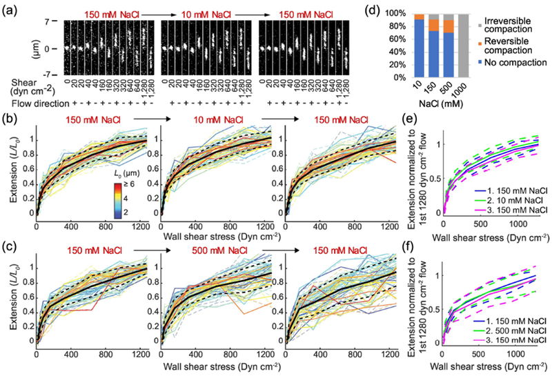
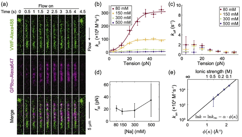
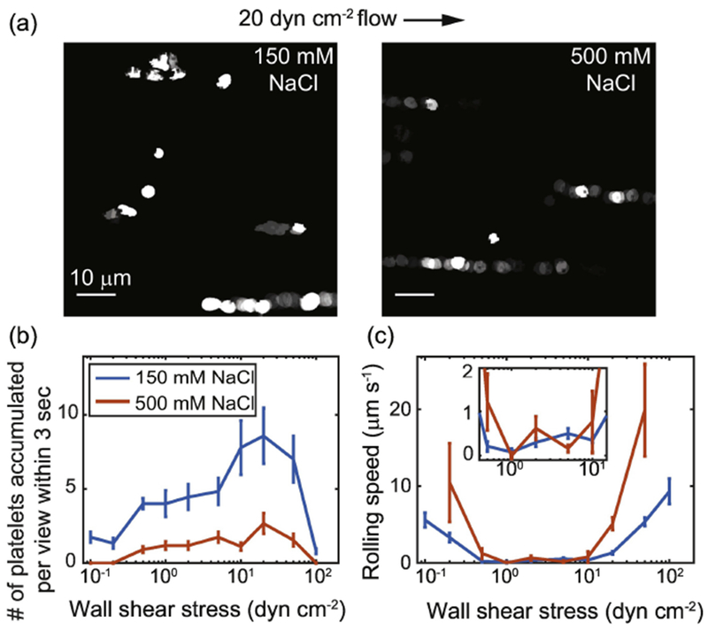
Von Willebrand factor (VWF), a large multimeric blood protein, senses changes in shear stress during bleeding and responds by binding platelets to plug ruptures in the vessel wall. Molecular mechanisms underlying this dynamic process are difficult to uncover using standard approaches due to the challenge of applying mechanical forces while monitoring structure and activity. By combining single-molecule fluorescence imaging with high-pressure, rapidly switching microfluidics, we reveal the key role of electrostatic steering in accelerating the binding between flow-activated VWF and GPIbα, and in rapidly immobilizing platelets under flow. We measure the elongation and tension-dependent activation of individual VWF multimers under a range of ionic strengths and pH levels, and find that the association rate is enhanced by 4 orders of magnitude by electrostatic steering. Under supraphysiologic salt concentrations, strong electrostatic screening dramatically decreases platelet binding to VWF in flow, revealing the critical role of electrostatic attraction in VWF-platelet binding during bleeding.
Keywords: hemostasis; mechanosensing; microfluidics; single-molecule fluorescence; single-molecule force spectroscopy.
Copyright © 2019 The Author(s). Published by Elsevier Ltd.. All rights reserved.
Figures
Similar articles
-
Flow-induced elongation of von Willebrand factor precedes tension-dependent activation.Nat Commun. 2017 Aug 23;8(1):324. doi: 10.1038/s41467-017-00230-2. Nat Commun. 2017. PMID: 28831047 Free PMC article.
-
A biophysical view on von Willebrand factor activation.J Cell Physiol. 2018 Feb;233(2):799-810. doi: 10.1002/jcp.25887. Epub 2017 May 16. J Cell Physiol. 2018. PMID: 28256724 Review.
-
Specific electrostatic interactions between charged amino acid residues regulate binding of von Willebrand factor to blood platelets.J Biol Chem. 2017 Nov 10;292(45):18608-18617. doi: 10.1074/jbc.M117.797456. Epub 2017 Sep 18. J Biol Chem. 2017. PMID: 28924049 Free PMC article.
-
von Willebrand factor self-association on platelet GpIbalpha under hydrodynamic shear: effect on shear-induced platelet activation.Blood. 2010 Nov 11;116(19):3990-8. doi: 10.1182/blood-2010-02-269266. Epub 2010 Aug 9. Blood. 2010. PMID: 20696943 Free PMC article.
-
Role of fluid shear stress in regulating VWF structure, function and related blood disorders.Biorheology. 2015;52(5-6):319-35. doi: 10.3233/BIR-15061. Biorheology. 2015. PMID: 26600266 Free PMC article. Review.
Cited by
-
Interplay between inflammation and thrombosis in cardiovascular pathology.Nat Rev Cardiol. 2021 Sep;18(9):666-682. doi: 10.1038/s41569-021-00552-1. Epub 2021 May 6. Nat Rev Cardiol. 2021. PMID: 33958774 Free PMC article. Review.
-
Electrostatic Forces Mediate the Specificity of RHO GTPase-GDI Interactions.Int J Mol Sci. 2021 Nov 19;22(22):12493. doi: 10.3390/ijms222212493. Int J Mol Sci. 2021. PMID: 34830380 Free PMC article.
-
Biomechanical activation of blood platelets via adhesion to von Willebrand factor studied with mesoscopic simulations.Biomech Model Mechanobiol. 2023 Jun;22(3):785-808. doi: 10.1007/s10237-022-01681-3. Epub 2023 Jan 10. Biomech Model Mechanobiol. 2023. PMID: 36627458 Free PMC article.
-
Conformation of von Willebrand factor in shear flow revealed with stroboscopic single-molecule imaging.Blood. 2022 Dec 8;140(23):2490-2499. doi: 10.1182/blood.2022016969. Blood. 2022. PMID: 36040485 Free PMC article.
-
Autoinhibitory module underlies species difference in shear activation of von Willebrand factor.J Thromb Haemost. 2022 Nov;20(11):2686-2696. doi: 10.1111/jth.15837. Epub 2022 Aug 29. J Thromb Haemost. 2022. PMID: 36031939 Free PMC article.
References
-
- Sadler JE, Biochemistry and Genetics of von Willebrand Factor, Annu. Rev. Biochem 67 (1998) 395–424. - PubMed
-
- Sadler JE, Budde U, Eikenboom JCJ, Favaloro EJ, Hill FGH, Holmberg L, Ingerslev J, Lee CA, Lillicrap D, Mannucci PM, Mazurier C, Meyer D, Nichols WL, Nishino M, Peake IR, Rodeghiero F, Schneppenheim R, Ruggeri ZM, Srivastava A, Montgomery RR, Federici AB, Update on the pathophysiology and classification of von Willebrand disease: a report of the Subcommittee on von Willebrand Factor, J. Thromb. Haemost 4 (2006) 2103–2114. doi: 10.1111/j.1538-7836.2006.02146.x. - DOI - PubMed
Publication types
MeSH terms
Substances
Grants and funding
LinkOut - more resources
Full Text Sources
Other Literature Sources
Research Materials
Miscellaneous
