Review
. 2019 Jul;138(1):1-21.
doi: 10.1007/s00401-019-01977-2. Epub 2019 Feb 23.

α-Synuclein and astrocytes: tracing the pathways from homeostasis to neurodegeneration in Lewy body disease

Affiliations
Review

α-Synuclein and astrocytes: tracing the pathways from homeostasis to neurodegeneration in Lewy body disease

Zachary A Sorrentino et al. Acta Neuropathol. 2019 Jul.

Abstract

α-Synuclein is a soluble protein that is present in abundance in the brain, though its normal function in the healthy brain is poorly defined. Intraneuronal inclusions of α-synuclein, commonly referred to as Lewy pathology, are pathological hallmarks of a spectrum of neurodegenerative disorders referred to as α-synucleinopathies. Though α-synuclein is expressed predominantly in neurons, α-synuclein aggregates in astrocytes are a common feature in these neurodegenerative diseases. How and why α-synuclein ends up in the astrocytes and the consequences of this dysfunctional proteostasis in immune cells is a major area of research that can have far-reaching implications for future immunobiotherapies in α-synucleinopathies. Accumulation of aggregated α-synuclein can disrupt astrocyte function in general and, more importantly, can contribute to neurodegeneration in α-synucleinopathies through various pathways. Here, we summarize our current knowledge on how astrocytic α-synucleinopathy affects CNS function in health and disease and propose a model of neuroglial connectome altered by α-synuclein proteostasis that might be amenable to immune-based therapies.

Keywords: Astrocyte heterogeneity; Exosome; Glial cytoplasmic inclusion; Lewy body; NAC domain; Neurodegeneration; Therapy; Transmission; Tunneling nanotube; α-Synuclein; αSyn 3H11.


Figures
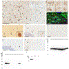
Figure 1.
Figure 1.. Astrocytic αSyn inclusions in DLB patients specifically detected by an antibody in the middle domain of αSyn.
a. Immunohistochemical staining with 3H11 antibody raised against residues 43–63 of αSyn with formic acid retrieval. αSyn is extensively detected within astrocytes in the hippocampus of a DLB patient. b. Higher magnification from Panel A showing astrocytes immunopositive for αSyn antibody 3H11. c. Immunofluorescent analysis with antibodies to GFAP (green) and αSyn 3H11 (red) demonstrating that αSyn is present in astrocytic processes in the DLB hippocampus. d. Detection of αSyn within classical LBs in the substantia nigra of a DLB patient with the 3H11 antibody. Inset shows magnified view of a typical intraneuronal LB. e. Detection of αSyn within GCI inclusions in the cerebellum of an MSA individual using the 3H11 antibody. Inset shows magnified view of typical GCI. f. αSyn antibody 3H11 fails to show any immunoreactivity in the hippocampus of an Alzheimer’s disease patient negative for pSer129 αSyn inclusions. g. Immunohistochemical staining of the DLB hippocampus from panel a using antibody EP1536Y against pSer129 αSyn; extensive astrocytic inclusions are not seen using this antibody. h. Immunohistochemical staining of the MSA cerebellum from panel e using antibody EP1536Y against pSer129 αSyn; 3H11 detects GCIs similarly to this antibody but no astrocytic staining is seen using either. i. A western blot of 200 ng recombinant αSyn proteins harboring various familial mutations probed with antibody 94–3A10 (residues 130–140) [45]. j. A western blot using the same proteins from panel G but probed with antibody 3H11; the antigenic region is residues ~47–55. k. Western blot showing that 3H11 specifically recognizes human αSyn but not βSyn or γSyn. Scale Bar: 100μm (A, D, E, F, G, H), 50μm (C).
Figure 2.
Figure 2.. Robust astrogliosis and astroglial αSyn accumulation in transgenic mouse models of α-synucleinopathy.
a. Immunofluorescent detection of GFAP in spinal sections from M83+/− transgenic mice overexpressing human [A53T] αSyn that were intramuscularly injected with preformed wild type mouse αSyn fibrils. In this model, pSer129-αSyn pathology and motor neuron death is apparent at 2 months post injection whereas increase in GFAP, representative of astrocyte activation, is observed at later time points. b. Immunoflourescent detection of GFAP (green) and pSer 129 αSyn (red) in the spine of terminal M83+/− transgenic mice described in panel a; in regions of neuronal death, astrocytes closely interact with extracellular aggregated αSyn. c. Immunoflourescent detection of GFAP (green) and pSer 129-αSyn (red) in the midbrain of M20 transgenic mice overexpressing human αSyn that were seeded with preformed αSyn fibrils in the striatum showing the presence of abundant astrocytic αSyn pathology. Scale Bar: 500μm (B), 100μm (C).
Figure 3.
Figure 3.. αSyn mediated alterations in neuro-glial homeostasis in health and disease.
Accumulation of αSyn leads to Lewy pathology through a variety of cellular processes. In neurons and astrocytes, αSyn has been implicated in dysfunction of lysosomes (LYS) and mitochondria (MITO), which enhances ROS production and toxic sequela. Neuronal αSyn can be transported into neighboring astrocytes via tunneling nanotubes (TNT), receptors such as Toll like receptors (TLR), or several indirect methods such as internalization of exocytosed material from the synaptic space. We hypothesize that astrocytic lysosomes preferentially cleave full length αSyn into smaller toxic truncation products that might have additional pathological functions with astrocytes. Toxic accumulation of αSyn within astrocytes may damage their normal functions and have adverse results such as synaptic accumulation of neurotransmitters including glutamate and dopamine (DA) that are in part cleared by astrocytes through EAAT2 and DAT/NET respectively. Astrocytes normally protect against oxidative stress in the CNS through production of multiple products including ARE factors, KEAP1/NFR2, Glutathione, NURR1 and NQO1; loss of these factors due to toxic accumulation of αSyn may contribute to neurodegeneration. Astrocytic uptake of αSyn and oxidative stress may impinge on the NF-κB pathways that can result in increased inflammatory cytokines, such as IL-6 or TNFα that will affect the function of neighboring microglia or peripheral immune cells which represents another modality by which astrocytes may contribute to neurodegeneration. Additionally, common risk factor genes such as DJ-1, Parkin and Pink1 are highly expressed in astrocytes and their dysfunction may affect mitophagy in a parallel pathway to that of toxic αSyn resulting in similar degenerative features. Red arrows denote pathological processes, green arrows denote putative therapies or beneficial pathways and black arrows denote normal physiological processes.

References

    1. Abounit S, Bousset L, Loria F, Zhu S, de Chaumont F, Pieri L, Olivo-Marin J-C, Melki R, Zurzolo C (2016) Tunneling nanotubes spread fibrillar α-synuclein by intercellular trafficking of lysosomes. EMBO J 35:2120–2138. doi: 10.15252/embj.201593411 - DOI - PMC - PubMed
    1. Ahn T-B, Langston JW, Aachi VR, Dickson DW (2012) Relationship of neighboring tissue and gliosis to alpha-synuclein pathology in a fetal transplant for Parkinson’s disease. Am J Neurodegener Dis 1:49–59 - PMC - PubMed
    1. Allen NJ, Eroglu C (2017) Cell Biology of Astrocyte-Synapse Interactions. Neuron 96:697–708. doi: 10.1016/j.neuron.2017.09.056 - DOI - PMC - PubMed
    1. Allen Reish HE, Standaert DG (2015) Role of α-synuclein in inducing innate and adaptive immunity in Parkinson disease. J Parkinsons Dis 5:1–19. doi: 10.3233/JPD-140491 - DOI - PMC - PubMed
    1. Anderson JP, Walker DE, Goldstein JM, de Laat R, Banducci K, Caccavello RJ, Barbour R, Huang J, Kling K, Lee M, Diep L, Keim PS, Shen X, Chataway T, Schlossmacher MG, Seubert P, Schenk D, Sinha S, Gai WP, Chilcote TJ (2006) Phosphorylation of Ser-129 Is the Dominant Pathological Modification of α-Synuclein in Familial and Sporadic Lewy Body Disease. J Biol Chem 281:29739–29752. doi: 10.1074/jbc.M600933200 - DOI - PubMed

Publication types

Substances