. 2019 Mar;41(3):313-321.
doi: 10.1007/s00276-019-02196-w. Epub 2019 Feb 23.

Ultrasound-guided decompression surgery of the distal tarsal tunnel: a novel technique for the distal tarsal tunnel syndrome-part III

Affiliations

Ultrasound-guided decompression surgery of the distal tarsal tunnel: a novel technique for the distal tarsal tunnel syndrome-part III

Simone Moroni et al. Surg Radiol Anat. 2019 Mar.

Abstract

Background: The aim of this study was to provide a safe ultrasound-guided minimally invasive surgical approach for a distal tarsal tunnel release concerning nerve entrapments.

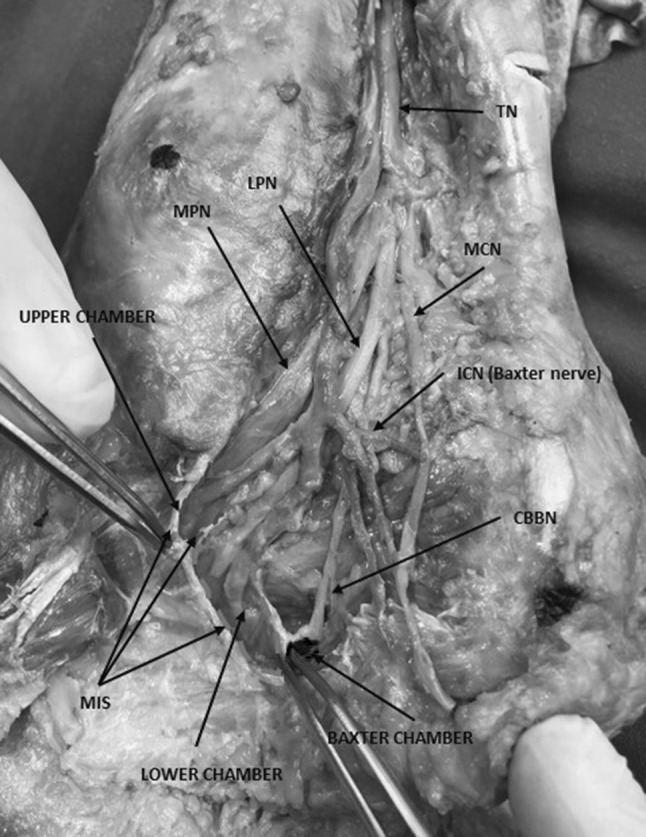
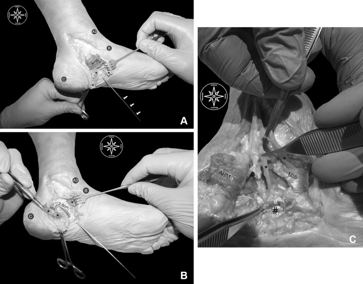
Methods and results: The study was carried out on ten fresh-frozen feet. All of them have been examined by high-resolution ultrasound at the distal tarsal tunnel. The surgical approach has been marked throughout the course of the medial intermuscular septum (MIS, the lateral fascia of the abductor hallucis muscle). After the previous steps, nerve decompression was carried out through a MIS release through a 2.5 mm (± 0.5 mm) surgical portal. As a result, an effective release of the MIS has been obtained in all fresh-frozen feet.

Conclusion: The results of our anatomic study indicate that this novel ultrasound-guided minimally invasive surgical approach for the release of the MIS might be an effective, safe and quick decompression technique treating selected patients with a distal tarsal tunnel syndrome.

Keywords: Heel pain; Minimally invasive; Nerve entrapment; Tarsal tunnel; Ultrasound; Ultrasound-guided.


Conflict of interest statement

No outside funding was received. Nothing to declare.

Figures

Fig. 1
Fig. 1
Overview of the main components of the proximal and distal tarsal tunnel and all important chambers. TN tibial nerve, mpn medial plantar nerve, lpn lateral plantar nerve; icn inferior calcaneal nerve (i.e., baxter nerve), cbbn calcaneal branch of the baxter nerve, mcn medial calcaneal nerve, ms medial septum, upper chamber blue rounded area bounded by blue dotted line, lower chamber green rounded area bounded by green dotted line; “Baxter chamber”: red rounded area bounded by red dotted line. (Color figure online)
Fig. 2
Fig. 2
Nerves (lpn, mpn, bn) entering separated tubes at the distal tarsal tunnel, perforating the medial septum. AHM abductor hallucis muscle, mpn medial plantar nerve, lpn lateral plantar nerve, bn Baxter’s nerve, cbbn calcaneal branch of the Baxter’s nerve, ms medial septum and its extension, red arrows nerves entering upper and lower calcaneal tubes
Fig. 3
Fig. 3
Instruments for the minimally invasive ultrasound-guided procedure. High-resolution ultrasound; dissection material; 20-gauge needle; syringe; V-shape; hooked knife
Fig. 4
Fig. 4
Ultrasound visualization of the terminal branches (lpn, mpn, bn) on the [BC] line. The figure on the right side shows an anatomical overview of the structures including the reference line “Dellon-McKinnon” (A1–B, malleolar-calcaneal) and the “Triangle of Heimkes” (A2–B–C). Lpn lateral plantar nerve, mpn medial plantar nerve, bn Baxter’s Nerve, FHL flexor hallucis longus muscle, QPM quadratus plantae muscle, AHM abductor hallucis muscle, mpv medial plantar vein, lpv lateral plantar vein, mpa medial plantar artery, lpa lateral plantar artery, black arrow heads superficial layer of the flexor retinaculum, white stars medial septum (deep fascia of the abductor hallucis muscle), black stars medial septum extension (“interfascicular septum”)
Fig. 5
Fig. 5
Presurgical mapping of the distal tarsal tunnel. A2-B line: a line from the anterior tip of the medial malleolus (anterior colliculus) to the center of the posterior calcaneal tuberosity; CB line: a line from the navicular tuberosity to the center of the posterior calcaneal tuberosity. Red dots from C perpendicular to the sole of the foot: reference for the surgical portal; white dots and lines: course of MPN and LPN through A2-B, BC and C projection lines; blue dots and line: skin projection of medial septum extension from its intersection with BC line and its mean proximal point calculated from our anatomical study part I; muscle belly: represents the full dorso-plantar height of the medial intermuscular septum; red line: surgical portal for the medial intermuscular septum release in the upper calcaneal tube; dotted tract of the surgical portal line represents the full cut length for the medial intermuscular septum release. (Color figure online)
Fig. 6
Fig. 6
Algorithm. BC-line a line from the navicular tuberosity to the center of the posterior calcaneal tuberosity, C projection perpendicular line from the navicular tuberosity to the sole of the foot, MIS medial intermuscular septum
Fig. 7
Fig. 7
a Dissection routine. A2: medial malleolus B: navicular tuberosity C: center of the posterior calcaneal tuberosity; AHM: exposed abductor hallucis muscle belly; asterisk subcutaneus fat; white arrows: buttoned probe entering the surgical portal and following the course through the surgical line. b Gross anatomical findings. A2 medial malleolus; B navicular tuberosity; C center of the posterior calcaneal tuberosity; asterisk abductor hallucis muscle belly over the medial intermuscular septum; 2.5 cm: the mean length of the retrograde cut performed over the MIS which exposes the buttoned probe entering the surgical portal and following the course through the surgical line. c Gross anatomical findings. MIS: medial intermuscular septum; AHM abductor hallucis muscle belly over the medial intermuscular septum; numbers sign plantar fascia; asterisk proof of undamaged medial and lateral plantar nerves. (Color figure online)

References

    1. Ahmad M, Tsang K, Mackenney PJ, Adedapo AO. Tarsal tunnel syndrome: a literature review. Foot Ankle Surg. 2012;18(3):149–152. doi: 10.1016/j.fas.2011.10.007. - DOI - PubMed
    1. Antoniadis G, Scheglmann K. Posterior tarsal tunnel syndrome: diagnosis and treatment. Dtsch Arztebl Int. 2008;105(45):776–781. - PMC - PubMed
    1. Association AD. Economic costs of diabetes in the U.S. in 2012. Diabetes Care. 2013;36(6):1033–1046. doi: 10.2337/dc12-2625. - DOI - PMC - PubMed
    1. Baxter DE, Pfeffer GB (1992) Treatment of chronic heel pain by surgical release of the first branch of the lateral plantar nerve. Clin Orthop Relat Res (279):229–236 - PubMed
    1. Boulton AJ, Vileikyte L, Ragnarson-Tennvall G, Apelqvist J. The global burden of diabetic foot disease. Lancet. 2005;366(9498):1719–1724. doi: 10.1016/S0140-6736(05)67698-2. - DOI - PubMed