. 2019 Mar;12(3):e005529.
doi: 10.1161/CIRCHEARTFAILURE.118.005529.

Recruitment of RNA Polymerase II to Metabolic Gene Promoters Is Inhibited in the Failing Heart Possibly Through PGC-1α (Peroxisome Proliferator-Activated Receptor-γ Coactivator-1α) Dysregulation

Affiliations

Recruitment of RNA Polymerase II to Metabolic Gene Promoters Is Inhibited in the Failing Heart Possibly Through PGC-1α (Peroxisome Proliferator-Activated Receptor-γ Coactivator-1α) Dysregulation

Santosh Bhat et al. Circ Heart Fail. 2019 Mar.

Abstract

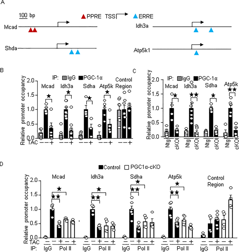
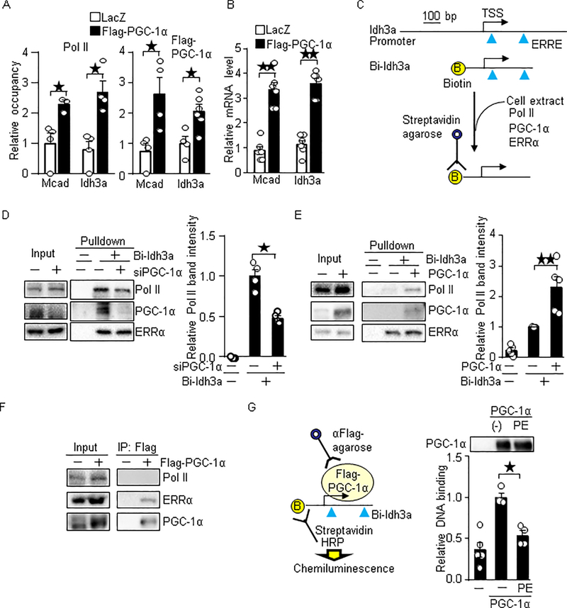
Background: Proper dynamics of RNA polymerase II, such as promoter recruitment and elongation, are essential for transcription. PGC-1α (peroxisome proliferator-activated receptor [PPAR]-γ coactivator-1α), also termed PPARGC1a, is a transcriptional coactivator that stimulates energy metabolism, and PGC-1α target genes are downregulated in the failing heart. However, whether the dysregulation of polymerase II dynamics occurs in PGC-1α target genes in heart failure has not been defined.

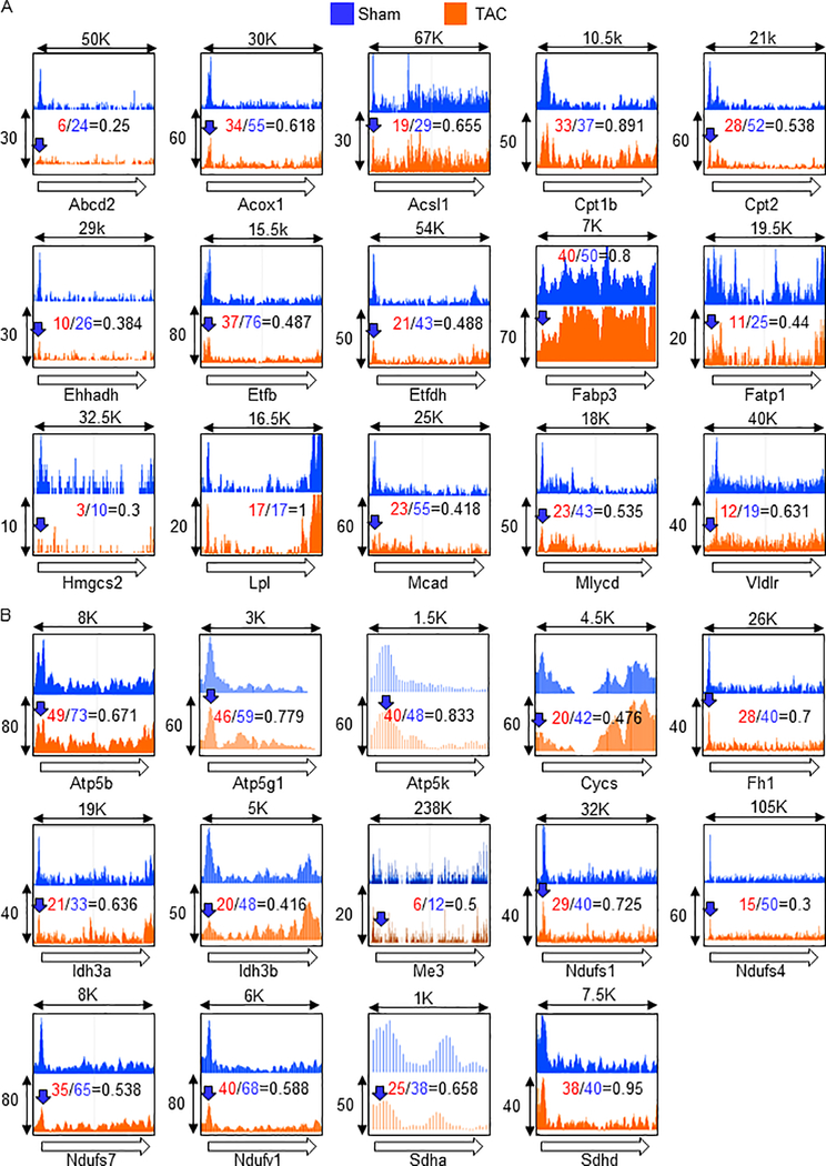
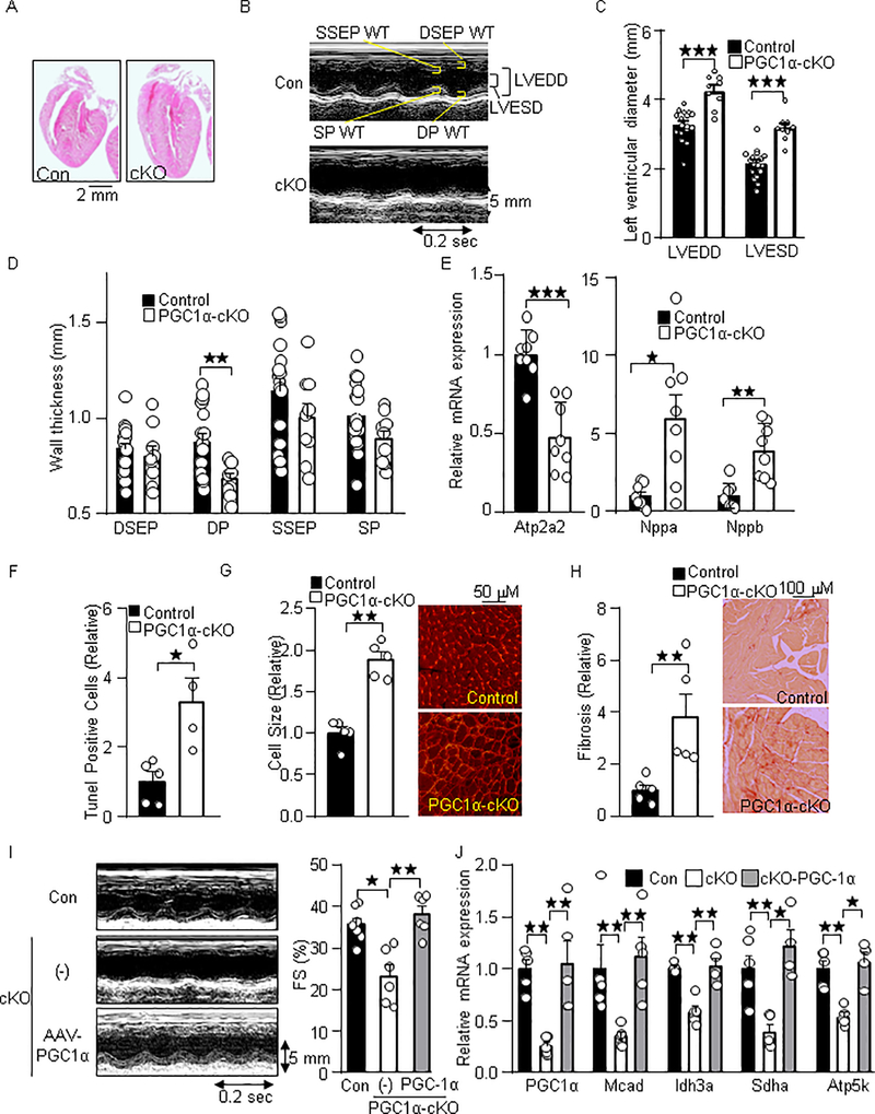
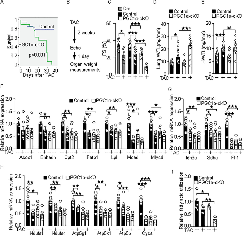
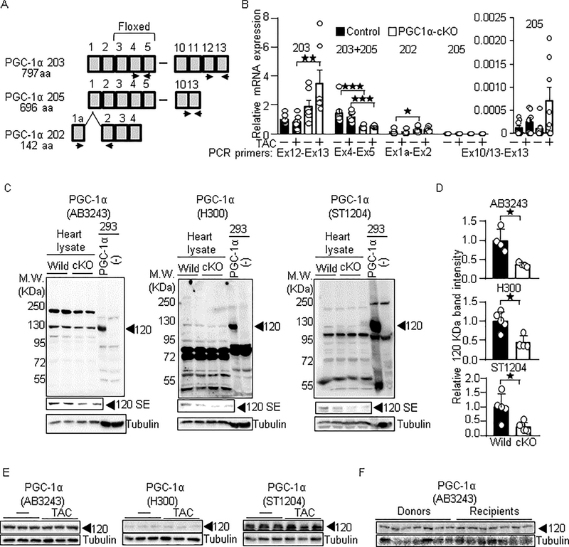
Methods and results: Chromatin immunoprecipitation-sequencing revealed that reduced promoter occupancy was a major form of polymerase II dysregulation on PGC-1α target metabolic gene promoters in the pressure-overload-induced heart failure model. PGC-1α-cKO (cardiac-specific PGC-1α knockout) mice showed phenotypic similarity to the pressure-overload-induced heart failure model in wild-type mice, such as contractile dysfunction and downregulation of PGC-1α target genes, even under basal conditions. However, the protein levels of PGC-1α were neither changed in the pressure-overload model nor in human failing hearts. Chromatin immunoprecipitation assays revealed that the promoter occupancy of polymerase II and PGC-1α was consistently reduced both in the pressure-overload model and PGC-1α-cKO mice. In vitro DNA binding assays using an endogenous PGC-1α target gene promoter sequence confirmed that PGC-1α recruits polymerase II to the promoter.

Conclusions: These results suggest that PGC-1α promotes the recruitment of polymerase II to the PGC-1α target gene promoters. Downregulation of PGC-1α target genes in the failing heart is attributed, in part, to a reduction of the PGC-1α occupancy and the polymerase II recruitment to the promoters, which might be a novel mechanism of metabolic perturbations in the failing heart.

Keywords: RNA polymerase II; chromatin; energy metabolism; heart failure.


Figures

Fig. 1.
Fig. 1.
Reduced promoter occupancy is a major form of Pol II dysregulation in metabolic gene promoters under PO conditions. ChIP-seq was performed with anti-Pol II antibody after 4 days of TAC. The Pol II occupancies in known or predicted PPAR (A) and ERR (B) target gene promoters were shown. Open arrows indicate direction of gene body, and therefore, origin of the arrow indicates promoter region, whereas arrow head indicates transcription termination site. Blue arrows indicate at least 15% reduction of a peak of Pol II occupancy in the promoter region under PO conditions. X-axis is length of genomic region (Kbp). Y-axis is relative ChIP coverage provided by Integrated Genome Browser.
Fig. 2.
Fig. 2.
Cardiac-specific loss of PGC-1α results in heart failure. (A) H&E stained images of the PGC-1α-cKO mice. (B) Representative M-mode echocardiography of the PGC-1α-cKO mice. (C) Enlarged left ventricular diameter and (D) reduced wall thickness (WT). LVEDD: LV end diastolic dimension, LVESD: LV end systolic dimension, DSEP: Diastolic septal, DP: Distolic posterior: SP: Systolic posterior, SSEP: systolic septal. (E) Heart failure marker genes expression in PGC-1α-cKO mice. The expression of indicated genes was examined by quantitative real time PCR (N=8). Atp2a2: ATPase, Ca++ transporting, cardiac muscle, slow twitch 2, Nppa: Natriuretic peptide A, and Nppb: Natriuretic peptide B. Histological analyses of myocardium in PGC-1α-cKO mice. Histochemical analyses were performed with (F) Tunel, (G) WGA, and (H) PSR staining. (I) Re-expression of PGC-1α with AAV normalizes cardiac systolic dysfunction in PGC-1α-cKO mice. Representative M-mode echocardiography (Left). Fractional shortening (Right). (J) Re-expression of PGC-1α with AAV normalizes PGC-1α target gene expression in PGC-1α-cKO mice. Echocardiographic measurements (I) and gene expression analysis (J) were performed after 3 weeks of transduction of AAV- PGC-1α and the control (GFP). The numbers of mice examined in each experimental group were: 6–17(C-D), 8 (E), 4–5 (F-H), 5–7 (I) and 4–6 (J). * p<0.05; ** p<0.01; *** p<0.001 as indicated.
Fig. 3.
Fig. 3.
PGC-1α-cKO mice are susceptible to PO-induced heart failure. (A) High mortality rate in PGC-1α-cKO mice under PO conditions. PGC-1α-cKO mice were subjected to TAC. Statistical analysis was performed with the Kaplan-Meier log rank test. (B) Timeline of echocardiography and organ weight measurements. Fractional shortening (FS) (C), lung congestion (D), cardiac hypertrophy (E), PGC-1α target genes expression (F-H), and fatty acid utilization activity (I) were examined in PGC-1α-cKO mice after 2 weeks of TAC. PGC-1α target genes involved in fatty acid metabolism (F), Krebs cycle (G) and mitochondrial ATP production (H). The numbers of mice examined in each experimental group were: 39(Wt)-29(cKO) (A), 7–17(C), 12–26 (D-E), 7–8 (F-H), and 4–9 (I). * p<0.05; ** p<0.01; *** p<0.001 as indicated.
Fig. 4.
Fig. 4.
PGC-1α is not significantly downregulated in the failing heart. (A-B) The expression of PGC-1α mRNA in PGC-1α-cKO mice. The splice isoforms of PGC-1α are shown in Ensembl (http://useast.ensembl.org/index.html). (A) Schematic representation of splice isoforms of PGC-1α and primer sets for detecting them. PGC-1α 203, canonical full length of PGC-1α, encodes 797 amino acids (aa) of protein. PGC-1α 205, lacking Ex11 to 12, encodes 696 aa of protein. PGC-1α 202 is composed of alternative Ex1 and Ex2 to 4, which encodes 142 aa of protein. (B) Relative copy number of indicated PGC-1α splice isoforms. Relative copy number of PGC-1α 203 in wild type mice under basal conditions is defined as 1. N=6–8. (C) PGC-1α specific signal in heart lysate. Western blot analyses were performed with heart lysate derived from PGC-1α-cKO and from HEK293 cells with exogenous PGC-1α expression. Anti-PGC-1α antibodies used for these analyses were Millipore AB3243, Santa Cruz H-300, and Calbiochem ST1204. SE: Short exposure. (D) Relative signal intensities of the signal at 120 KDa/tubulin shown in right panels. N=4–6. (E-F) The proteins levels of PGC-1α are not significantly downregulated in the TAC model (E) and human failing hearts (F). Donors: Healthy hearts. Recipients: Failing hearts. * p<0.05; ** p<0.01; *** p<0.001 as indicated.
Fig. 5.
Fig. 5.
PGC-1α dissociates target gene promoters in the failing heart. (A) Schematic representation of PGC-1α target gene promoters. (B-C) The promoter occupancy of PGC-1α in the target gene promoters in the PO model (B) and PGC-1α-cKO mice (C). The numbers of mice examined in each experimental group were: 4–6 (B and C) and 4–6 (D). * p<0.05; ** p<0.01 as indicated.
Fig. 6.
Fig. 6.
PGC-1α promotes Pol II recruitment. (A-B) Overexpression of PGC-1α promotes Pol II recruitment and target gene expression in cardiomyocytes. Flag-PGC-1α was overexpressed with adenovirus vector. (A) ChIP assays were performed with anti-Pol II and anti-Flag antibodies. N=3–8. (B) The expression levels of indicated genes were examined. (C) Schematic representation for the DNA binding assay. Biotin-labeled Idh3a promoter containing ERRE and TSS were incubated with cell lysate. The PGC-1α, ERRα and Pol II bound to the DNA was examined with Western blot analyses. (D) Knockdown of PGC-1α inhibits Pol II recruitment in vitro. N=4. (E) Overexpression of PGC-1α promotes Pol II recruitment. N=5. (F) The binding of PGC-1α to Pol II was not observed with co-immunoprecipitation assay. Flag-PGC-1α was overexpressed in cardiomyocytes with adenovirus vector. Co-immunoprecipitation assays were performed with anti-Flag antibody. (G) PGC-1α purified from phenylephrine (PE) treated cells has a lesser ability to bind to the promoter. Cardiomyocytes expressed Flag-PGC-1α were treated with 100 μM PE for 16 hours. Flag-PGC-1α was immunoprecipitated with anti-Flag-antibody. The immunocomplex was incubated with HEK293 cell lysate as a source of ERRs and general transcriptional machineries, biotin-labeled Idh3a promoter and HRP (horseradish peroxidase)-conjugated streptavidin. The binding of PGC-1α and biotin-labeled Idh3a promoter was measured by chemiluminescence. N=4–5. * p<0.05; ** p<0.01 as indicated.

References

    1. Sihag S, Cresci S, Li AY, Sucharov CC and Lehman JJ. PGC-1alpha and ERRalpha target gene downregulation is a signature of the failing human heart. J Mol Cell Cardiol. 2009;46:201–12. - PMC - PubMed
    1. Arany Z, Novikov M, Chin S, Ma Y, Rosenzweig A and Spiegelman BM. Transverse aortic constriction leads to accelerated heart failure in mice lacking PPAR-gamma coactivator 1alpha. Proc Natl Acad Sci U S A. 2006;103:10086–91. - PMC - PubMed
    1. Lu Z, Xu X, Hu X, Fassett J, Zhu G, Tao Y, Li J, Huang Y, Zhang P, Zhao B and Chen Y. PGC-1 alpha regulates expression of myocardial mitochondrial antioxidants and myocardial oxidative stress after chronic systolic overload. Antioxidants & redox signaling. 2010;13:1011–22. - PMC - PubMed
    1. Hu X, Xu X, Huang Y, Fassett J, Flagg TP, Zhang Y, Nichols CG, Bache RJ and Chen Y. Disruption of sarcolemmal ATP-sensitive potassium channel activity impairs the cardiac response to systolic overload. Circ Res. 2008;103:1009–17. - PMC - PubMed
    1. Karamanlidis G, Garcia-Menendez L, Kolwicz SC Jr., Lee CF and Tian R. Promoting PGC-1alpha-driven mitochondrial biogenesis is detrimental in pressure-overloaded mouse hearts. Am J Physiol Heart Circ Physiol. 2014;307:H1307–16. - PMC - PubMed

Publication types

MeSH terms

Substances