Review
. 2019:115:247-295.
doi: 10.1016/bs.apcsb.2018.10.008. Epub 2018 Dec 5.

cAMP-mediated regulation of melanocyte genomic instability: A melanoma-preventive strategy

Affiliations
Review

cAMP-mediated regulation of melanocyte genomic instability: A melanoma-preventive strategy

Nathaniel C Holcomb et al. Adv Protein Chem Struct Biol. 2019.

Abstract

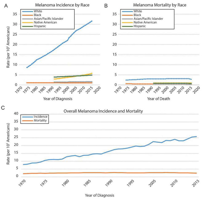
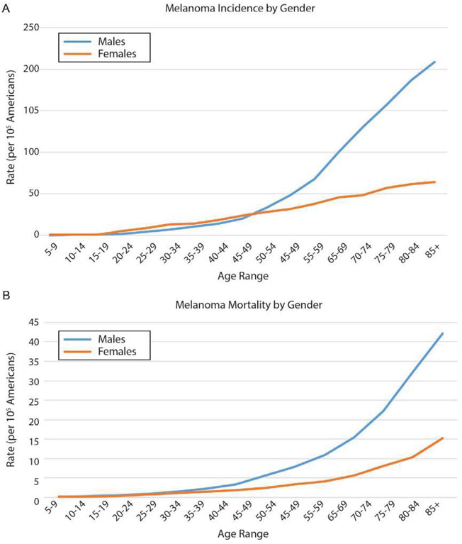
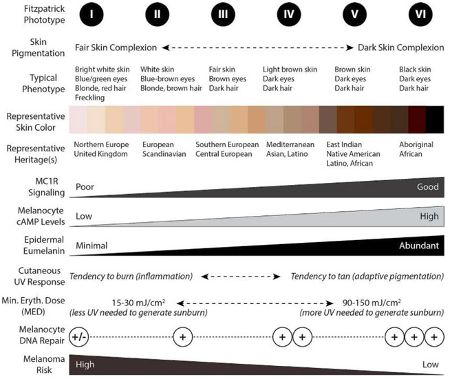
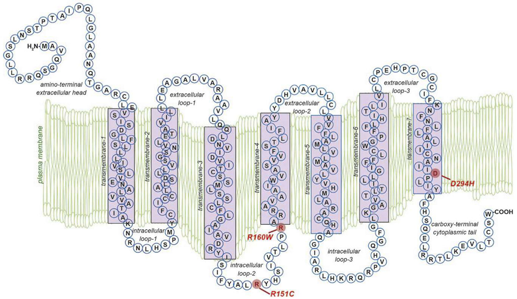
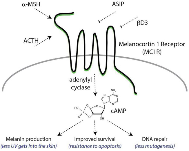
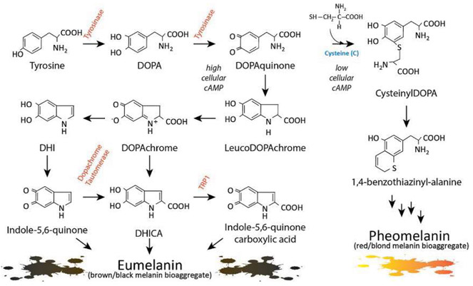
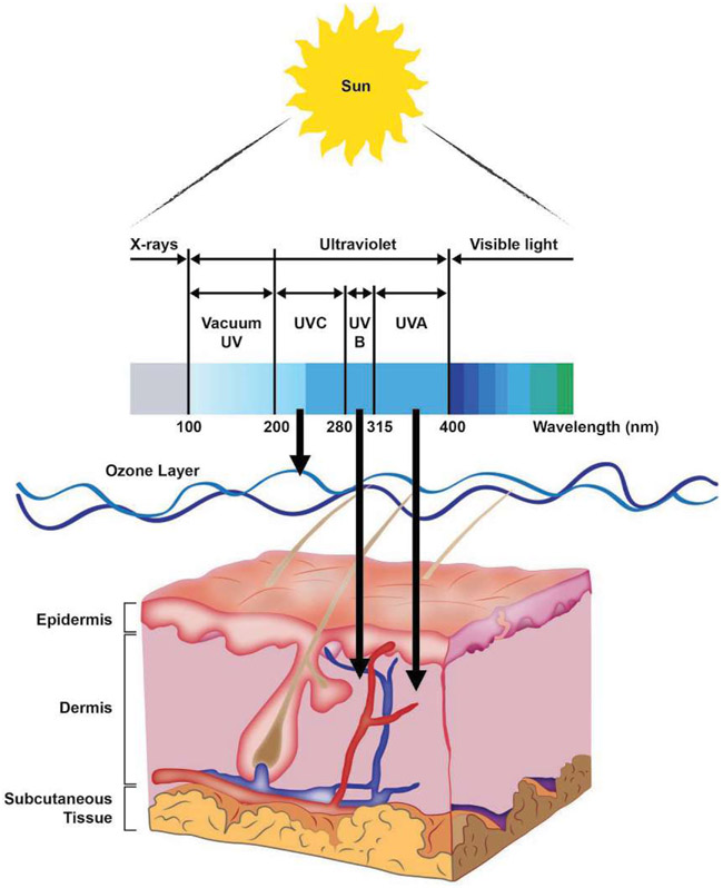
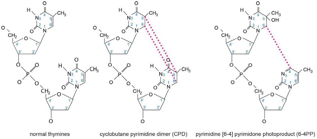
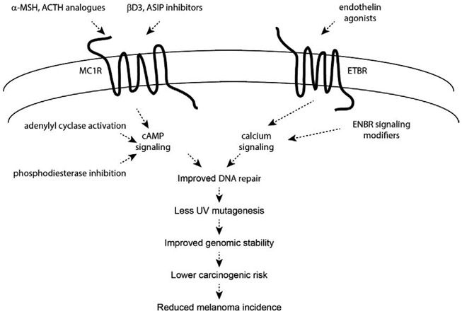
Malignant melanoma of the skin is the leading cause of death from skin cancer and ranks fifth in cancer incidence among all cancers in the United States. While melanoma mortality has remained steady for the past several decades, melanoma incidence has been increasing, particularly among fair-skinned individuals. According to the American Cancer Society, nearly 10,000 people in the United States will die from melanoma this year. Individuals with dark skin complexion are protected damage generated by UV-light due to the high content of UV-blocking melanin pigment in their epidermis as well as better capacity for melanocytes to cope with UV damage. There is now ample evidence that suggests that the melanocortin 1 receptor (MC1R) is a major melanoma risk factor. Inherited loss-of-function mutations in MC1R are common in melanoma-prone persons, correlating with a less melanized skin complexion and poorer recovery from mutagenic photodamage. We and others are interested in the MC1R signaling pathway in melanocytes, its mechanisms of enhancing genomic stability and pharmacologic opportunities to reduce melanoma risk based on those insights. In this chapter, we review melanoma risk factors, the MC1R signaling pathway, and the relationship between MC1R signaling and DNA repair.

Keywords: ATR; MC1R; Melanin; Melanoma; Mutation; Nucleotide excision repair; Risk; UV; cAMP.


Conflict of interest statement

Conflicts of interest

The authors would like to state that there are no conflicts of interest that arose during the preparation of this manuscript.

Figures

Fig. 1
Fig. 1
US incidence and mortality of cutaneous malignant melanoma by race, 1975–2015. (A) Melanoma incidence has been increasing steadily over the past four decades among non-Hispanic Caucasians, while all other ethnicities show very moderate, if any, growth in melanoma incidence. (B) Melanoma mortality has remained steady in each ethnic group in the time frame shown, with the mortality being highest among whites. (C) Overall, melanoma incidence has been increasing steadily since the early 1970s, almost exclusively a consequence of increased incidence among whites. Mortality has been effectively unchanged in the same period. Source: NCI SEER (Surveillance, Epidemiology, and End Results, http://seer.cancer.gov); rates are per 105 individuals.
Fig. 2
Fig. 2
US incidence and mortality of cutaneous malignant melanoma by gender, 2011–2015, all races and all ages. (A) Melanoma incidence increases with age in both males and females, but the rate of increase in males is considerably higher than the rate seen in females. For females, melanoma incidence increases steadily from the late teens onward, while for males the incidence is actually less than it is for females until the fourth decade of life when the incidence balloons and quickly separates from the female melanoma incidence. (B) Similarly, melanoma mortality is equivalent between younger males and females, and while male and female melanoma mortality begin to increase around the mid-40s, the rate of increase is considerably greater in males than it is in females. Source: NCI SEER (Surveillance, Epidemiology, and End Results, http://seer.cancer.gov); rates are per 105 individuals.
Fig. 3
Fig. 3
Relationship between skin complexions, Fitzpatrick scale score, epidermal melanin composition, quality of MC1R signaling, UV tanning/burn response, DNA repair and melanoma risk. Skin complexion can be described by Fitzpatrick phototype, with individuals of least pigmentation having phototype I and persons of darkest complexion having phototype VI. Though many genes determine pigmentation, skin complexion and UV responses are heavily regulated by the MC1R signaling and epidermal eumelanin composition. Robust MC1R signaling leads to induction of cAMP in melanocytes which promotes eumelanin production responsible for a vigorous tanning response (adaptive pigmentation) and better protection from subsequent UV insults. MC1R signaling also enhances melanocyte genomic stability by improving the efficiency of DNA repair. Thus, melanoma risk is heavily influenced by skin pigmentation and MC1R signaling.
Fig. 4
Fig. 4
Physical structure of the MC1R protein. The MC1R is a 7-transmembrane Gs protein-coupled receptor (GPCR). Extracellular and transmembrane domains engage MC1R ligands while intracellular and transmembrane domains regulate adenylyl cyclase interactions and signaling. Residues identified in red include the common polymorphisms of the “RHC” phenotype that significantly reduce MC1R function and lead to reduced cAMP signaling and preferential production of the red/blonde pheomelanin pigment.
Fig. 5
Fig. 5
Complexity of native MC1R signaling. MC1R signaling is regulated by cognate agonists and antagonists. Activators of MC1R include the high-affinity melanocortin ligands alpha melanocyte stimulating hormone (α-MSH) and adrenocorticotropic hormone (ACTH). α-MSH and ACTH are generated in response to UV-induced DNA damage in keratinocytes. MC1R agonists stimulate MC1R to activate adenylyl cyclase and generate c-AMP. Downstream responses to c-AMP signaling include increased synthesis of melanin pigment, increased cellular survival through resistance to apoptosis, and increased efficiency of DNA repair to reduce mutagenic potential of UV-induced photodamage. MC1R signaling is antagonized by agouti signaling protein (ASIP) and β-defensin 3 (βD3).
Fig. 6
Fig. 6
Melanin synthesis. There are two main types of melanin: the dark brown/black UV-protective eumelanin and the red/blonde sulfated pheomelanin pigment. Each is derived from progressive cyclization and oxidation of the amino acid tyrosine. Tyrosinase, the rate-limiting enzyme for melanogenesis, catalyzes the first two stages of melanin biosynthesis. When MC1R is functional and cAMP levels are high, melanocytes produce eumelanin preferentially (bottom left section). In contrast, when MC1R is dysfunctional and cAMP levels are low, cysteine is incorporated and pheomelanin is made instead (right section). Inherited deficiencies of melanogenic enzymes such as tyrosinase, dopachrome tautomerase, and tyrosinase-related protein-1 are associated with hypopigmentary disorders of albinism.
Fig. 7
Fig. 7
UV radiation and the skin. The ultraviolet (UV) region of the electromagnetic spectrum of light can be divided into three components, UV-A, UV-B and UV-C, based on photon wavelength. The most energetic of these is UV-C, which has the shortest wavelength. However, the ozone layer of the atmosphere absorbs essentially all UV-C, and as a result ambient solar UV exposure is predominantly UV-A (90–95%) and UV-B (5–10%). Longer wavelength UV-A penetrates deeply into the skin, reaching well into the dermis. In contrast, UV-B affects primarily the epidermis. UVA is efficient at generating reactive oxygen species that damage DNA through oxidative base modifications. UV-B is directly absorbed by pyrimidine bases in DNA to produce photoproducts. Mutations and cancer can result from UV-generated changes to DNA if left unrepaired.
Fig. 8
Fig. 8
Chemical structures of UV photoproducts. When UV-light strikes DNA at dipyrimidine nucleotides, such as the thymine dimer shown on the left, there are two common DNA lesions formed. First, the 5′ and 6′ positions of the pyrimidine ring structures can form a cyclobutane ring between the adjacent thymines, generating a DNA lesion referred to as a cyclobutane pyrimidine dimer (CPD) as shown in the middle panel. This lesion is the less helically distorting, and therefore more poorly repaired, of the two adducts shown, and is the most commonly generated adduct when DNA is exposed to UV-light. The second most commonly generated DNA adduct is the pyrimidine [6–4] pyrimidone photoproduct (6–4PP), a lesion formed when the 6′ carbon of one thymine covalently binds the 4′ carbon of an adjacent thymine, as shown in the panel on the right. The 6–4PP is considerably more helically distorting than the CPD lesion, and is therefore more readily recognized by NER, more rapidly repaired, and consequently less mutagenic than a CPD lesion.
Fig. 9
Fig. 9
Potential fates of UV-damaged DNA. UV-photodamage, in this case a CPD lesion, predominantly occur at TCG and CCG sequences when the middle cytosine nucleotide is methylated as shown. Whether the CPD will generate a mutation depends on whether it will be resolved before the next round of cellular replication. The CPD may be repaired efficiently by NER, resulting in a return to the initial sequence in an error free manner. Additionally, damage tolerant polymerases such as DNA polymerase η may synthesize across the CPD adduct in an error free manner, allowing more time for NER machinery to remove the adduct. However, damage tolerant polymerases have lower fidelity than traditional replicative polymerases, and when a damage tolerant polymerase adds a nucleotide incorrectly across from a methylated cytosine that is part of a CPD adduct, the most commonly incorporated nucleotide is A. After misincorporation of this A, NER would use it as the template to replace the adducted C, and instead would incorporate T, generating a C-T transition mutation. Finally, methylated cytosines that are part of a CPD lesion are prone to deamination. This deamination produces a T, and if the deaminated T in the CPD is bypassed “correctly” by DNA polymerase η, a C to T transition will occur. NER is still required to remove the CPD adduct, but the mutation is already fixed.
Fig. 10
Fig. 10
Overview of nucleotide excision repair (NER). Helically distorting DNA damage, such as that produced by UV light is repaired by the NER pathway, which can be subdivided into global genome NER (GG-NER) and transcription-coupled NER (TC-NER). GG-NER occurs anywhere in the genome and is initiated by recognition of helical-distorting DNA damage such as a cyclobutane pyrimidine dimer (CPD). In GG-NER, XPC, as part of a multiprotein complex including RAD23B, serves as the primary recognition factor. XPC is further assisted by the UV damage-binding protein 2 (DDB2) to specifically recognize CPD adducts. Without DDB2, CPDs are poorly recognized by XPC. In contrast, TC-NER is initiated by the stalling of RNA polymerase at a CPD present on the template strand of an actively transcribed gene and involves the cofactors CSA and CSB. TC-NER and GG-NER only differ in their first step, damage recognition; the pathways converge downstream into a common NER mechanism beginning with pre-incision complex formation. In both GG-NER and TC-NER, TFIIH (a multi-complex protein containing the XPB and XPD helicases as well as eight other subunits) is recruited to the site of damage. Using its XPB and XPD helicase components, TFIIH unwinds the DNA around the photolesion, initiating strand separation to enable the recruitment of other NER factors to form a “pre-incision complex.” XPA, replication protein A (RPA), XPG, XPF, and ERCC1 are each recruited to the complex, and at some point, initiation factors exit the region. Next, there is incision of the photolesion-containing strand some distance away from the DNA lesion. Strand incision is accomplished by XPF-ERCC1 working in complex to cleave the strand 5′ to the damage and by XPG 3′ to the damage. Evidence suggests that 5′ strand cleavage may actually occur before 3′ strand cleavage, and in fact, DNA polymerase may begin filling in the gap from the 5′ side before the 3′ incision step. After 5′ and 3′ strand incision, a 24-32mer oligonucleotide harboring the photolesion is generated that is removed (associated with TFIIH) from chromatin. The resultant gap is then filled in by DNA polymerases together with PCNA, RFC, and RPA using the undamaged sister strand to ensure fidelity of repair. Finally, DNA ligation is achieved by DNA ligases I, III or XRCC1 to complete the process.
Fig. 11
Fig. 11
Model of cAMP-enhanced NER. With UV exposure, ATR (ataxia telangiectasia and rad3-related) is activated and physically interacts with AKAP12 (A kinase anchoring protein 12). ATR phosphorylates AKAP12 on the S732 moiety, an event required for nuclear translocation of the complex. With MC1R (melanocortin 1 receptor) interactions with high-affinity agonists (e.g., alpha melanocyte stimulating hormone; α-MSH), AC (adenylyl cyclase) is activated and the second messenger cAMP is formed. cAMP-dependent protein kinase (protein kinase A; PKA) is activated, interacts with AKAP12 and ATR and phosphorylates ATR on the S435 residue. This post-translational modification accelerates and enhances association of the complex with the core NER factor XPA (xeroderma pigmentosum A protein) and together the ATR/AKAP12/XPA translocates to photodamage in the chromatin. Activation of this pathway results in more robust 5′ strand incision to optimize NER.
Fig. 12
Fig. 12
Melanoma-preventive strategies based on enhancing melanization and DNA repair in melanocytes. Individuals with inherited MC1R signaling defects are at increased risk of melanoma because of ineffective epidermal melanization and sub-optimal nucleotide excision repair (NER), leading to more UV penetration into the skin and less capacity to reverse mutagenic photodamage. Discovering that melanocyte NER can be improved by MC1R and ETBR signaling pathways offers opportunities to regulate DNA repair and genomic stability pharmacologically. MC1R and ETBR signaling could be targeted by agonists or inhibitors of antagonists, but may not be effective in persons carrying defective MC1R receptors. Alternatively, signaling pathways could be pharmacologically manipulated (e.g., adenylyl cyclase activation or phosphodiesterase inhibition in the case of cAMP signaling). If effective approaches can be developed to improve melanocyte UV resistance, these would theoretically reduce melanoma risk by reducing the mutagenic burden of UV.

Similar articles

Cited by

References

    1. Abdel-Malek ZA, Ruwe A, Kavanagh-Starner R, Kadekaro AL, Swope V, Haskell-Luevano C, et al. (2009). Alpha-MSH tripeptide analogs activate the melanocortin 1 receptor and reduce UV-induced DNA damage in human melanocytes. Pigment Cell & Melanoma Research, 22(5), 635–644. 10.1111/j.1755-148X.2009.00598.x. - DOI - PubMed
    1. Abdel-Malek ZA, Scott MC, Furumura M, Lamoreux ML, Ollmann M, Barsh GS, et al. (2001). The melanocortin 1 receptor is the principal mediator of the effects of agouti signaling protein on mammalian melanocytes. Journal of Cell Science, 114, 1019–1024 Pt. 5. - PubMed
    1. Abdel-Malek Z, Scott MC, Suzuki I, Tada A, Im S, Lamoreux L, et al. (2000). The melanocortin-1 receptor is a key regulator of human cutaneous pigmentation. Pigment Cell Research, 13(Suppl. 8), 156–162. - PubMed
    1. Abdel-Rahman MH, Pilarski R, Massengill JB, Christopher BN, Noss R, & Davidorf FH (2011). Melanoma candidate genes CDKN2A/p16/INK4A, p14ARF, and CDK4 sequencing in patients with uveal melanoma with relative high-risk for hereditary cancer predisposition. Melanoma Research, 21(3), 175–179. 10.1097/CMR.0b013e328343eca2. - DOI - PubMed
    1. Adimoolam S, & Ford JM (2002). p53 and DNA damage-inducible expression of the xeroderma pigmentosum group C gene. Proceedings of the National Academy of Sciences of the United States of America, 99(20), 12985–12990. - PMC - PubMed

Publication types