. 2019 Apr 2;27(4):660-668.e4.
doi: 10.1016/j.str.2019.01.012. Epub 2019 Feb 21.

A Conserved Basic Patch and Central Kink in the Nipah Virus Phosphoprotein Multimerization Domain Are Essential for Polymerase Function

Affiliations

A Conserved Basic Patch and Central Kink in the Nipah Virus Phosphoprotein Multimerization Domain Are Essential for Polymerase Function

Jessica F Bruhn et al. Structure. .

Abstract

Nipah virus is a highly lethal zoonotic pathogen found in Southeast Asia that has caused human encephalitis outbreaks with 40%-70% mortality. NiV encodes its own RNA-dependent RNA polymerase within the large protein, L. Efficient polymerase activity requires the phosphoprotein, P, which tethers L to its template, the viral nucleocapsid. P is a multifunctional protein with modular domains. The central P multimerization domain is composed of a long, tetrameric coiled coil. We investigated the importance of structural features found in this domain for polymerase function using a newly constructed NiV bicistronic minigenome assay. We identified a conserved basic patch and central kink in the coiled coil that are important for polymerase function, with R555 being absolutely essential. This basic patch and central kink are conserved in the related human pathogens measles and mumps viruses, suggesting that this mechanism may be conserved.

Keywords: Nipah virus; RNA-dependent RNA polymerase; X-ray crystallography; coiled coil; differential scanning calorimetry; henipavirus; minigenome; oligomerization; phosphoprotein.


Conflict of interest statement

DECLARATION OF INTERESTS

The authors declare no competing interests.

Figures

Figure 1.
Figure 1.. Nipah Virus Phosphoprotein Organization
(A) NiV P is comprised of modular domains (boxes) connected by disordered linkers (lines). The N-terminal region (1–38) facilitates chaperoning of N0 (Yabukarski et al., 2014). Residues 475–578 comprise the P multimerization domain (PMD). Residues 660–709 comprise the C-terminal X domain (XD), which binds the nucleocapsid (Chan et al., 2004). (B) The crystal structure of the wild-type NiV PMD (PDB ID: 4N5B) is shown here in cartoon form with the N-terminal cap in yellow and the coiled-coiled region in blue. (C) The amino acid sequence of the PMD is listed in capital letters with alpha helices designated by shaded boxes (yellow for the N-terminal cap and blue for the coiled-coil domain). The register of the heptad repeats comprising the coiled-coil region is annotated with lower-case letters. Knob residues (a and d) are indicated in bold. Residues mutated in this study are marked with triangles and residues deleted in this study are marked with a black bar.
Figure 2.
Figure 2.. The N-terminal Water Pocket
(A) A water-filled pocket was observed in the center of the NiV P coiled-coil domain towards its N-terminus (PDB ID: 4N5B) (Bruhn et al., 2014). This water pocket is mediated by glycine 519 facing the interior of the coil, which creates empty space. This is further facilitated by hydrophilic residues S515 above and N522 below G519 which interact with the water molecules. Distances between side-chain oxygens and water molecules are indicated. (B) A sequence alignment across henipavirus P proteins surrounding the water pocket demonstrates that this pocket is probably conserved between Nipah and Hendra viruses, but is not conserved across the other viruses. Water pocket-mediating residues are in bold and conserved residues are highlighted. The heptad register is also indicated. Viruses included are Nipah virus (NiV), Hendra virus (HeV), Cedar virus (CedPV), Bat paramyxovirus Eid_hel/GH-M74a/GHA/2009 Kumasi virus (KV) (Drexler et al., 2009), and Mojang virus (MojV). (C) The high-resolution crystal structure of the mutant G519N is shown here. This mutation did not disrupt the overall fold of this protein and there are no waters present in the interior of the coiled coil. (D) An image of the electron density around reside 519 showing that there are no water molecules present. (2mFo-DFc map, σ = 1.0).
Figure 3.
Figure 3.. The Central Kink
A kink is observed in the center of the tetramerization domain of three of the four paramyxovirus P proteins that have been structurally characterized to date. (A) These kinked structures are shown for comparison: NiV (PDB ID: 4N5B), mumps virus (MuV) (PDB ID: 4EIJ), and measles virus (MeV) (PDB ID: 3ZDO). In NiV P, this kink is mediated by an insertion of three amino acids that break the otherwise perfect heptad repeat, as well as a proline that allows for a bend in the helix. This kink is indicated with an arrow. (B) A sequence alignment of this region demonstrates that this kink is conserved across most henipaviruses. Conserved residues are highlighted in orange. The inserted residues, 541–543, and proline 544 are shown in bold, and the heptad register is annotated. Viruses included are Nipah virus (NiV), Hendra virus (HeV), Cedar virus (CedPV), Bat paramyxovirus Eid.hel/GH45/2008 Kumasi virus (KV) (Drexler et al., 2009)and Mojang virus (MojV). (C) A deletion of three residues, including the proline, (Δ542–544) allows for the continuation of a perfect heptad repeat. This perfect heptad is annotated. (D) The crystal structure of Δ542–544 reveals that this kink has been removed without altering the overall architecture of this domain.
Figure 4.
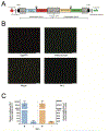
Figure 4.. Construction and characterization of a NiV bicistronic minigenome (bMG)
(A) Schematic of the NiV bicistronic minigenome (bMG) produced in cells expressing T7 polymerase. Large gray squares indicate respective NiV N and P gene 5′ and 3′ untranslated regions (UTRs) flanking the reporter protein coding sequences. NiV genomic leader, trailer, and intergenic (IG) regions are indicated by arrows. Gray hourglass shapes indicate Hepatitis Delta virus (HDV) and Hammerhead ribozymes. Black arrowheads indicate porcine techovirus-1 2A (p2A) ribosomal skipping site inserted between luciferase and fluorescent proteins in each respective transcription unit (TU). T7 promoter and terminator sequences are indicated by a green arrow and a red octagon respectively. (B) Fluorescence micrographs of BSRT7/5 cells transfected with NiV bMG and wild-type NiV N, P, and L expression plasmids at 48 h post-transfection. The top left panel shows red fluorescence protein (TagRFP) expression from TU1. The top right panel shows green fluorescent protein (mNeonGreen) expression from TU2. The bottom left panel shows a merged image of the top two panels showing co-localization of red and green fluorescence signals (indicated by a lighter yellow-green color). The right panel shows the absence of fluorescent protein expression in a control experiment lacking the NiV L plasmid. (C) Relative luminescent units (RLU) expressed from Gaussia luciferase (TU1, left side) and Nanoluciferase (TU2, right side) of the NiV bMG at 48 h post-transfection with wild-type NiV P. Error bars delineate standard deviation of the mean of biological triplicate samples.
Figure 5.
Figure 5.. Importance of Conserved Structural Feature in the NiV PMD for Polymerase Function
(A) Wild-type and mutant NiV P proteins were tested in NiV bMG assays. Activity was normalized to wild-type P. DRK is a triple mutant that includes D554A, R555A, and K559A. Activity on the left side of graph indicate percent Gaussia luciferase activity from TU1, while the right side of the graph indicates percent Nanoluciferase activity from TU2. The graph represents a compilation of four independent experiments performed either in biological duplicate or triplicate. Error bars delineate standard deviation from the mean. P values express statistical significance in comparison to values derived from wild-type NiV P samples. (B) A western blot of cell lysates from the bMG assay with detection of wild-type and mutants NiV P, and GAPDH as a control.
Figure 6.
Figure 6.. The Conserved Central Basic Patch
All PMDs of paramyxovirus P proteins that have been structurally characterized to date contain a central basic patch. (A) The electrostatic surfaces of these protein domains were calculated using APBS (Konecny et al., 2012) and are colored according to charge, with blue representing positive charge and red representing negative charge: NiV (PDB ID: 4N5B), Sendai virus (SeV) (PDB ID: 1EZJ), MuV (PDB ID: 4EIJ), and MeV (PDB ID: 3ZDO). The NiV basic patch is indicated with a box. (B) A zoomed-in view of the central basic patch is drawn here. Important charged residues mutated in this study are labeled and shown as sticks. (C) A sequence alignment of this basic patch is compared across henipaviruses. The heptad repeat is indicated and residues facing the interior of the coil are colored grey. Charged residues are indicated with blue or red boxes according to charge. Mutated residues are indicated with triangles. Viruses included are Nipah virus (NiV), Hendra virus (HeV), Cedar virus (CedPV), Bat paramyxovirus Eid_hel/GH-M74a/GHA/2009 Kumasi virus (KV) (Drexler et al., 2009), and Mojang virus (MojV).
Figure 7.
Figure 7.. Thermal Stability of Structural Mutants G519N and Δ542–544
Normalized molar heat capacity (Cp) is plotted over a range of 25 to 130°C for wild type (Bruhn et al., 2014), G519N and Δ542–544 NiV PMD.

References

    1. Adams PD, Afonine PV, Bunkoczi G, Chen VB, Davis IW, Echols N, Headd JJ, Hung LW, Kapral GJ, Grosse-Kunstleve RW, et al. (2010). PHENIX: a comprehensive Python-based system for macromolecular structure solution. Acta Crystallogr D Biol Crystallogr 66, 213–221. - PMC - PubMed
    1. Blanc E, Roversi P, Vonrhein C, Flensburg C, Lea SM, and Bricogne G (2004). Refinement of severely incomplete structures with maximum likelihood in BUSTER-TNT. Acta crystallographica Section D, Biological crystallography 60, 2210–2221. - PubMed
    1. Bowman MC, Smallwood S, and Moyer SA (1999). Dissection of individual functions of the Sendai virus phosphoprotein in transcription. J Virol 73, 6474–6483. - PMC - PubMed
    1. Bruhn JF, Barnett KC, Bibby J, Thomas JM, Keegan RM, Rigden DJ, Bornholdt ZA, and Saphire EO (2014). Crystal structure of the nipah virus phosphoprotein tetramerization domain. J Virol 88, 758–762. - PMC - PubMed
    1. Buchholz UJ, Finke S, and Conzelmann KK (1999). Generation of bovine respiratory syncytial virus (BRSV) from cDNA: BRSV NS2 is not essential for virus replication in tissue culture, and the human RSV leader region acts as a functional BRSV genome promoter. J Virol 73, 251–259. - PMC - PubMed

Publication types

MeSH terms

LinkOut - more resources