. 2019 Mar 25;48(6):827-839.e9.
doi: 10.1016/j.devcel.2019.01.020. Epub 2019 Feb 21.

Assessment and Maintenance of Unigametic Germline Inheritance for C. elegans

Affiliations

Assessment and Maintenance of Unigametic Germline Inheritance for C. elegans

Karen L Artiles et al. Dev Cell. .

Abstract

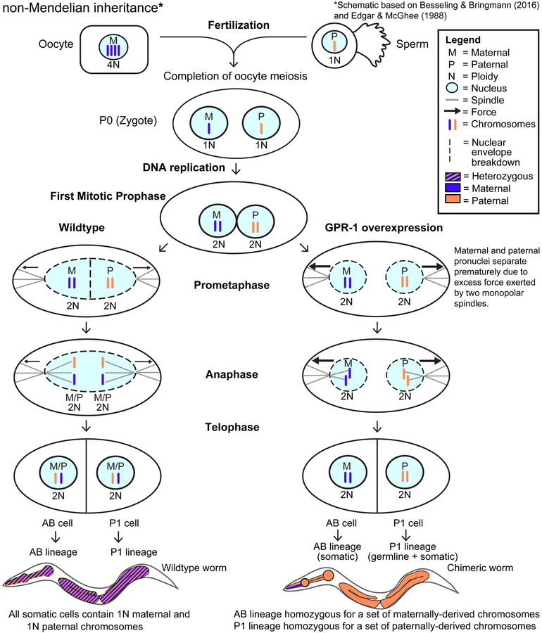
The recent work of Besseling and Bringmann (2016) identified a molecular intervention for C. elegans in which premature segregation of maternal and paternal chromosomes in the fertilized oocyte can produce viable animals exhibiting a non-Mendelian inheritance pattern. Overexpression in embryos of a single protein regulating chromosome segregation (GPR-1) provides a germline derived clonally from a single parental gamete. We present a collection of strains and cytological assays to consistently generate and track non-Mendelian inheritance. These tools allow reproducible and high-frequency (>80%) production of non-Mendelian inheritance, the facile and simultaneous homozygosis for all nuclear chromosomes in a single generation, the precise exchange of nuclear and mitochondrial genomes between strains, and the assessments of non-canonical mitosis events. We show the utility of these strains by demonstrating a rapid assessment of cell lineage requirements (AB versus P1) for a set of genes (lin-2, lin-3, lin-12, and lin-31) with roles in C. elegans vulval development.

Keywords: C. elegans; genetic engineering; inheritance; mitosis; mosaic; non-Mendelian; synthetic biology.


Conflict of interest statement

Declaration of Interests

The authors declare no competing interests.

Figures

Figure 1
Figure 1. Overexpression of the microtubule force regulator GPR-1 induces non-Mendelian inheritance.
Diagram of zygotic division in wildtype (Edgar and McGhee, 1988) and GPR-1 overexpressing (OE) cells (Besseling and Bringmann, 2016). In wildtype, fertilization combines the oocyte and sperm to form the zygote (P0 cell). As meiosis completes, 3N DNA content is lost from the maternal pro-nucleus by polar body extrusion. The prophase zygote contains two pronuclei, containing 1N paternal (P, orange) or 1N maternal (M, blue) chromosomes, respectively. During S phase, chromosomes are duplicated, resulting in 2N DNA content in each pronucleus in the form of sister chromatids. In wildtype zygotes, the duplicated chromosomes are arranged on the metaphase plate. During anaphase, sister chromatids are separated resulting in one copy of each parental chromosome being inherited by each daughter cell. This zygotic division yields the two founder cells of the P1 and AB lineages. As expected from Mendelian inheritance, every somatic cell in wildtype animals contains a full complement of paternal (1N) and maternal (1N) chromosomes. In contrast, in the GPR-1 overexpression strain additional force is exerted on pronuclei which results in premature separation during prometaphase. This results in maternal and paternal chromosomes remaining at opposite poles thus preventing proper chromatid segregation. GPR-1(OE) worms that have undergone abnormal chromosome partitioning become chimeric, with a P1 lineage that is homozygous for paternal chromosomes and an AB lineage homozygous for maternal chromosomes. Because the P1 lineage gives rise to the germline, all oocytes and sperm from a GPR-1(OE) animal contain identical, paternally-derived chromosomes.
Figure 2
Figure 2. Developing an easily tractable system for non-Mendelian chromosomal inheritance by stable GPR-1 overexpression and pharyngeal muscle fluorescence.
(A) A silencing-resistant GPR-1 transgene, expressed under a germline-specific promoter (Pmex-5), was tagged with GFP at the N-terminus. PATC-rich DNA segments, which can counteract germline silencing, were incorporated into the transgene and are indicated as black bars (Frøkjaer-Jensen et al., 2016). A PATC-rich 3’ UTR from smu-1 was also included. (B) Lineages of pharyngeal muscle (PM) cells, adapted from Wormbook (Mango, 2007). PM1, PM2 muscle rings are derived entirely from the AB cell lineage (blue). Muscle ring PM3 consists of three lobes, two derived from the AB-lineage (blue) and one from the P1-lineage (orange). PM4 and PM5 consist of multinucleate cells resulting from the fusion of both AB and P1-derived cells (yellow). PM6 and PM7 consist entirely of P1-derived cells (orange). (C) Simplified schematic of fluorescently expressing AB, P1, and fused cells as viewed under a dissection microscope. Note that the fluorescence intensity in fused cells depends on the proportion of nuclei from the AB versus P1 lineage. For example, in the AB-derived pattern, expression is limited to 1/6 nuclei in PM4 and 2/6 nuclei in PM5, resulting in partial expression and dim fluorescence. The fainter fluorescence in these compartments can be difficult to visualize. Also, PM1 is not indicated in the schematic as it is hard to observe at low magnification fluorescence microscopy.
Figure 3
Figure 3. Pharyngeal myo-2p::mCherry patterns in GPR-1(OE) crosses
(A) We crossed GPR-1 overexpressing hermaphrodites (GPR-1(OE), PD1594) to males with pharyngeal mCherry (myo-2p::mCherry, VS21) and characterized resulting classes of hermaphroditic F1 cross-progeny by fluorescence. Chromosome schematics indicate classic Mendelian segregation with maternal and paternal DNA “mixing” (left) and non-Mendelian segregation with no DNA mixing (right). Chromosomes are indicated by grey bars and genotype is indicated by grey (wildtype) or colored (blue = GPR-1(OE), red = myo-2p::Cherry) boxes. F1 segregation (see Table 2A: Mendelian = 18% (± 5%), Non-Mendelian (Maternal DNA-> P1 lineage) = 2% (± 1%), and Non-Mendelian (Paternal DNA-> P1 lineage) = 80% (± 6%), mean ± SEM (N = 4 crosses)). F2 self-progeny of four Mendelian heterozygous F1s were also classified and quantified (Table 2B: full pharynx 35% ± 4%, P1 lineage only 19% ± 2%, AB lineage only 21% ± 2%, no fluorescence 25% ± 3%). (B-C) Photomicrographs of mCherry expression in the pharynx of F1 hermaphrodite cross progeny: (B) full pharyngeal expression (C) Top: AB-derived pharyngeal expression. We note that faint mCherry expression in PM4 and PM5 is expected, as only 3 of 12 nuclei are derived from AB lineage. Bottom: P1-derived pharyngeal expression. (D) A typical cross using a GPR-1(OE) toolkit strain to analyze expression of Your Favorite Mutation/gene (YFM) in chimeric worms exemplified here by using myo-2p::mCherry as YFM. We crossed pharyngeal GFP marked GPR-1 overexpressing hermaphrodites (myo-2p::GFP; GPR-1(OE), PD2220) with myo-2p::mCherry homozygous males (YFM). Left: Hermaphroditic “Mendelian” cross progeny uniformly expressing both GFP and mCherry throughout their pharynxes (18%). Right: Hermaphroditic “non-Mendelian” cross progeny, which have presumably undergone an abnormal zygotic division resulting in maternally derived chromosomes in the AB cell lineage and paternally derived chromosomes in the P1 cell lineage (80%), expressing GFP in all AB-derived pharyngeal tissue. Half of these chimeric animals also expressed paternally inherited mCherry exclusively in their P1-derived pharyngeal tissue. Expected % of hermaphroditic cross progeny of each class are shown. (E) Fluorescence microscopy showing the pharyngeal pattern of F1 non-Mendelian cross progeny from (D). Top. mCherry is expressed throughout the P1-derived pharyngeal cells. GFP is expressed throughout the AB-derived pharyngeal cells. Bottom. Merged fluorescent images and DIC image overlay.
Figure 4
Figure 4. Analysis of vulval development mutants
(A) Vulval development relies in part on communication between the Anchor Cell (AC) and the Vulval Precursor Cells (VPCs) via EGF-like and Notch-like signaling pathways. (B-D) Cell lineage diagrams illustrating how (B) VPCs and the AC/VU precursors are derived from the two lineages (AB and P1) resulting from division of the zygote. (C) Cell genotypes in AB/P1 mosaics with a mutation restricted to the P1 lineage will include mutant AC/VU precursors and wild-type VPCs and (D) conversely, cell genotypes in AB/P1 mosaics with a mutation restricted to the AB lineage will include wild-type AC/VU precursors and mutant VPCs (E) A summary of phenotypes observed when the listed vulval development mutation was present in either the P1 lineage (including AC/VU precursors) or the AB lineage (including VPCs). Gene, allele used, previously inferred/expected role, expected lineage of action, and phenotype observation (aggregate number of worms exhibiting stated phenotype/number examined @23°C and corresponding percentage) is listed. Additional data and details, including the crosses used to generate these chimeric worms, are described in Figure S1 and Table S1.
Figure 5
Figure 5. Nuclear/mitochondria genome exchange and one-step isogenesis through non-canonical genetics.
(A) Experimental schematic. We generated nuclear/mitochondrial genome hybrid strains by crossing fluorescently marked GPR-1(OE) strains (either PD2217 or PD2218) with a wild strain (PX174) containing many polymorphisms relative to the laboratory N2 strain. Non-Mendelian F1 progeny were selected based on AB-specific expression of a maternally derived pharyngeal fluorescence reporter (either myo-2::GFP or myo-2::mCherry). Hybrid strains were generated by picking self-progeny. Hybrid strains (PD2231, PD2232, PD2233, PD2234), predicted to have a nuclear genome entirely derived from the male parent and a mitochondrial genome entirely derived from the maternal parent were sequenced along with the parent strains (PX174, PD2217, PD2218). Note that sperm-derived paternal mitochondria, which are rapidly eliminated by autophagy after fertilization (Sato and Sato, 2011), were omitted from the schematic for clarity. (B) SNPs unique to strain PX174 (relative to N2 reference strain VC2010) were determined using a Kmer-based approach. For each parental and hybrid strain, the number of sequencing reads matching the paternal (orange) and maternal (blue) base at each SNP position along the chromosome are plotted.

Comment in

References

    1. Adalsteinsson BT, and Ferguson-Smith AC (2014). Epigenetic control of the genome-lessons from genomic imprinting. Genes (Basel) 5, 635–655. - PMC - PubMed
    1. Anderegg C, and Markert CL (1986). Successful rescue of microsurgically produced homozygous uniparental mouse embryos via production of aggregation chimeras. Proc. Natl. Acad. Sci. U.S.A. 83, 6509–6513. - PMC - PubMed
    1. Bagijn MP, Goldstein LD, Sapetschnig A, Weick E-M, Bouasker S, Lehrbach NJ, Simard MJ, and Miska EA (2012). Function, targets, and evolution of Caenorhabditis elegans piRNAs. Science 337, 574–578. - PMC - PubMed
    1. Batista PJ, Ruby JG, Claycomb JM, Chiang R, Fahlgren N, Kasschau KD, Chaves DA, Gu W, Vasale JJ, Duan S, et al. (2008). PRG-1 and 21U-RNAs Interact to Form the piRNA Complex Required for Fertility in C. elegans. Molecular Cell 31, 67–78. - PMC - PubMed
    1. Bean CJ, Schaner CE, and Kelly WG (2004). Meiotic pairing and imprinted X chromatin assembly in Caenorhabditis elegans. Nat. Genet 36, 100–105. - PMC - PubMed

Publication types

MeSH terms