. 2019 Oct 15;18(4):276-285.
doi: 10.2463/mrms.tn.2018-0066. Epub 2019 Feb 25.

Improvement of Signal Inhomogeneity Induced by Radio-frequency Transmit-related Phase Error for Single-step Quantitative Susceptibility Mapping Reconstruction

Affiliations

Improvement of Signal Inhomogeneity Induced by Radio-frequency Transmit-related Phase Error for Single-step Quantitative Susceptibility Mapping Reconstruction

Hirohito Kan et al. Magn Reson Med Sci. .

Abstract

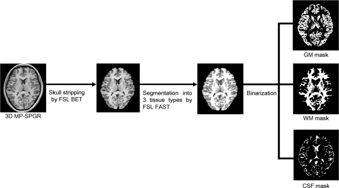
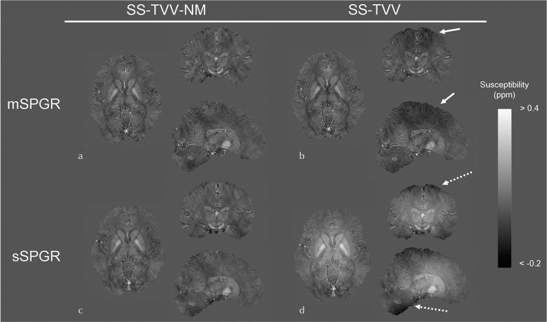
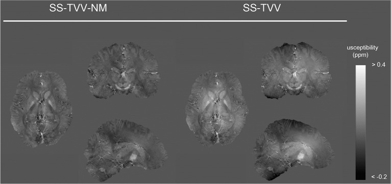
To mitigate the susceptibility inhomogeneity induced by radio-frequency transmit phase error through the whole brain in quantitative susceptibility mapping (QSM) using single-echo gradient echo sequence, we developed a novel single-step QSM reconstruction algorithm and compared it with a previous algorithm in five healthy volunteers. The proposed algorithm had effectively suppressed the susceptibility inhomogeneity through the whole brain and achieved acceptable quality, similar to that of the susceptibility map calculated from a multi-echo gradient echo sequence.

Keywords: quantitative susceptibility mapping; single-step quantitative susceptibility mapping; susceptibility inhomogeneity.


Conflict of interest statement

Conflicts of Interest

Masahiro Takizawa is an employee of Hitachi Ltd. The other authors have no conflicts of interest.

Figures

Fig. 1
Fig. 1
Flowchart of the generation of the GM, WM, and CSF masks. 3D magnetization-prepared gradient echo sequences, which provide strong T1 contrast, were obtained. A 3D dataset was constructed with skull-stripping and then segmented into three tissue types (WM, GM, and CSF). The segmented masks were binarized for VOI analyses through the whole brain. WM, white matter; GM, gray matter; CSF, cerebrospinal fluid; VOI, volume of interest; MP-SPGR, magnetization-prepared spoiled gradient echo; FSL BET, brain extraction tool; FSL FAST, FMRIB’s automated segmentation tool.
Fig. 2
Fig. 2
Location of slice position to investigate the effects of slice position-dependent susceptibility inhomogeneity. The standard deviations of the susceptibility value were measured in the centrum semiovale level (Slice 1), basal ganglia level (Slice 2), and cerebellum level (Slice 3).
Fig. 3
Fig. 3
Locations of the ROIs. The ROIs were placed on CN, PU, GP, IC, SN, RN, and DN. ROI, region of interest. CN, caudate nucleus; PU, putamen; GP, globus pallidus; IC, internal capsule; SN, substantia nigra; RN, red nucleus; DN, dentate nucleus.
Fig. 4
Fig. 4
Susceptibility maps estimated by SS-TVV-NM and SS-TVV from mSPGR and sSPGR, respectively. A slight signal inhomogeneity was observed in the map estimated by SS-TVV from mSPGR (dashed arrow). Moreover, a large shading was observed by the combination of SS-TVV and sSPGR (dashed arrow). mSPGR, multiple spoiled gradient echo sequence; sSPGR, single-echo spoiled gradient echo sequence; SS-TVV-NM, single-step total variation with variable kernel size and norm minimization within the volume of interest.
Fig. 5
Fig. 5
Susceptibility maps estimated from multi-shot gradient EPI (SPGR-EPI) sequence by SS-TVV-NM and SS-TVV. The obtained susceptibility maps had similar tendencies in terms of signal inhomogeneity of the results of sSPGR. SS-TVV-NM, single-step total variation with variable kernel size and norm minimization within the volume of interest.

References

    1. Ayton S, Fazlollahi A, Bourgeat P, et al. Cerebral quantitative susceptibility mapping peredicts amyloid-β-related cognitive decline. Brain 2017; 140:2112–2119. - PubMed
    1. Acosta-Cabronero J, Cardenas-Blanco A, Betts MJ, et al. The whole-brain pattern of magnetic susceptibility perturbations in Parkinson’s disease. Brain 2017; 140:118–131. - PubMed
    1. Kan H, Arai N, Kasai H, Kunitomo H, Hirose Y, Shibamoto Y. Quantitative susceptibility mapping using principles of echo shifting with a train of observations sequence on 1.5T MRI. Magn Reson Imaging 2017; 42:37–42. - PubMed
    1. Bilgic B, Ye H, Wald LL, Setsompop K. Simultaneous time interleaved multislice (STIMS) for rapid susceptibility weighted acquisition. Neuroimage 2017; 155:577–586. - PMC - PubMed
    1. Langkammer C, Schweser F, Shmueli K, et al. Quantitative susceptibility mapping: report from the 2016 reconstruction challenge. Magn Reson Med 2018; 79:1661–1673. - PMC - PubMed

MeSH terms