. 2019 May 7;47(8):4068-4085.
doi: 10.1093/nar/gkz076.

Depletion of the RNA binding protein HNRNPD impairs homologous recombination by inhibiting DNA-end resection and inducing R-loop accumulation

Affiliations

Depletion of the RNA binding protein HNRNPD impairs homologous recombination by inhibiting DNA-end resection and inducing R-loop accumulation

Luigi Alfano et al. Nucleic Acids Res. .

Abstract

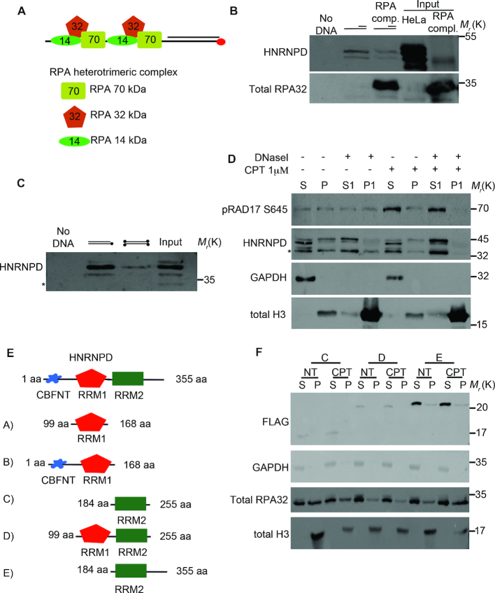
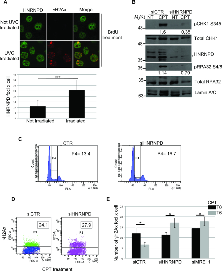
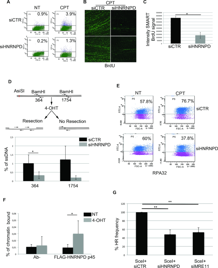
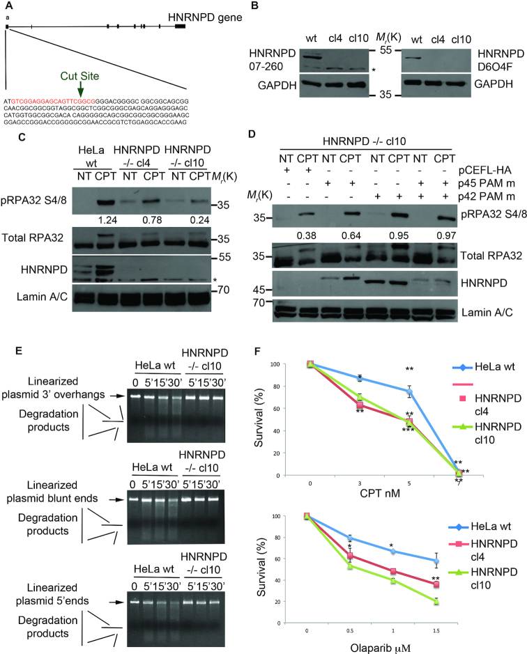
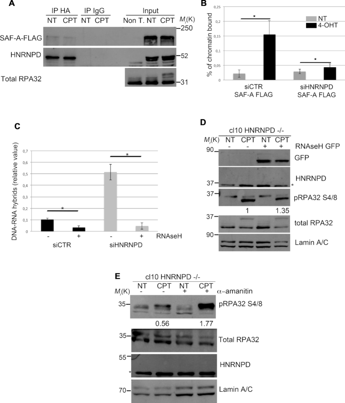
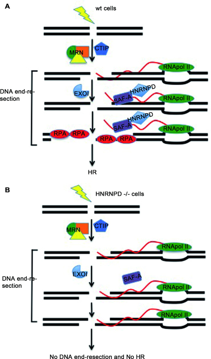
DNA double strand break (DSB) repair through homologous recombination (HR) is crucial to maintain genome stability. DSB resection generates a single strand DNA intermediate, which is crucial for the HR process. We used a synthetic DNA structure, mimicking a resection intermediate, as a bait to identify proteins involved in this process. Among these, LC/MS analysis identified the RNA binding protein, HNRNPD. We found that HNRNPD binds chromatin, although this binding occurred independently of DNA damage. However, upon damage, HNRNPD re-localized to γH2Ax foci and its silencing impaired CHK1 S345 phosphorylation and the DNA end resection process. Indeed, HNRNPD silencing reduced: the ssDNA fraction upon camptothecin treatment; AsiSI-induced DSB resection; and RPA32 S4/8 phosphorylation. CRISPR/Cas9-mediated HNRNPD knockout impaired in vitro DNA resection and sensitized cells to camptothecin and olaparib treatment. We found that HNRNPD interacts with the heterogeneous nuclear ribonucleoprotein SAF-A previously associated with DNA damage repair. HNRNPD depletion resulted in an increased amount of RNA:DNA hybrids upon DNA damage. Both the expression of RNase H1 and RNA pol II inhibition recovered the ability to phosphorylate RPA32 S4/8 in HNRNPD knockout cells upon DNA damage, suggesting that RNA:DNA hybrid resolution likely rescues the defective DNA damage response of HNRNPD-depleted cells.


Figures

Figure 1.
Figure 1.
Proteomic screen and HNRNPD chromatin binding ability. (A) Schematic representation of the proteomic screen by using a synthetic DNA structure coated with the heterotrimeric RPAwt complex, including the 70, 32 and 14 kDa isoforms, which was used as bait for the proteomic screen after being challenged with HeLa nuclear extracts. (B) DNA pull down assay of the synthetic DNA structure coated or not with the recombinant RPA complex produced in E. coli (input) followed by western blot analysis with the indicated antibodies. HeLa nuclear extract was incubated for 30 min with 0,3 mg/ml of RNase A on ice, followed by centrifugation to remove debris. (Sequences are listed in Supplementary Table S1). (C) DNA pull-down of the schematically indicated biotinylated DNA structures (Sequences are listed in Supplementary Table S1). Biotin is represented as black dots. Western blot analysis was performed with the indicated antibodies. The asterisk indicates a non-specific band (which appears using the Millipore antibody). (D) Chromatin enriched purification was performed upon 2 h treatment with 1μM of CPT followed by western blot analysis. DNase I treatment of the first pellet fraction (P) was performed, when indicated, with 80U of enzyme for 30 min at 30°C originating a new supernatant and pellet fraction (S1 and P1 respectively). All the purification steps were performed upon incubation for 30 min with 0,3 mg/ml of RNase A on ice, followed by centrifugation to remove debris. H3 and GAPDH were used as markers for the chromatin and soluble fractions, respectively. RAD17 S645 was used as a DNA damage control. (E) Schematic representation of HNRNPD deletion mutants. (F) HeLa cells were transfected with the indicated DNA, followed by 48 h of incubation. Chromatin enriched purification was performed upon 2 h treatment with 1μM of CPT followed by western blot analysis. All the purification steps were performed upon incubation for 30 min with 0.3 mg/ml of RNase A on ice, followed by centrifugation to remove debris. H3 and GAPDH were used as markers for the chromatin and soluble fractions, respectively. RPA32 was used as a DNA damage control.
Figure 2.
Figure 2.
HNRNPD regulates the DNA damage response. (A) HeLa cells were incubated with 10 μM BrdU for 16 h followed by exposure to 50 J/m2 of UVC to induce localized DSBs, and cells were allowed to incubate for 1 h before fixation. Immunofluorescence was performed with the indicated antibodies. Lower panel: ImageJ analysis of laser irradiation images to quantify the number of HNRNPD foci × cell before and after irradiation. Data represent the mean ± standard deviation (s.d.). (n = 3 independent experiments). *P-value <0.05. (B) Western blot analysis with the indicated antibodies of HeLa cells, 48 h after transfection with siHNRNPD or siCTR followed by 2-h treatment with 1 μM CPT. GAPDH was assessed as a normalization control. The values of band density corresponding to pCHK1 S345 and pRPA32 S4/8, normalized to the total protein levels, are reported. (C) Cell cycle profile of HeLa cells, 48 h after transfection with siHNRNPD or siCTR, was analyzed through flow cytometry upon propidium iodide (PI) staining. (D) γH2Ax values were measured by FACS analysis in HeLa cells, transfected with the indicated siRNAs for 48 h, then treated with 1 μM CPT for 2 h. The insets report the percentage of γH2Ax positive cells. (E) HeLa cells were transfected with the indicated siRNAs for 48 h, treated with 1μM CPT for 2 h (T0) followed by drug washout and incubation in CPT-free medium for additional 6 h (T6). γH2AX foci were measured at T0 and T6 with the ImageJ software analysis. Data represent the mean ± s.d. (n = 4 independent experiments). *P-value <0.05.
Figure 3.
Figure 3.
HNRNPD regulates the DNA end-resection process. (A) FACS analysis of ssDNA formation in HeLa cells transfected with siCTR or siHNRNPD for 24 h, then pulse-labelled with 10 μM BrdU for additional 24 h and treated with 1 μM CPT for 2 h. HeLa cells were prepared for cytofluorimetric analysis in non-denaturing conditions in order to quantify the percentage of positive ssDNA cells. The untreated condition (no DNA damage) reveals the background level given in each cell type by the FITC-antibody detecting BrdU (in this case BrdU is entrapped within the double helix and cannot be recognized by the antibody). Such background values, subtracted to the values obtained following CPT treatment, indicate a ∼3-fold decrease in single strand DNA upon HNRNPD silencing. (B) To perform the SMART technique, HeLa cells (both siCTR and siHNRNPD) were pulse-labelled with 10 μM BrdU for 24 h then treated with 1 μM CPT for 2 h before spreading onto Silane Prep Slides. Immunofluorescence was performed with the BrdU antibody. Two representative images for each cell type (siCTR and siHNRNPD) are shown. (C) BrdU signal intensity of SMART technique analyzed by ImageJ software. Data represent the mean ± standard deviation (s.d.). (n = 2 independent experiments). *P-value <0.05. (D) HeLa ER-AsiSI were transfected with siCTR or siHNRNPD for 48 h followed by treatment with 300 nM of 4-OHT. The Real-time qPCR was performed on the genomic DNA with the indicated primers amplifying regions including the positions at 364 and 1754 bp downstream from the DSB, as depicted. The values of %ssDNA are reported as the mean ± s.d. (n = 4 independent experiments) and were calculated as follows: %ssDNA = 1/[(2ΔCt-1) + 0.5] × 100 (83). **P-value <0.01; *P-value <0.05. (E) HeLa cell lines were transfected with the indicated siRNAs for 48 h treated or not with 1 μM CPT for 2 h; before fixative, cells were pre-extracted with the CSK + 0.1% Triton X100 + 0.3 μg/ml RNase A followed by incubation with indicated antibody. RPA32 values were analyzed through flow cytometry. (F) ChIP experiments of HeLa ER-AsiSI, treated or mock treated with 300 nM 4-OHT for 1 h, were performed by using either an HNRNPD antibody or control IgGs (Ab-). The HNRNPD chromatin binding ability was measured, as a percentage of immunoprecipitated input, from qPCR values of an 80 bp amplicon including the AsiSI site. Data represent the mean ± s.d. (n = 4 independent experiments). *P-value <0.05. (G) HeLa DR-GFP cells were transfected with the plasmid encoding the SceI restriction enzyme in presence of siCTR, siHNRNPD or siMRE11 followed by FACS analysis measurement of GFP levels used to calculate %HR frequency compared with siCTR which was set as 100%. Data represent the mean ± s.d. (n = 3 independent experiments), **P-value <0.01.
Figure 4.
Figure 4.
HNRNPD knockout affects the DNA damage response and sensitizes cells to CPT and olaparib treatment. (A) Schematic representation of the CRISPR/Cas9 strategy used to develop the HNRNPD ko cells. The sgRNA guide is indicated in red. (B) Western Blot analysis of HeLa HNRNPD ko clones with either the Millipore (07-260, left panel), or the Cell signalling (D6O4F, right panel), antibody. The asterisk indicates a non-specific band, which was detected by the Millipore antibody. GAPDH was used as a loading control. (C) Western blot analysis of DNA damage response markers following treatment with 1 μM CPT for 2 h in HeLa wt cells and HNRNPD knockout cell clones. Lamin A/C was used as a loading control. The values of band density corresponding to pRPA32 S4/8, normalized to the total protein levels, are reported. (D) HeLa HNRNPD–/– cl10 cells were transfected with the pCEFL-HA HNRNPD isoforms with the PAM mutated sequence for 48 h followed by 1μM CPT for additional 2 h followed by western blot analysis with the indicated antibodies. Different expression levels are likely dependent on either a different transfection efficiency or by plasmids co-transfection. Lamin A/C was used as a loading control. The values of band density corresponding to pRPA32 S4/8, normalized to the total protein levels, are reported. (E) DNA end-resection assay in vitro with the nuclear protein extracts from HeLa wt and HNRNPD cl10 cell lines. Linearized plasmid with either 3′, blunt or 5′ overhangs were incubated with protein extracts for the indicated time points and run on a 0.8% agarose gel + EtBr. (F) HeLa and HNRNPD cell clones were treated with the indicated drugs at crescent concentrations for ten days followed by staining with crystal violet. Asterisks indicate: ***P-value <0.001; **P-value <0.01; *P-value <0.05, respectively.
Figure 5.
Figure 5.
HNRNPD regulates R-loops through the localization of SAF-A protein. (A) HeLa cells were transfected with either SAF-A FLAG or HNRNPD-HA, incubated for 48 h and treated or not for 2 h with 1μM CPT. Protein lysates were incubated 30 min on ice with 0,3 μg/ml of RNase A and 50 μg/ml of EtBr followed by centrifugation to remove the debris. Immunoprecipitation was conducted with anti-HA antibody over night at +4°C. An antibody against total RPA32 was used as a DNA damage control. (B) HeLa ER-AsiSI were transfected with the indicated siRNAs in combination with the plasmid coding for SAF-A-FLAG for 48 h and treated or mock treated with 300 nM 4-OHT for 1 h. ChIP analysis was performed by using either an anti FLAG antibody or control IgGs (Ab–). SAF-A chromatin binding ability was measured as a percentage of immunoprecipitated input, from qPCR values of a 200 bp amplicon upstream to AsiSI cut site. Data represent the mean ± s.d. (n = 3 independent experiments). *P-value <0.05. (C) HeLa cells were transfected with the indicated siRNAs for 48 h followed by induction of DSBs through the AsiSI enzyme. The immunoprecipitation was carried out with the anti-DNA:RNA hybrid S9.6 antibody over night at +4°C. Data represent the mean ± s.d. (n = 3 independent experiments). *P-value <0.05. (D) HeLa HNRNPD ko cell line cl10 was transfected with RNaseH GFP coding plasmid and incubated 48 h followed by 1 μM CPT treatment and western blot analysis. Lamin A/C was used as a normalization control. Densitometric analysis of pRPA32 S4/8 activation was carried out through the ImageJ software. (E) HeLa HNRNPD ko cl10 was treated with 10 μM of α-amanitin for 6 h followed by co-treatment with 1 μM CPT (where indicated) for additional 2 h followed by western blot analysis. Lamin A/C was used as a normalization control. Densitometric analysis of pRPA32 S4/8 activation was carried out through the ImageJ software.
Figure 6.
Figure 6.
Schematic representation of the hypothesized HNRNPD mechanism of action during homologous recombination. (A) In HeLa wild type cells, upon DNA damage, HNRNPD is relocated to DNA damage sites. Upon damage also SAF-A is located near the DNA lesion and both proteins are associated with a proper removal of the R-loop structures during the DNA end-resection process. (B) In HNRNPD silenced cells, SAF-A does not localize onto damaged sites and R-loops accumulate impairing the DNA end-resection and ultimately the homologous recombination process.

References

    1. Jasin M., Richardson C.. Frequent chromosomal translocations induced by DNA double-strand breaks. Nature. 2000; 405:697–700. - PubMed
    1. Aparicio T., Baer R., Gautier J.. DNA double-strand break repair pathway choice and cancer. DNA Repair (Amst). 2014; 19:169–175. - PMC - PubMed
    1. Jackson S.P., Bartek J.. The DNA-damage response in human biology and disease. Nature. 2009; 461:1071–1078. - PMC - PubMed
    1. Ciccia A., Elledge S.J.. The DNA damage Response: Making it safe to play with knives. Mol. Cell. 2010; 40:179–204. - PMC - PubMed
    1. Lieber M.R. The mechanism of human nonhomologous DNA end joining. J. Biol. Chem. 2008; 283:1–5. - PubMed

MeSH terms

Substances