Review
. 2019 May 7;47(8):3811-3827.
doi: 10.1093/nar/gkz120.

Poly(ADP-ribosyl)ation by PARP1: reaction mechanism and regulatory proteins

Affiliations
Review

Poly(ADP-ribosyl)ation by PARP1: reaction mechanism and regulatory proteins

Elizaveta E Alemasova et al. Nucleic Acids Res. .

Abstract

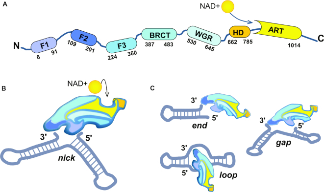
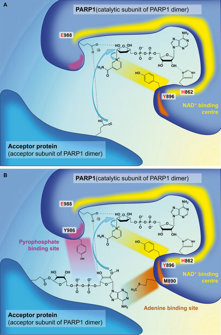
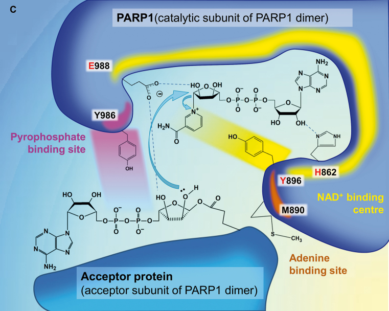
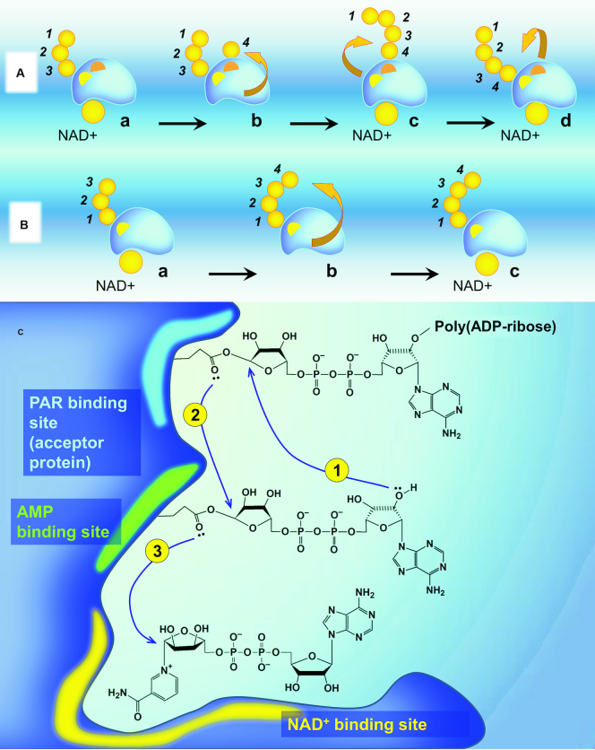
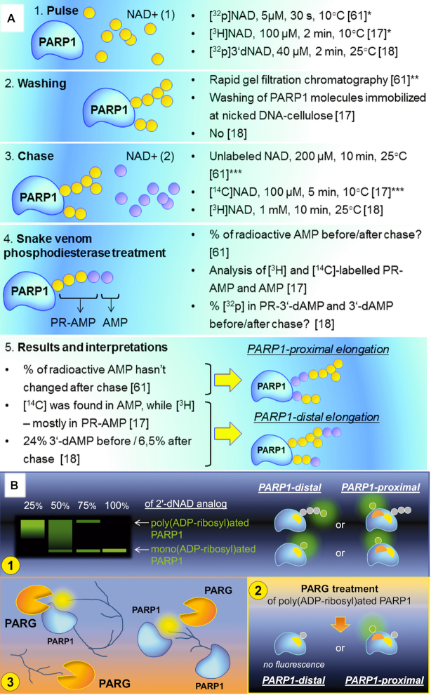
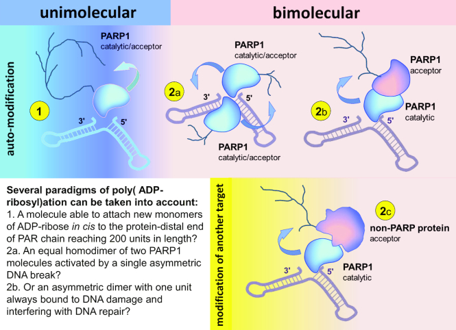
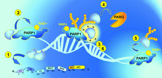
Poly(ADP-ribosyl)ation (PARylation) is posttranslational modification of proteins by linear or branched chains of ADP-ribose units, originating from NAD+. The central enzyme for PAR production in cells and the main target of poly(ADP-ribosyl)ation during DNA damage is poly(ADP-ribose) polymerase 1 (PARP1). PARP1 ability to function as a catalytic and acceptor protein simultaneously made a considerable contribution to accumulation of contradictory data. This topic is directly related to other questions, such as the stoichiometry of PARP1 molecules in auto-modification reaction, direction of the chain growth during PAR elongation and functional coupling of PARP1 with PARylation targets. Besides DNA damage necessary for the folding of catalytically active PARP1, other mechanisms appear to be required for the relevant intensity and specificity of PARylation reaction. Indeed, in recent years, PARP research has been enriched by the discovery of novel PARP1 interaction partners modulating its enzymatic activity. Understanding the details of PARP1 catalytic mechanism and its regulation is especially important in light of PARP-targeted therapy and may significantly aid to PARP inhibitors drug design. In this review we summarize old and up-to-date literature to clarify several points concerning PARylation mechanism and discuss different ways for regulation of PAR synthesis by accessory proteins reported thus far.


Figures

Figure 1.
Figure 1.
PARP1 interaction with DNA is necessary for organisation of the catalytic centre. (A) Domain structure of PARP1. F1-F3—zinc fingers 1–3 (F1F2 operates in DNA recognition, F3 is necessary for allosteric activation); BRCT—BRCA1 C-terminal domain (is dispensable for PARP1 activation, contains auto-modification sites); WGR—Trp-Gly-Arg domain (indispensable for the transfer of activation from F1F2 to the catalytic domain); HD—helical subdomain of the catalytic domain (auto-inhibitory); ART—(ADP-ribosyl)transferase domain (contains the active site and a fold, conserved in all PARP family members). (B) DNA-induced PARP1 folding. 1) F1F2 binds DNA nick in only one orientation (F2 on the 3′ stem, F1 on the 5′ stem (15)), directing the assembly of remaining PARP1 molecule; 2) F3 binds to the F3 binding surface created by F1 and DNA. Owing to versatile interaction between F1 and F3, a single point mutation at the interaction surface (W246A) completely abolishes activation of the full-length PARP1 (15). For the same reason, PARP1 cleavage at the F2-F3 linker by caspase 3 during apoptosis results in PARP1 inactivation (15) despite other mixtures of PARP1 fragments being able to restore the enzymatic activity; 3) WGR binds to the surface composed by DNA, F1 and F3. BRCT-WGR linker remains flexible and is able to reach the active centre of PARP1 during auto-modification of the enzyme; 4) PARP1 catalytic domain interacts with the surface organised by WGR and F3 (15), HD subdomain is unfolded, allowing productive NAD+ binding by PARP1 ART (29). (C) PARP1 activation by different DNA structures. Initial recognition of 3′ stem by F2 results in DNA distortions and exposure of 5′ site (15). Subsequent scanning for this site by flexibly linked F1 zinc finger permits PARP1 to effectively recognise DNA single-strand breaks with different gap lengths and double-strand breaks (15). It is possible that the recognition of other non-B DNA structures, like DNA hairpins, crosses and loops (118), can occur via an analogous mechanism.
Figure 2.
Figure 2.
PARP1 catalytic mechanism. (A) Initiation. (B) Elongation. (C) Branching. The key amino acid residues of the donor site (‘H-Y-E triad': His862, Tyr896, E988) are shown in red. Important residues of the acceptor site, Met890 and Tyr986, of which mutations were especially indicative for understanding the mechanism of PAR synthesis, are also presented. Mutation M890V reduces PARP1 activity more than 200-fold (39) because of a clash between the side chain of Val890 and that of Tyr896 or the N1-atom of ADP, resulting in displacement of the accepting ribose from the PAR-binding site (37). Met890 in PARP3 acceptor site is replaced by an arginine (Arg408 in PARP3) forming a salt bridge with Asp455 (119). It is possible that this amino acid change results in decreased length of PAR polymers synthesized by PARP3 compared to PARP1 and PARP2 (36). Mutation Y986H increases the affinity of the PAR-binding site to the pyrophosphate moiety (37), resulting in 15-fold higher branching compared to wild-type PARP1 (39). Mutations E988D and E988Q reduced PARP1 elongation activity 20- and 2800-fold, correspondingly, indicating the importance of the Glu988 side chain carboxylate in this reaction (41). More information on different mutations in the PARP1 active centre and their consequences can be found in the original works (37,39,41).
Figure 2.
Figure 2.
PARP1 catalytic mechanism. (A) Initiation. (B) Elongation. (C) Branching. The key amino acid residues of the donor site (‘H-Y-E triad': His862, Tyr896, E988) are shown in red. Important residues of the acceptor site, Met890 and Tyr986, of which mutations were especially indicative for understanding the mechanism of PAR synthesis, are also presented. Mutation M890V reduces PARP1 activity more than 200-fold (39) because of a clash between the side chain of Val890 and that of Tyr896 or the N1-atom of ADP, resulting in displacement of the accepting ribose from the PAR-binding site (37). Met890 in PARP3 acceptor site is replaced by an arginine (Arg408 in PARP3) forming a salt bridge with Asp455 (119). It is possible that this amino acid change results in decreased length of PAR polymers synthesized by PARP3 compared to PARP1 and PARP2 (36). Mutation Y986H increases the affinity of the PAR-binding site to the pyrophosphate moiety (37), resulting in 15-fold higher branching compared to wild-type PARP1 (39). Mutations E988D and E988Q reduced PARP1 elongation activity 20- and 2800-fold, correspondingly, indicating the importance of the Glu988 side chain carboxylate in this reaction (41). More information on different mutations in the PARP1 active centre and their consequences can be found in the original works (37,39,41).
Figure 3.
Figure 3.
The direction of PAR synthesis. (A) A ‘head-out' mechanism of PAR elongation (the proximal addition model). PARP1 is first shown (a) with a partially formed PAR chain attached to the PAR binding site (yellow) and with NAD+ bound non-covalently in the NAD+ binding site. When NAD+ loses nicotinamide, its ADP-ribose occupies the AMP binding site (orange) (b). Then the PAR chain is transferred to the 2′-OH of the new ADP-ribose monomer and temporally located at the monomer site (c) before being translocated back to the PAR binding site (d) (57). (B) A ‘tail-out' (distal addition) mechanism. The 2′-OH of the ADP-ribose unit distal to PARP1 performs the nucleophilic attack on the NAD+ molecule held in the NAD+ binding site. (C) A hypothetical mechanism of PARP1-proximal elongation. (1) The transfer of the growing PAR chain to the 2′-OH of the new ADP-ribose monomer located at the monomer (AMP-binding) site; (2) Translocation of elongated PAR polymer back to the PAR binding site; (3) Occupation of AMP-binding site by NAD+ ADP-ribose (new monomer).
Figure 4.
Figure 4.
What attempts to establish the direction of PAR elongation were made? (A) Pulse-and-chase experiments (17,18,57). PR-AMP—phosphoribosyl-AMP. Potential problems of certain steps are indicated: (*) Not all PARP1 molecules or PAR acceptor sites were modified during the pulse stage; (**) the removal of the unincorporated NAD+ used in the pulse reaction was not full; (***) potential PAR branching at the pulse stage. (B) (1) PARP1 modification by ‘chain-terminating' 2′-deoxy NAD+ analogue in the presence of natural NAD+ (19). (2) We propose that additional experiments with PARG treatment would be helpful to distinguish possible situations shown in the panel (Figure 4B, (1)). PAR-binding site in PARP1 is shown with yellow, AMP-binding site is shown in orange. (3) If PARP1 elongates PAR chains according to the protein-distal model, it could compete with PARG for the distal end of the polymer by binding the terminal ADP-ribose moiety within the active site. Non-covalent interaction of PAR and PARP1 active site is shown in yellow.
Figure 5.
Figure 5.
Poly(ADP-ribosyl)ation in dimers and monomers: terminology and different paradigms. (1) Unimolecular (monomolecular, intramolecular, in cis modification). One molecule of PARP1 interacts with DNA break, becomes catalytically active and modifies itself (as a monomer). In this case one PARP1 polypeptide serves as a catalyst and an acceptor of poly(ADP-ribosyl)ation at the same time. (2) Bimolecular (intermolecular, hetero- or in trans modification) occurs in protein dimers: (2a) ‘Homodimers' of PARP1. The dimer of two PARP1 molecules is formed by protein-protein interactions. Binding of the first molecule to the DNA break induces its interdomain rearrangement, resulting in both activation of this molecule and symmetric self-assembly of the second PARP1 molecule driven by rearrangement of the protein-protein interaction surface. The active PARP1 homodimer consists of two identical subunits, both functioning as a catalyst and acceptor of PARylation simultaneously. (2b) ‘Heterodimers' (‘asymmetric homodimers') of two PARP1 molecules. DNA-bound PARP1 subunit is active and functions only as a catalyst. The second PARP1 molecule is inactive and functions only as an acceptor of poly(ADP-ribose). (2c) ‘Heterodimers' of PARP1 and other proteins. DNA-bound PARP1 molecule acts as a catalyst. Another (non-PARP1) protein is a target for poly(ADP-ribosyl)ation by PARP1.
Figure 6.
Figure 6.
PARP1 regulation by other proteins (ideas for possible mechanisms). (1) DNA-binding proteins: could inhibit PARP1 activity based on its displacement from the DNA or stimulate the enzyme by prevention of catalytically ineffective PARP1 binding with single-stranded DNA / by modulation of DNA conformation (DNA melting). (2) Proteins that physically interact with PARP1 may influence PARP1 allosteric activation. (3) Basic PAR-binding proteins: can screen the negative charge of growing PAR chains, stabilizing catalytically active PARP1-DNA complexes during PAR elongation. (4) PAR-binding proteins are also able to protect PAR from degradation by PARG and increase the life span of poly(ADP-ribose). (5) PARylation targets: could permit PARP1 to remain longer in the active non-PARylated state by providing another (non-PARP1) platform for modification. (6) PAR-binding proteins may facilitate PARylation reactions by inducing molecular crowding and raising the effective concentrations of the reaction participants.

References

    1. Perina D., Mikoč A., Ahel J., Ćetković H., Žaja R., Ahel I.. Distribution of protein poly(ADP-ribosyl)ation systems across all domains of life. DNA Repair (Amst.). 2014; 23:4–16. - PMC - PubMed
    1. Slade D., Dunstan M.S., Barkauskaite E., Weston R., Lafite P., Dixon N., Ahel M., Leys D., Ahel I.. The structure and catalytic mechanism of a poly(ADP-ribose) glycohydrolase. Nature. 2011; 477:616–620. - PMC - PubMed
    1. Faraone-Mennella M.R., Gambacorta A., Nicolaus B., Farina B.. Purification and biochemical characterization of a poly(ADP-ribose) polymerase-like enzyme from the thermophilic archaeon Sulfolobus solfataricus. Biochem. J. 1998; 335:441–447. - PMC - PubMed
    1. Hassa P.O., Hottiger M.O.. The diverse biological roles of mammalian PARPS, a small but powerful family of poly-ADP-ribose polymerases. Front. Biosci. 2008; 13:3046–3082. - PubMed
    1. Kleine H., Poreba E., Lesniewicz K., Hassa P.O., Hottiger M.O., Litchfield D.W., Shilton B.H., Lüscher B.. Substrate-assisted catalysis by PARP10 limits its activity to mono-ADP-ribosylation. Mol. Cell. 2008; 32:57–69. - PubMed

Publication types

MeSH terms