Review
. 2019 Feb 8:10:66.
doi: 10.3389/fendo.2019.00066. eCollection 2019.

The Role of Maternal Thyroid Hormones in Avian Embryonic Development

Affiliations
Review

The Role of Maternal Thyroid Hormones in Avian Embryonic Development

Veerle M Darras. Front Endocrinol (Lausanne). .

Abstract

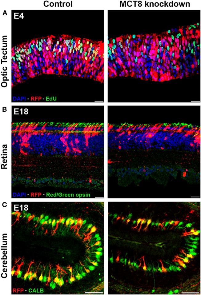
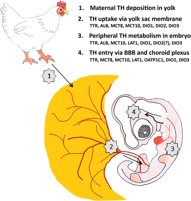
During avian embryonic development, thyroid hormones (THs) coordinate the expression of a multitude of genes thereby ensuring that the correct sequence of cell proliferation, differentiation and maturation is followed in each tissue and organ. Although THs are needed from the start of development, the embryonic thyroid gland only matures around mid-incubation in precocial birds and around hatching in altricial species. Therefore, maternal THs deposited in the egg yolk play an essential role in embryonic development. They are taken up by the embryo throughout its development, from the first day till hatching, and expression of TH regulators such as distributor proteins, transporters, and deiodinases in the yolk sac membrane provide the tools for selective metabolism and transport starting from this level. TH receptors and regulators of local TH availability are expressed in avian embryos in a dynamic and tissue/cell-specific pattern from the first stages studied, as shown in detail in chicken. Maternal hyperthyroidism via TH supplementation as well as injection of THs into the egg yolk increase TH content in embryonic tissues while induction of maternal hypothyroidism by goitrogen treatment results in a decrease. Both increase and decrease of maternal TH availability were shown to alter gene expression in early chicken embryos. Knockdown of the specific TH transporter monocarboxylate transporter 8 at early stages in chicken cerebellum, optic tectum, or retina allowed to reduce local TH availability, interfering with gene expression and confirming that development of the central nervous system (CNS) is highly dependent on maternal THs. While some of the effects on cell proliferation, migration and differentiation seem to be transient, others result in persistent defects in CNS structure. In addition, a number of studies in both precocial and altricial birds showed that injection of THs into the yolk at the start of incubation influences a number of parameters in posthatch performance and fitness. In conclusion, the data presently available clearly indicate that maternal THs play an important role in avian embryonic development, but how exactly their influence on cellular and molecular processes in the embryo is linked to posthatch fitness needs to be further explored.

Keywords: TH transporter; bird; deiodinase; development; thyroid hormone.


Figures

Figure 1
Figure 1
Impact of TH deficiency observed at early and later stages of embryonic chicken CNS development. (A) Electroporation of empty vector (control) or MCT8-RNAi vector in the optic tectum at E3 followed by EdU pulse-labeling 1 h before sampling at E4. The strong reduction in the number of proliferating (S phase) transfected cells (yellow) in the knockdown condition illustrates one of the early effects of TH deficiency on CNS development. (B) Electroporation of empty vector (control) or MCT8-RNAi vector in the retina at E4 followed by IHC staining for red/green opsin at E18. The lower amount of red/green expressing cones in the mature retina in the knockdown condition at E18 is the combined result of a reduced retinal progenitor cell proliferation and a shift in commitment toward short wavelength sensitive cones at the expense of long/medium wavelength sensitive cones occurring at earlier stages. The picture also shows a reduced thickness of the retina and a disorganization of the sublaminae in the inner plexiform layer in the knockdown condition. (C) Electroporation of empty vector (control) or MCT8-RNAi vector in the cerebellar anlage at E3 followed by IHC staining for calbindin (CALB) at E18. The clear reduction in dendritic tree complexity of the Purkinje cells in the knockdown condition may be due to diminished expression of LHX1, LHX5, and RORα, observed at earlier stages. Scale bars represent 20 μm for optic tectum and retina and 100 μm for cerebellum.
Figure 2
Figure 2
Maternal TH supply to the developing brain of a 4-day-old chicken embryo is regulated at 4 different levels. The factors controlling TH transport and metabolism (TH distributor proteins, TH transporters, deiodinases) shown to be present at E4 at the different levels are mentioned. ALB: albumin, BBB: blood-brain-barrier, DIO1-3: deiodinase 1-3, LAT1: L-type amino acid transporter 1, MCT8-10: monocarboxylate transporter 8-10, OATP1C1: organic anion transporting protein 1C1, TTR: transthyretin.

References

    1. Thommes RC. Ontogenesis of thyroid function and regulation in the developing chick embryo. J Exp Zool Suppl. (1987) 1:273–9. - PubMed
    1. McNabb FM. Comparative thyroid development in precocial Japanese quail and altricial ring doves. J Exp Zool Suppl. (1987) 1:281–90. - PubMed
    1. Olson JM, McNabb FMA, Jablonski MS, Ferris DV. Thyroid development in relation to the development of endothermy in the red-winged blackbird (Agelaius phoeniceus). Gen Comp Endocrinol. (1999) 116:204–12. 10.1006/gcen.1999.7363 - DOI - PubMed
    1. McNabb FM. The hypothalamic-pituitary-thyroid (HPT) axis in birds and its role in bird development and reproduction. Crit Rev Toxicol. (2007) 37:163–93. 10.1080/10408440601123552 - DOI - PubMed
    1. Vancamp P, Darras VM. Dissecting the role of regulators of thyroid hormone availability in early brain development: merits and potential of the chicken embryo model. Mol Cell Endocrinol. (2017) 459:71–8. 10.1016/j.mce.2017.01.045 - DOI - PubMed

LinkOut - more resources