

Comparative AAV-eGFP Transgene Expression Using Vector Serotypes 1-9, 7m8, and 8b in Human Pluripotent Stem Cells, RPEs, and Human and Rat Cortical Neurons
- PMID: 30800164
- PMCID: PMC6360060
- DOI: 10.1155/2019/7281912
Comparative AAV-eGFP Transgene Expression Using Vector Serotypes 1-9, 7m8, and 8b in Human Pluripotent Stem Cells, RPEs, and Human and Rat Cortical Neurons
Abstract
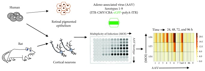
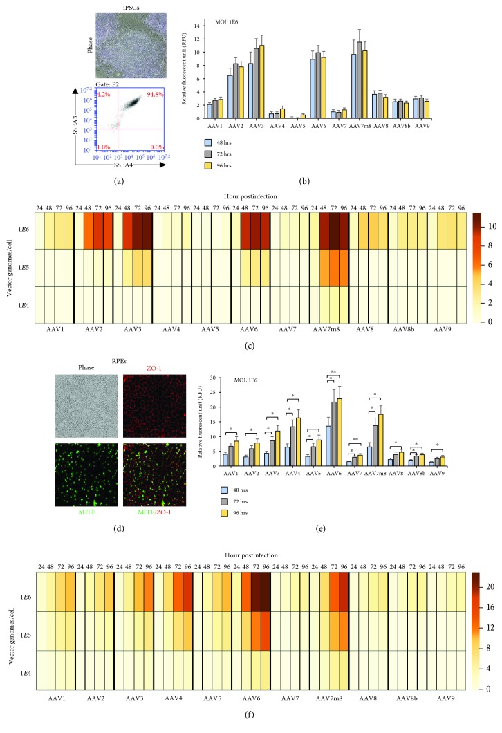
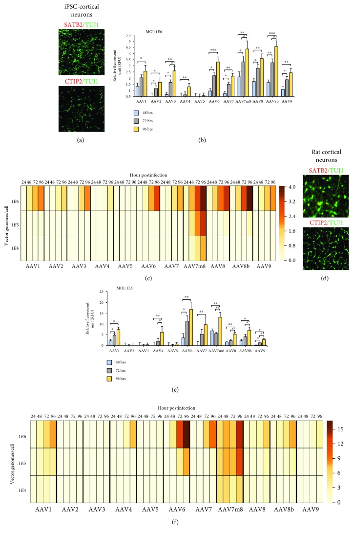
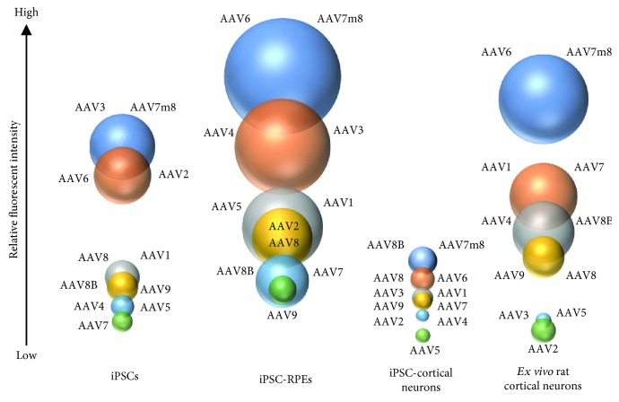
Recombinant adeno-associated virus (rAAV), produced from a nonpathogenic parvovirus, has become an increasing popular vector for gene therapy applications in human clinical trials. However, transduction and transgene expression of rAAVs can differ across in vitro and ex vivo cellular transduction strategies. This study compared 11 rAAV serotypes, carrying one reporter transgene cassette containing a cytomegalovirus immediate-early enhancer (eCMV) and chicken beta actin (CBA) promoter driving the expression of an enhanced green-fluorescent protein (eGFP) gene, which was transduced into four different cell types: human iPSC, iPSC-derived RPE, iPSC-derived cortical, and dissociated embryonic day 18 rat cortical neurons. Each cell type was exposed to three multiplicity of infections (MOI: 1E4, 1E5, and 1E6 vg/cell). After 24, 48, 72, and 96 h posttransduction, GFP-expressing cells were examined and compared across dosage, time, and cell type. Retinal pigmented epithelium showed highest AAV-eGFP expression and iPSC cortical the lowest. At an MOI of 1E6 vg/cell, all serotypes show measurable levels of AAV-eGFP expression; moreover, AAV7m8 and AAV6 perform best across MOI and cell type. We conclude that serotype tropism is not only capsid dependent but also cell type plays a significant role in transgene expression dynamics.
Figures
Similar articles
-
Glial fibrillary acidic protein promoter determines transgene expression in satellite glial cells following intraganglionic adeno-associated virus delivery in adult rats.J Neurosci Res. 2018 Mar;96(3):436-448. doi: 10.1002/jnr.24183. Epub 2017 Sep 23. J Neurosci Res. 2018. PMID: 28941260 Free PMC article.
-
Transduction of Adeno-Associated Virus Vectors Targeting Hair Cells and Supporting Cells in the Neonatal Mouse Cochlea.Front Cell Neurosci. 2019 Jan 24;13:8. doi: 10.3389/fncel.2019.00008. eCollection 2019. Front Cell Neurosci. 2019. PMID: 30733670 Free PMC article.
-
Spinal nociceptive circuit analysis with recombinant adeno-associated viruses: the impact of serotypes and promoters.J Neurochem. 2017 Sep;142(5):721-733. doi: 10.1111/jnc.14124. Epub 2017 Aug 4. J Neurochem. 2017. PMID: 28700081
-
Optimal different adeno-associated virus capsid/promoter combinations to target specific cell types in the common marmoset cerebral cortex.Mol Ther Methods Clin Dev. 2024 Sep 13;32(4):101337. doi: 10.1016/j.omtm.2024.101337. eCollection 2024 Dec 12. Mol Ther Methods Clin Dev. 2024. PMID: 39391837 Free PMC article.
-
Expressing Transgenes That Exceed the Packaging Capacity of Adeno-Associated Virus Capsids.Hum Gene Ther Methods. 2016 Feb;27(1):1-12. doi: 10.1089/hgtb.2015.140. Hum Gene Ther Methods. 2016. PMID: 26757051 Free PMC article. Review.
Cited by
-
The Potential of Induced Pluripotent Stem Cells to Test Gene Therapy Approaches for Neuromuscular and Motor Neuron Disorders.Front Cell Dev Biol. 2021 Apr 13;9:662837. doi: 10.3389/fcell.2021.662837. eCollection 2021. Front Cell Dev Biol. 2021. PMID: 33937264 Free PMC article. Review.
-
AAV-Mediated Gene Delivery to 3D Retinal Organoids Derived from Human Induced Pluripotent Stem Cells.Int J Mol Sci. 2020 Feb 3;21(3):994. doi: 10.3390/ijms21030994. Int J Mol Sci. 2020. PMID: 32028585 Free PMC article.
-
High-energy X-ray irradiation-induced functionalization of Ni(OH)₂ for enhanced supercapacitor electrodes.Sci Rep. 2025 Aug 9;15(1):29194. doi: 10.1038/s41598-025-13250-6. Sci Rep. 2025. PMID: 40783640 Free PMC article.
-
Protocol to generate PDMS topographical patterns for hiPSC-derived or rat primary neuronal cultures.STAR Protoc. 2025 Aug 1;6(3):104010. doi: 10.1016/j.xpro.2025.104010. Online ahead of print. STAR Protoc. 2025. PMID: 40753576 Free PMC article.
-
Base editing strategies to convert CAG to CAA diminish the disease-causing mutation in Huntington's disease.Elife. 2024 Jun 13;12:RP89782. doi: 10.7554/eLife.89782. Elife. 2024. PMID: 38869243 Free PMC article.
References
-
- Bennett J., Wellman J., Marshall K. A., et al. Safety and durability of effect of contralateral-eye administration of AAV2 gene therapy in patients with childhood-onset blindness caused by RPE65 mutations: a follow-on phase 1 trial. The Lancet. 2016;388(10045):661–672. doi: 10.1016/S0140-6736(16)30371-3. - DOI - PMC - PubMed
-
- Russell S., Bennett J., Wellman J. A., et al. Efficacy and safety of voretigene neparvovec (AAV2-hRPE65v2) in patients with RPE65-mediated inherited retinal dystrophy: a randomised, controlled, open-label, phase 3 trial. The Lancet. 2017;390(10097):849–860. doi: 10.1016/S0140-6736(17)31868-8. - DOI - PMC - PubMed
Grants and funding
LinkOut - more resources
Full Text Sources
Other Literature Sources
