

Development of a low-cost and portable smart fluorometer for detecting breast cancer cells
- PMID: 30800488
- PMCID: PMC6377908
- DOI: 10.1364/BOE.10.000399
Development of a low-cost and portable smart fluorometer for detecting breast cancer cells
Abstract
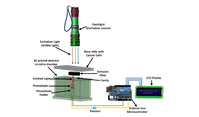
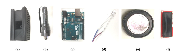
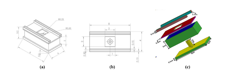
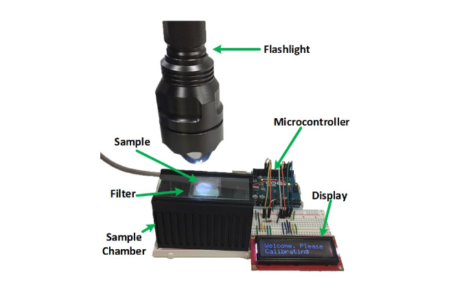
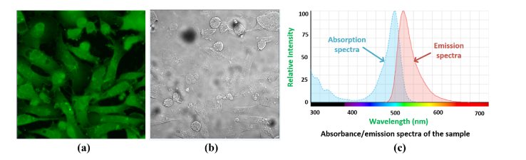
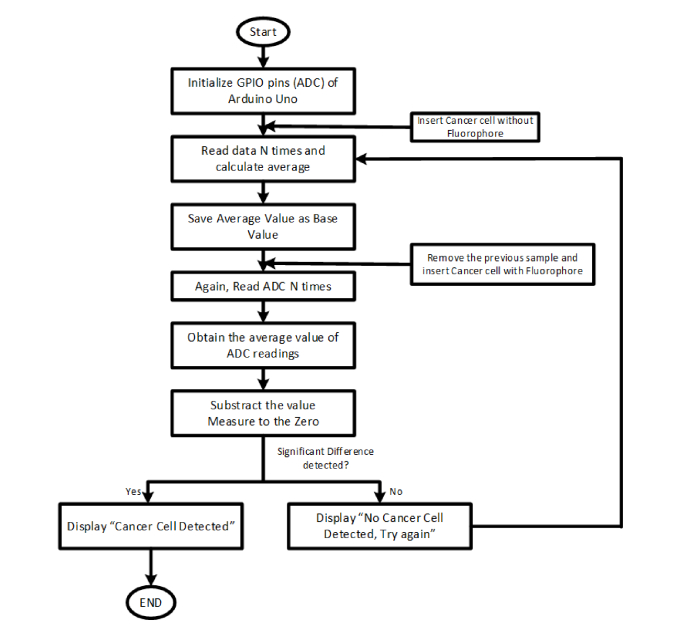
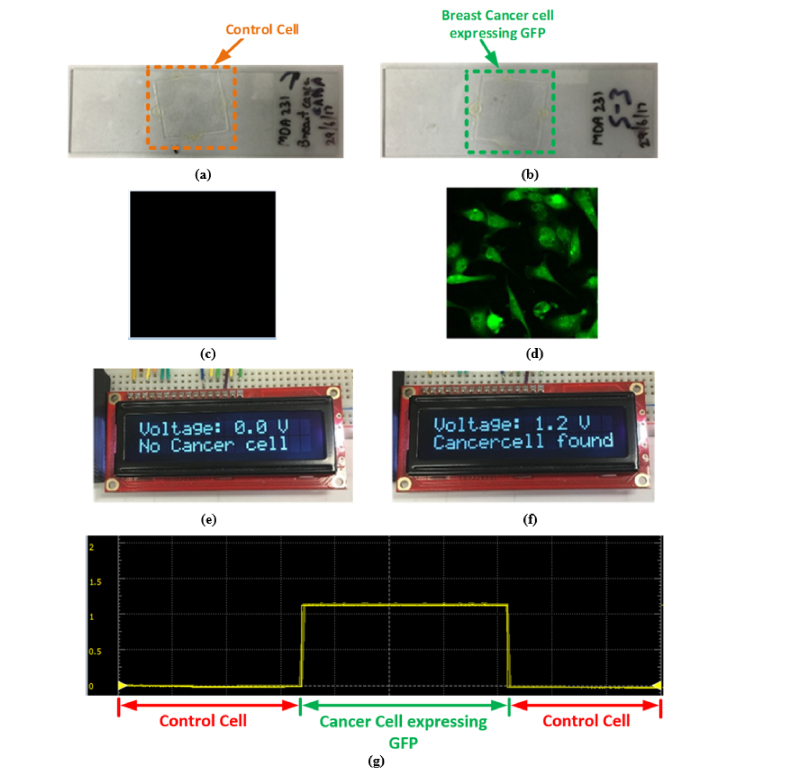
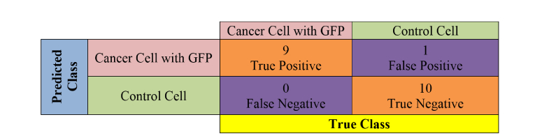
Instruments that allow the detection of fluorescence signal are invaluable tools for biomedical and clinical researchers. The technique is widely used in cell biology to microscopically detect target proteins of interest in mammalian cells. Importantly, fluorescence microscopy finds major applications in cancer biology where cancer cells are chemically labelled for detection. However, conventional fluorescence detection instruments such as fluorescence imaging microscopes are expensive, not portable and entail potentially high maintenance costs. Here we describe the design, development and applicability of a low-cost and portable fluorometer for the detection of fluorescence signal emitted from a model breast cancer cell line, engineered to stably express the green fluorescent protein (GFP). This device utilizes a flashlight which works in the visible range as an excitation source and a photodiode as the detector. It also utilizes an emission filter to mainly allow the fluorescence signal to reach the detector while eliminating the use of an excitation filter and dichroic mirror, hence, making the device compact, low-cost, portable and lightweight. The custom-built sample chamber is fabricated with a 3D printer to house the detector circuitry. We demonstrate that the developed fluorometer is able to distinguish between the cancer cell expressing GFP and the control cell. The fluorometer we developed exhibits immense potential for future applicability in the selective detection of fluorescently-labelled breast cancer cells.
Conflict of interest statement
The authors declare that there are no conflicts of interest related to this article.
Figures
References
-
- “IX51 Inverted Microscope from Olympus | Biocompare.com,” https://www.biocompare.com/19419-Inverted-Microscopes/396657-IX51-Invert....
-
- “Meiji MT6000H Fluorescence Microscope with X-Y Stage & Focus Automation - New York Microscope Co.,” https://www.microscopeinternational.com/product/meiji-mt6000h-fluorescen....
-
- Miller A. R., Davis G. L., Oden Z. M., Razavi M. R., Fateh A., Ghazanfari M., Abdolrahimi F., Poorazar S., Sakhaie F., Olsen R. J., Bahrmand A. R., Pierce M. C., Graviss E. A., Richards-Kortum R., “Portable, Battery-Operated, Low-Cost, Bright Field and Fluorescence Microscope,” PLoS One 5(8), e11890 (2010).10.1371/journal.pone.0011890 - DOI - PMC - PubMed
LinkOut - more resources
Full Text Sources
Other Literature Sources