

Interstitial magnetic thermotherapy dosimetry based on shear wave magnetomotive optical coherence elastography
- PMID: 30800498
- PMCID: PMC6377902
- DOI: 10.1364/BOE.10.000539
Interstitial magnetic thermotherapy dosimetry based on shear wave magnetomotive optical coherence elastography
Abstract
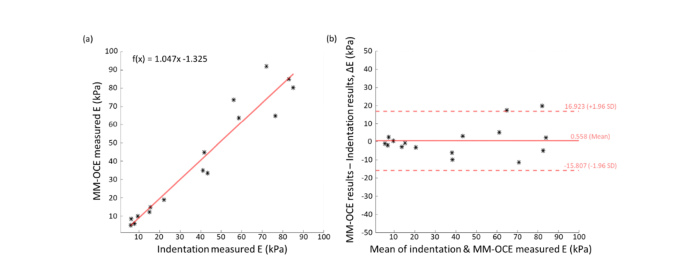
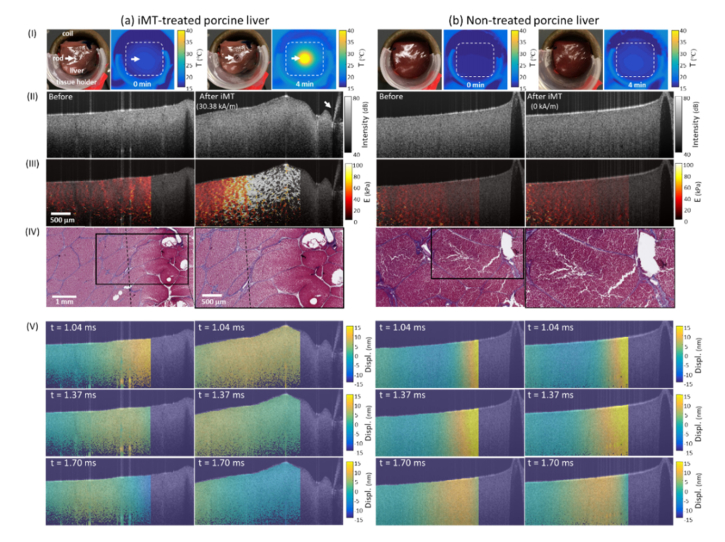
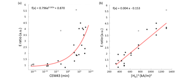
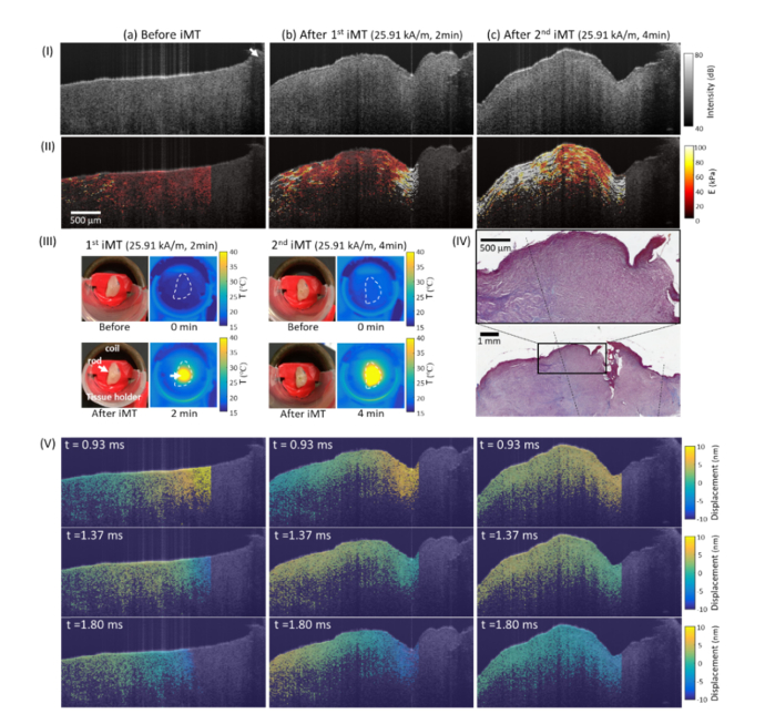
While magnetic thermoseeds are often utilized in interstitial magnetic thermotherapy (iMT) to enable localized tumor ablation, we propose to extend their use as the perturbative source in magnetomotive optical coherence elastography (MM-OCE) so that the heat-induced elasticity alterations can be 'theranostically' probed. MM-OCE measurements were found to agree with indentation results. Tissue stiffening was visualized on iMT-treated porcine liver and canine soft tissue sarcoma specimens, where histology confirmed thermal damages. Additionally, the elasticity was found to increase exponentially and linearly with the conventional thermal dosage metrics and the deposited thermal energy, respectively. Collectively, a physiologically-meaningful, MM-OCE-based iMT dosimetry is feasible.
Conflict of interest statement
The authors declare that there are no conflicts of interest related to this article.
Figures
Similar articles
-
Magnetomotive Optical Coherence Elastography for Magnetic Hyperthermia Dosimetry Based on Dynamic Tissue Biomechanics.IEEE J Sel Top Quantum Electron. 2016 Jul-Aug;22(4):6802816. doi: 10.1109/JSTQE.2015.2505147. Epub 2015 Dec 17. IEEE J Sel Top Quantum Electron. 2016. PMID: 28163565 Free PMC article.
-
Biomechanical sensing of in vivo magnetic nanoparticle hyperthermia-treated melanoma using magnetomotive optical coherence elastography.Theranostics. 2021 Mar 23;11(12):5620-5633. doi: 10.7150/thno.55333. eCollection 2021. Theranostics. 2021. PMID: 33897871 Free PMC article.
-
Strain and elasticity imaging in compression optical coherence elastography: The two-decade perspective and recent advances.J Biophotonics. 2021 Feb;14(2):e202000257. doi: 10.1002/jbio.202000257. Epub 2020 Nov 3. J Biophotonics. 2021. PMID: 32749033 Review.
-
Mechanical contrast in spectroscopic magnetomotive optical coherence elastography.Phys Med Biol. 2015 Sep 7;60(17):6655-68. doi: 10.1088/0031-9155/60/17/6655. Epub 2015 Aug 13. Phys Med Biol. 2015. PMID: 26271056 Free PMC article.
-
Optical coherence elastography in ophthalmology.J Biomed Opt. 2017 Dec;22(12):1-28. doi: 10.1117/1.JBO.22.12.121720. J Biomed Opt. 2017. PMID: 29275544 Free PMC article. Review.
Cited by
-
Magnetic particles in motion: magneto-motive imaging and sensing.Theranostics. 2022 Jan 24;12(4):1783-1799. doi: 10.7150/thno.54056. eCollection 2022. Theranostics. 2022. PMID: 35198073 Free PMC article. Review.
-
Inactivation and sensitization of Pseudomonas aeruginosa by microplasma jet array for treating otitis media.NPJ Biofilms Microbiomes. 2021 Jun 2;7(1):48. doi: 10.1038/s41522-021-00219-2. NPJ Biofilms Microbiomes. 2021. PMID: 34078901 Free PMC article.
-
Recent advances in optical elastography and emerging opportunities in the basic sciences and translational medicine [Invited].Biomed Opt Express. 2022 Dec 16;14(1):208-248. doi: 10.1364/BOE.468932. eCollection 2023 Jan 1. Biomed Opt Express. 2022. PMID: 36698669 Free PMC article. Review.
-
Imaging-guided precision hyperthermia with magnetic nanoparticles.Nat Rev Bioeng. 2025 Mar;3(3):245-260. doi: 10.1038/s44222-024-00257-3. Epub 2024 Nov 7. Nat Rev Bioeng. 2025. PMID: 40260131 Free PMC article.
-
Single-shot two-dimensional spectroscopic magnetomotive optical coherence elastography with graphics processing unit acceleration.Opt Lett. 2020 Aug 1;45(15):4124-4127. doi: 10.1364/OL.397900. Opt Lett. 2020. PMID: 32735239 Free PMC article.
References
-
- Dewhirst M., Stauffer P. R., Das S., Craciunescu O. I., Vujaskovic Z., “Hyperthermia,” in Clinical Radiation Oncology, Gunderson L. L., Tepper J. E., eds. (Elsevier, 2015), pp. 381–398.
Grants and funding
LinkOut - more resources
Full Text Sources