. 2019 Feb 11:1:95-104.
doi: 10.1038/s42256-019-0019-2.

Deep learning cardiac motion analysis for human survival prediction

Affiliations

Deep learning cardiac motion analysis for human survival prediction

Ghalib A Bello et al. Nat Mach Intell. .

Abstract

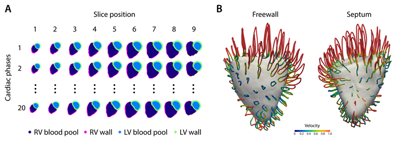
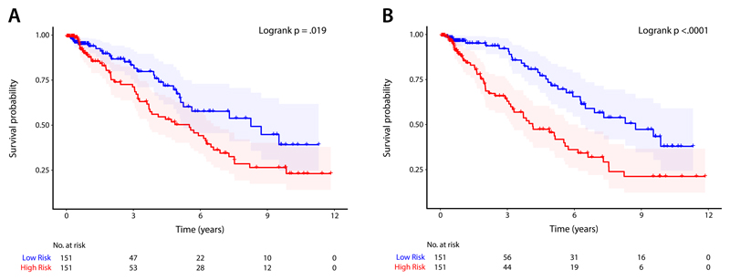
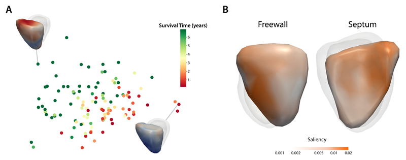
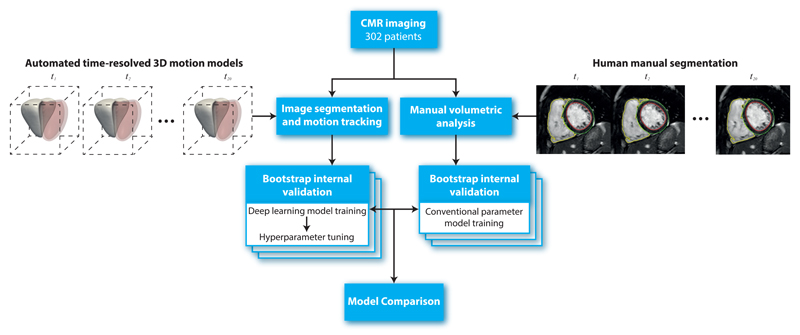
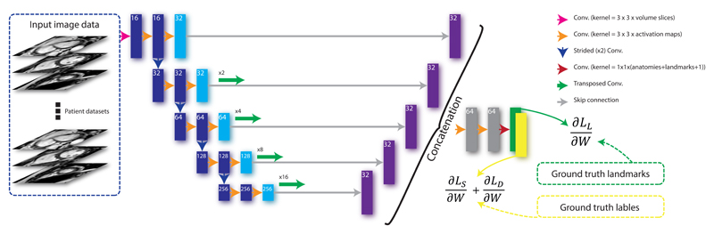
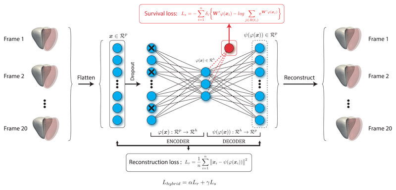
Motion analysis is used in computer vision to understand the behaviour of moving objects in sequences of images. Optimising the interpretation of dynamic biological systems requires accurate and precise motion tracking as well as efficient representations of high-dimensional motion trajectories so that these can be used for prediction tasks. Here we use image sequences of the heart, acquired using cardiac magnetic resonance imaging, to create time-resolved three-dimensional segmentations using a fully convolutional network trained on anatomical shape priors. This dense motion model formed the input to a supervised denoising autoencoder (4Dsurvival), which is a hybrid network consisting of an autoencoder that learns a task-specific latent code representation trained on observed outcome data, yielding a latent representation optimised for survival prediction. To handle right-censored survival outcomes, our network used a Cox partial likelihood loss function. In a study of 302 patients the predictive accuracy (quantified by Harrell's C-index) was significantly higher (p = .0012) for our model C=0.75 (95% CI: 0.70 - 0.79) than the human benchmark of C=0.59 (95% CI: 0.53 - 0.65). This work demonstrates how a complex computer vision task using high-dimensional medical image data can efficiently predict human survival.

Keywords: Heart Failure; Hypertension, Pulmonary; Machine Learning; Magnetic Resonance Imaging, Cine; Motion; Survival.


Conflict of interest statement

Competing interests The authors declare no competing financial interests.

Figures

Figure 1
Figure 1
A) An example of an automatic cardiac image segmentation of each short-axis cine image from apex (slice 1) to base (slice 9) across 20 temporal phases. Data were aligned to a common reference space to build a population model of cardiac motion. B) Trajectory of right ventricular contraction and relaxation averaged across the study population plotted as looped pathlines for a sub-sample of 100 points on the heart (magnification factor of x4). Colour represents relative myocardial velocity at each phase of the cardiac cycle. A surface-shaded model of the heart is shown at end-systole. These dense myocardial motion fields for each patient were used as an input to the prediction network. LV, left ventricular; RV, right ventricular.
Figure 2
Figure 2
Kaplan-Meier plots for A) a conventional parameter model using a composite of manually-derived volumetric measures, and B) a deep learning prediction model (4Dsurvival) whose input was time-resolved three dimensional models of cardiac motion. For both models, patients were divided into low- and high-risk groups by median risk score. Survival function estimates for each group (with 95% confidence intervals) are shown. For each plot, the Logrank test was performed to compare survival curves between risk groups (conventional parameter model: χ2 = 5.5, p = .019; 4Dsurvival: χ2 = 15.6, p < .0001)
Figure 3
Figure 3
A) A 2-dimensional projection of latent representations of cardiac motion in the 4Dsurvival network labelled by survival time. A visualisation of RV motion is shown for two patients with contrasting risks. B) Saliency map showing regional contributions to survival prediction by right ventricular motion. Absolute regression coefficients are expressed on a log-scale.
Figure 4
Figure 4
Flowchart to show the design of the study. In total 302 patients with CMR imaging had both manual volumetric analysis and automated image segmentation (right ventricle shown in solid white, left ventricle in red) across 20 temporal phases (t = 1, .., 20). Internal validity of the predictive performance of a conventional parameter model and a deep learning motion model was assessed using bootstrapping. CMR, cardiac magnetic resonance.
Figure 5
Figure 5
The architecture of the segmentation algorithm. A fully convolutional network takes each stack of cine images as an input, applies a branch of convolutions, learns image features from fine to coarse levels, concatenates multi-scale features and finally predicts the segmentation and landmark location probability maps simultaneously. These maps, together with the ground truth landmark locations and label maps, are then used in the loss function (see Equation 1) which is minimised via stochastic gradient descent.
Figure 6
Figure 6
The prediction network (4Dsurvival) is a denoising autoencoder that takes time-resolved cardiac motion meshes as its input (right ventricle shown in solid white, left ventricle in red). For the sake of simplicity two hidden layers, one immediately preceding and the other immediately following the central layer (latent code layer), have been excluded from the diagram. The autoencoder learns a task-specific latent code representation trained on observed outcome data, yielding a latent representation optimised for survival prediction that is robust to noise. The actual number of latent factors is treated as an optimisable parameter.

References

    1. Wang L, Zhao G, Cheng L, Pietikäinen M. Machine learning for vision-based motion analysis: Theory and techniques. Springer; 2010.
    1. Mei T, Zhang C. Deep learning for intelligent video analysis. 2017 URL https://www.microsoft.com/en-us/research/publication/deep-learning-intel...
    1. Liang F, Xie W, Yu Y. Beating heart motion accurate prediction method based on interactive multiple model: An information fusion approach. BioMed Res Int. 2017;2017:9. - PMC - PubMed
    1. Savarese G, Lund LH. Global public health burden of heart failure. Cardiac Fail Rev. 2017;3:7–11. - PMC - PubMed
    1. Galie N, et al. 2015 ESC/ERS guidelines for the diagnosis and treatment of pulmonary hypertension: The Joint Task Force for the Diagnosis and Treatment of Pulmonary Hypertension of the European Society of Cardiology (ESC) and the European Respiratory Society (ERS): Endorsed by: Association for European Paediatric and Congenital Cardiology (AEPC), International Society for Heart and Lung Transplantation (ISHLT) Eur Hear J. 2016;37:67–119. - PubMed