Review
. 2019 May;57(5):e23288.
doi: 10.1002/dvg.23288. Epub 2019 Mar 13.

Developmental biology of the meninges

Affiliations
Review

Developmental biology of the meninges

Krishnakali Dasgupta et al. Genesis. 2019 May.

Abstract

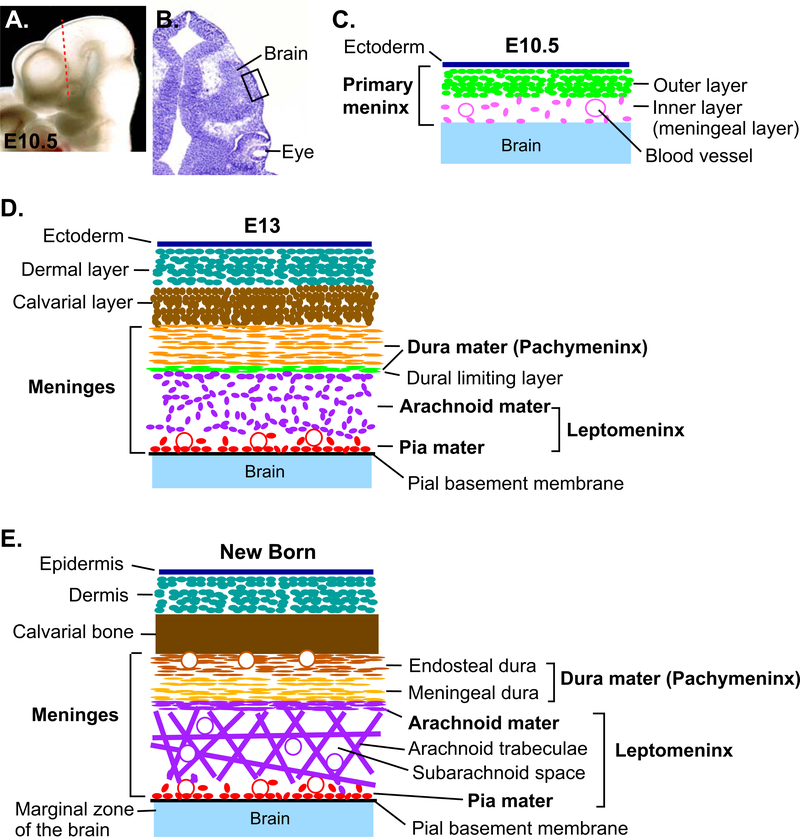
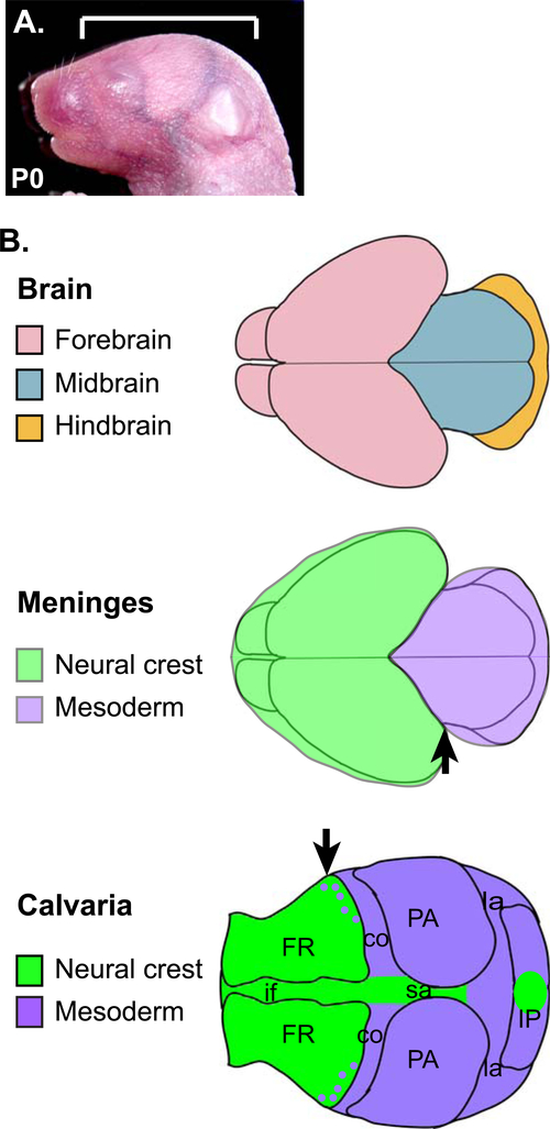
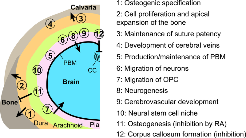
The meninges are membranous layers surrounding the central nervous system. In the head, the meninges lie between the brain and the skull, and interact closely with both during development. The cranial meninges originate from a mesenchymal sheath on the surface of the developing brain, called primary meninx, and undergo differentiation into three layers with distinct histological characteristics: the dura mater, the arachnoid mater, and the pia mater. While genetic regulation of meningeal development is still poorly understood, mouse mutants and other models with meningeal defects have demonstrated the importance of the meninges to normal development of the calvaria and the brain. For the calvaria, the interactions with the meninges are necessary for the progression of calvarial osteogenesis during early development. In later stages, the meninges control the patterning of the skull and the fate of the sutures. For the brain, the meninges regulate diverse processes including cell survival, cell migration, generation of neurons from progenitors, and vascularization. Also, the meninges serve as a stem cell niche for the brain in the postnatal life. Given these important roles of the meninges, further investigation into the molecular mechanisms underlying meningeal development can provide novel insights into the coordinated development of the head.

Keywords: brain development; calvaria; craniofacial development; head mesenchyme; meninges.


Figures

Figure 1.
Figure 1.. Development of the meninges
(A) Lateral view of the head of E10.5 mouse embryo. (B) A coronal section through the head of E10.5 mouse embryo at a position shown in A (dotted line). The section was stained with cresyl violet to show the morphology. (C) A schematic of the tissue layers from the boxed area in B. After the primary meninx is established around the brain at ~E9.5, the mesenchyme becomes divided into an outer dense layer and an inner reticular layer (meningeal layer) at ~E10.5. (D) Around E13, the meninges begin to differentiate into the pachymeninx (dura mater) and the leptomeninx (arachnoid mater and pia mater) separated by dural limiting layer. (E) Later, the dura mater appears as two tightly attached layers, the endosteal dura and the meningeal dura. The endosteal dura serves as periosteum of the inner surface of the calvarial bone. The cavitation of the leptomeninges generates arachnoid trabeculae made of collagen fibers and the fibroblasts, and the subarachnoid space inside the trabeculae.
Figure 2.
Figure 2.. Contribution of the cells from the neural crest and the mesoderm to the meninges and the calvaria
A) Lateral view of the head of a newborn mouse. The bracket indicates the area depicted in B. B) Dorsal views of the head showing the three components of the head. Note the difference in the rostro-caudal positions of the neural crest-mesoderm boundary in the meninges and in the calvaria (arrows). co: coronal suture, FR: frontal bone, if: interfrontal suture, IP: interparietal bone, la: lambdoidal suture, PA: parietal bone, sa: sagittal suture.
Figure 3.
Figure 3.. Influences of the meninges to development of the calvaria and the brain
A schematic representation of a coronal section of the head of mouse embryos at late gestation stages (>E12.5). The left-dorsal quadrant of the head is shown. The positions of the circled numbers indicate the meningeal component thought to be responsible for each interaction. For the numbers straddling the arachnoid mater and the pia mater, there is not enough evidence to assign the function to only one layer or the other, and both layers are likely involved. CC: corpus callosum, OPC: oligodendrocyte precursor cells, PBM: pial basement membrane, RA: retinoic acid.

References

    1. Adeeb N, Mortazavi MM, Tubbs RS, & Cohen-Gadol AA (2012). The cranial dura mater: a review of its history, embryology, and anatomy. Childs Nerv Syst, 28(6), 827–837. doi:10.1007/s00381-012-1744-6 - DOI - PubMed
    1. Aldinger KA, Lehmann OJ, Hudgins L, Chizhikov VV, Bassuk AG, Ades LC, … Millen KJ (2009). FOXC1 is required for normal cerebellar development and is a major contributor to chromosome 6p25.3 Dandy-Walker malformation. Nature Genetics, 41(9), 1037–U1116. doi:10.1038/ng.422 - DOI - PMC - PubMed
    1. Anazi S, Maddirevula S, Faqeih E, Alsedairy H, Alzahrani F, Shamseldin HE, … Alkuraya FS (2017). Clinical genomics expands the morbid genome of intellectual disability and offers a high diagnostic yield. Mol Psychiatry, 22(4), 615–624. doi:10.1038/mp.2016.113 - DOI - PubMed
    1. Angelov DN, & Vasilev VA (1989). Morphogenesis of rat cranial meninges. A light- and electron-microscopic study. Cell Tissue Res, 257(1), 207–216. - PubMed
    1. Antila S, Karaman S, Nurmi H, Airavaara M, Voutilainen MH, Mathivet T, … Alitalo K (2017). Development and plasticity of meningeal lymphatic vessels. Journal of Experimental Medicine, 214(12), 3645–3667. doi:10.1084/jem.20170391 - DOI - PMC - PubMed

Publication types

LinkOut - more resources