. 2019 May;110(5):1653-1664.
doi: 10.1111/cas.13979. Epub 2019 Mar 12.

Therapeutic efficacy evaluation of radioimmunotherapy with 90 Y-labeled anti-podoplanin antibody NZ-12 for mesothelioma

Affiliations

Therapeutic efficacy evaluation of radioimmunotherapy with 90 Y-labeled anti-podoplanin antibody NZ-12 for mesothelioma

Hitomi Sudo et al. Cancer Sci. 2019 May.

Abstract

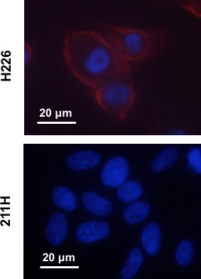
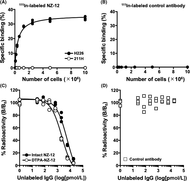
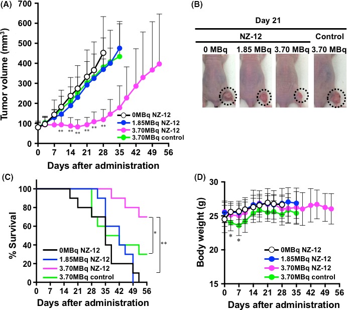
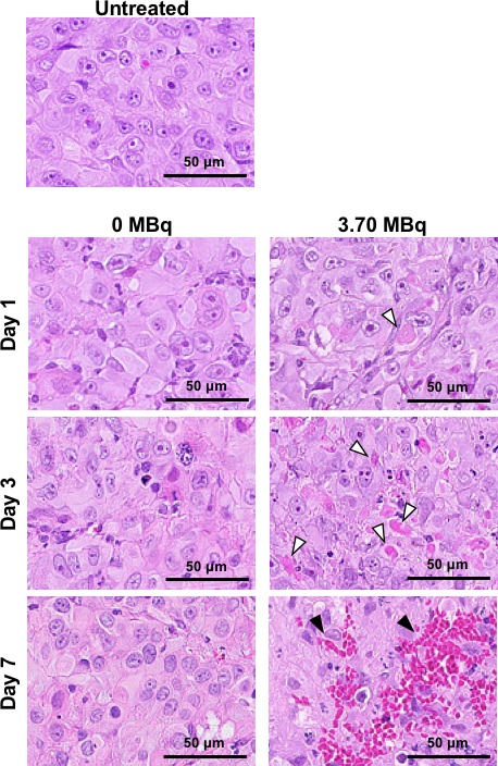
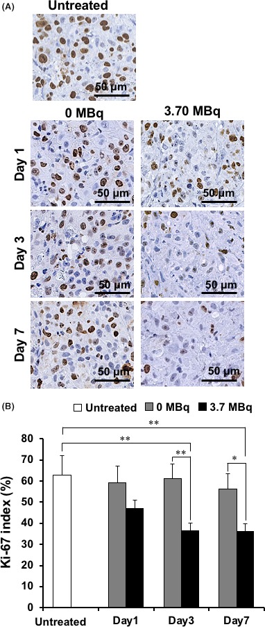
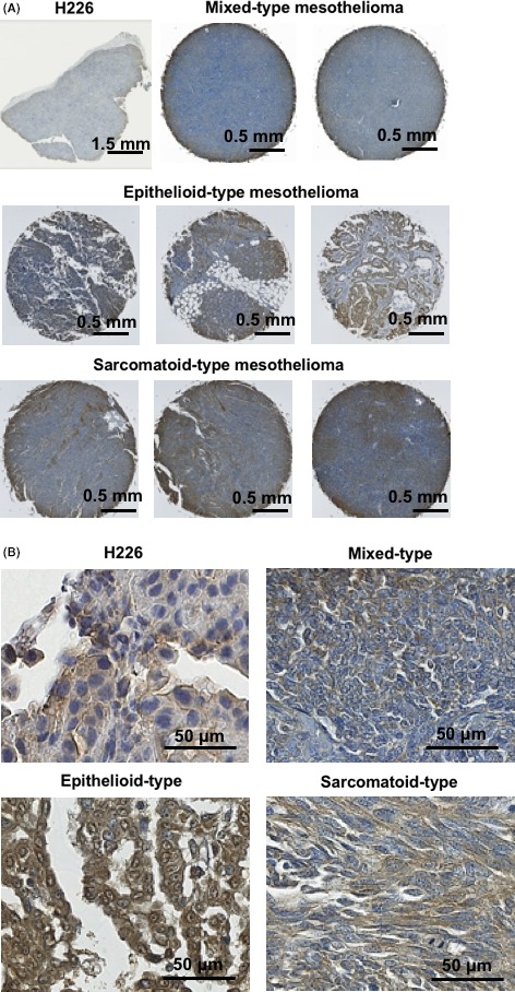
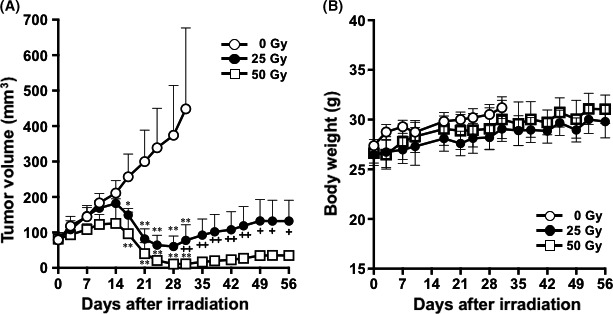
Podoplanin is a type I transmembrane sialomucin-like glycoprotein that is highly expressed in malignant mesothelioma. The rat-human chimeric antibody NZ-12 has high affinity for human podoplanin and antibody-dependent cellular cytotoxicity and is applicable for radioimmunotherapy (RIT) to enhance the antitumor effect. In the present study, we evaluated the in vivo and in vitro properties of radiolabeled NZ-12 and the antitumor effect of RIT with 90 Y-labeled NZ-12 in an NCI-H226 (H226) malignant mesothelioma xenograft mouse model. 111 In-labeled NZ-12 bound specifically to H226 cells with high affinity, and accumulation was high in H226 tumors but low in major organs. RIT with 90 Y-labeled NZ-12 significantly suppressed tumor growth and prolonged survival without body weight loss and obvious adverse effects. Higher podoplanin expression levels were observed in human mesothelioma specimens, suggesting higher tumor accumulation of 90 Y-labeled NZ-12 in patients compared with the H226 tumor xenografts. Our findings suggest that 90 Y-labeled NZ-12 is a promising RIT agent as a new therapeutic option for malignant mesothelioma that warrants further clinical studies to evaluate the dosimetry and efficacy in patients.

Keywords: antibody; malignant mesothelioma; podoplanin; radiation; radioimmunotherapy.


Conflict of interest statement

Authors declare no conflicts of interest for this article.

Figures

Figure 1
Figure 1
Immunofluorescence staining of H226 (podoplanin‐positive) and 211H (podoplanin‐negative) cells using anti‐podoplanin antibody LpMab‐17 (red). The nuclei were counterstained with DAPI (blue)
Figure 2
Figure 2
In vitro characterization of radiolabeled antibody NZ‐12. A, Cell binding assay of 111In‐labeled NZ‐12 with H226 (black circles) and 211H cells. (white circles). B, Cell binding assay of 111In‐labeled control antibody (IgG) with H226. C, Competitive inhibition assay for intact NZ‐12 (black circles) and CHX‐A′′‐DTPA‐conjugated NZ‐12 (white circles) with H226 cells. D, Competitive inhibition assay for control antibody (IgG)
Figure 3
Figure 3
Therapeutic efficacy of 90Y‐labeled antibodies in nude mice bearing H226 tumors. A, Tumor growth curves after injection with 90Y‐labeled NZ‐12 and control antibody. Data indicate mean and SD (n = 10). **< .01 vs 0 MBq of 90Y‐labeled NZ‐12 (intact NZ‐12 only). B, Representative images of mice at day 21 after injection with 90Y‐labeled NZ‐12 and control antibody. Dashed circles indicate tumors. C, Kaplan‐Meier survival curves based on the endpoints of tumor volume of 500 mm3. **< .01, 0 MBq and 1.85 MBq of 90Y‐labeled NZ‐12 vs 3.70 MBq of 90Y‐labeled NZ‐12; *< .05, 3.70 MBq of 90Y‐labeled control antibody vs 3.70 MBq of 90Y‐labeled NZ‐12. D, Body weight changes after injection with 90Y‐labeled NZ‐12 and control antibody. Data indicate mean and SD (n = 10). *< .05 vs 0 MBq of 90Y‐labeled NZ‐12
Figure 4
Figure 4
Histological analysis of H226 tumors after injection with 0 MBq (intact NZ‐12 only) and 3.70 MBq of 90Y‐labeled NZ‐12. Hematoxylin‐and‐eosin‐stained sections of H226 tumors at days 1, 3, and 7 after injection. White arrowheads indicate necrosis and black arrowheads indicate hemorrhage
Figure 5
Figure 5
Tumor cell proliferation analysis by Ki‐67 immunostaining. A, Ki‐67‐stained sections of H226 tumors at days 1, 3, and 7 after injection with 0 MBq (intact NZ‐12 only) and 3.70 MBq of 90Y‐labeled NZ‐12. B, Quantification of proliferating (Ki‐67 positive) cells. Data are mean and SD. **< .01, *< .05
Figure 6
Figure 6
Podoplanin expression analysis in human mesothelioma specimens. A, Representative images of immunohistochemical staining with LpMab17. B, High magnification images of (A)
Figure 7
Figure 7
Therapeutic efficacy of external‐beam radiation with X‐rays in nude mice bearing H226 tumors. A, Tumor growth curves after irradiation with X‐rays. Data indicate mean and SD (n = 5). **< .01 and *< .05 vs 0 Gy. ++ < .01 and + < .05, 25 Gy vs 50 Gy. B, Body weight changes after irradiation with X‐rays. Data indicate mean and SD (n = 5)

Similar articles

Cited by

References

    1. Robinson BW, Lake RA. Advances in malignant mesothelioma. N Engl J Med. 2005;353(15):1591‐1603. - PubMed
    1. Sterman DH, Albelda SM. Advances in the diagnosis, evaluation, and management of malignant pleural mesothelioma. Respirology. 2005;10(3):266‐283. - PubMed
    1. Taioli E, Wolf AS, Camacho‐Rivera M, et al. Determinants of survival in malignant pleural mesothelioma: a surveillance, epidemiology, and end results (SEER) study of 14,228 patients. PLoS One. 2015;10(12):e0145039. - PMC - PubMed
    1. Vogelzang NJ, Rusthoven JJ, Symanowski J, et al. Phase III study of pemetrexed in combination with cisplatin versus cisplatin alone in patients with malignant pleural mesothelioma. J Clin Oncol. 2003;21(14):2636‐2644. - PubMed
    1. Kindler HL, Ismaila N, Armato S 3rd, et al. Treatment of malignant pleural mesothelioma: American Society of Clinical Oncology Clinical Practice Guideline. J Clin Oncol. 2018;36(13):1343‐1373. - PMC - PubMed

MeSH terms