. 2019 Feb 25;15(2):e1008006.
doi: 10.1371/journal.pgen.1008006. eCollection 2019 Feb.

The C-terminal region of Net1 is an activator of RNA polymerase I transcription with conserved features from yeast to human

Affiliations

The C-terminal region of Net1 is an activator of RNA polymerase I transcription with conserved features from yeast to human

Katharina Hannig et al. PLoS Genet. .

Abstract

RNA polymerase I (Pol I) synthesizes ribosomal RNA (rRNA) in all eukaryotes, accounting for the major part of transcriptional activity in proliferating cells. Although basal Pol I transcription factors have been characterized in diverse organisms, the molecular basis of the robust rRNA production in vivo remains largely unknown. In S. cerevisiae, the multifunctional Net1 protein was reported to stimulate Pol I transcription. We found that the Pol I-stimulating function can be attributed to the very C-terminal region (CTR) of Net1. The CTR was required for normal cell growth and Pol I recruitment to rRNA genes in vivo and sufficient to promote Pol I transcription in vitro. Similarity with the acidic tail region of mammalian Pol I transcription factor UBF, which could partly functionally substitute for the CTR, suggests conserved roles for CTR-like domains in Pol I transcription from yeast to human.


Conflict of interest statement

The authors have declared that no competing interests exist.

Figures
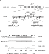
Fig 1
Fig 1. Schematic representation of the ribosomal DNA (rDNA) locus and the Net1 protein of S. cerevisiae.
(A) On top: schematic representation of the yeast rDNA locus on the right arm of chromosome XII (CHR XII), consisting of 100–300 repeats (cartoon on the top). In the middle: enlargement of one rDNA repeat depicting 35S rDNA transcription units, intergenic sequences 1 and 2 (IGS1,2), the 5S rDNA, an autonomous replication sequence (ARS), and a bi-directional Pol II-dependent promoter (E-pro). Positions of restriction sites (E, EcoRI; X, XcmI) used in ChEC and psoralen crosslinking experiments are depicted. At the bottom: cis elements at the 5’- (promoter) and 3’- (E-element) region of the 35S rDNA, including the core element (CE), the upstream element (UE), replication fork barrier element (RFB), and the Pol I transcription termination site (Term). Arrows at promoter regions point in the direction of transcription. B) Schematic representation of the Net1 protein, and the C-terminal and N-terminal truncated version of the protein analyzed in this study (Net1ΔCTR and CTR, respectively). Amino acids included in the different proteins are depicted on the right. Different domains within the Net1 sequence interacting with Cdc14, Fob1 and Sir2 are depicted at the bottom, and represented in the cartoon as dotted, and striped rectangles, respectively. Black bars denote the positions of two pfam motifs found in Net1. The positions of acknowledged phosphorylation sites are shown on top of the cartoon representing the full-length Net1 protein.
Fig 2
Fig 2. Yeast strains expressing Net1 with a C-terminal deletion are affected in growth but produce a nucleolar protein which still interacts with Cdc14 and Sir2.
(A, B) Diploid yeast strains (W13533, W13534, W13535, W13536, W13537, W13538, W13539, W11979) were sporulated yielding haploid progenies carrying a deletion of NET1 (net1Δ), or alleles for expression of (truncated) Net1GFP fusion proteins encompassing the amino acids of the full-length Net1 as indicated on the left. All progenies expressed Nop56mCherry fusion protein as nucleolar marker. A) Serial dilutions of cell suspensions of the haploid progenies of the diploid strains were spotted on XYD plates and incubated at 25°C for 2d before a photograph was taken. Cartoons of the respective Net1GFP proteins are depicted on the right according to Fig 1B. B) Growth of individual haploid progenies of the diploid strains in YPD media at 30°C was determined using a TECAN plate reader system. Haploid wildtype strains y3290 and y3298 were cultured in parallel (NET1). The mean doubling time and standard deviation error was calculated from two to three independent biological and two independent technical replicates. The number (n) of independent measurements for the respective genotype is indicated on top of the bars. See S1 Dataset for raw data. C) Diploid yeast strains (W13776, W13777, W13778, W13779) were sporulated yielding haploid progenies expressing either Cdc14myc or Sir2myc and the indicated Net1FLAG, Net1ΔCTRFLAG, or the untagged wild-type Net1 protein. Whole-cell extracts (WCE) of the haploid strains were subjected to immuno-precipitation (IP) with anti-FLAG M2 antibody agarose (αFLAG-IP). Proteins were analyzed in a western blot with anti-Myc and anti-FLAG antibodies (αMyc,αFLAG). A fluorograph of the western blot membrane is shown. The size of molecular weight markers in kDa is indicated on the left. Positions of the tagged proteins are indicated on the right. D-E) Diploid yeast strains (W13533, W13534) were sporulated, yielding haploid progenies expressing Nop56mCherry and either Net1GFP, or Net1ΔctrGFP. Diploid parental strains (D, F) and haploid progenies (E, G) were subjected to live cell fluorescence microscopy. Data was collected for differential interference contrast (DIC, first lane of panels), mCherry, and GFP fluorescence (second, and third lane of panels, respectively). mCherry and GFP signals were merged in the fourth lane of panels. A scale bar (5μm) is shown in the first lane of panels.
Fig 3
Fig 3. Expression of the CTR in trans rescues growth defects and Nop56 delocalization in net1Δctr strains.
(A, B) Diploid yeast strains (W12509, W12533, W13762) were sporulated. Haploid progenies carried a deletion of NET1 (net1Δ), or alleles for expression of the indicated truncated Net1 proteins. Where indicated (+) strains co-expressed chromosomally encoded GFPCTR. Growth analyses on plates and in liquid culture were performed with individual haploid progenies of the diploid strains as described in the legend to Fig 2A and 2B. Yeast strains carrying a NET1 wild-type allele (WT, K699 (A), and y3290 and y3298 (B)) were included as controls. C, D) The diploid yeast strain W15407 was sporulated, yielding haploid progenies carrying either a NET1 wild-type allele (C) or a net1Δctr allele (D), and expressing Nop56mCherry and GFPCTR. These haploid strains were subjected to live cell fluorescence microscopy as described in the legend to Fig 2D.
Fig 4
Fig 4. 35S rDNA promoter association of RENT complex components is impaired in net1Δctr strains.
Haploid yeast strains expressing the indicated proteins C-terminally fused to MNHA were subjected to ChEC, or ChIP experiments. A, C) Crude nuclei suspensions were subjected to ChEC in the presence of calcium for the indicated times (min ChEC), DNA was isolated, cut with XcmI, separated in a 1% agarose gel and subjected to Southern blot analysis by indirect end-labeling using the radiolabeled probes rDNp, IGS2. An autoradiograph of the membrane is shown. Cartoons at the sites depict a map of the detected genomic fragments including probe hybridization sites and important cis elements described in the legend to Fig 1A. Arrows at the top of the cartoons point to the uncut full-length XcmI fragments. An asterisk marks the site of a DNA double-strand break occurring at the 3’ end of the RFB in the absence of ChEC. B, D) fragmented chromatin was subjected to ChIP with the indicated MNHA tagged fusion proteins using anti-HA (3F10) antibody. DNA co-precipitating with the tagged proteins and from input fractions was isolated and analyzed by quantitative PCR with different primer pairs amplifying distinct regions (1–6) within the rDNA locus indicated in the cartoon at the bottom. Bar graphs depict the efficiency of co-precipitation of the fragments as percentages of the input DNA (% ChIP). Mean values and standard deviation errors were derived from three independent ChIP experiments analyzed each in triplicate qPCR reactions (n = 9). A, B) ChEC and ChIP analyses with strains (y3041, y3042, y3043, y3044) expressing either Net1-MNHA or Net1ΔCTR-MNHA (ΔCTR-MNHA). C, D) ChEC and ChIP analyses with strains (y3058, y3068, y3250), carrying a NET1 or a net1Δctr allele, and expressing Cdc14-MNHA, in the absence or presence of a chromosomally integrated FLAGCTR expression cassette.
Fig 5
Fig 5
Robust Pol I association with 35S rRNA genes requires the CTR. (A, B) Haploid yeast strains (y3288, y3289, y3299, y3302, y3595, y3597, y3599, y3601) carrying a NET1 or a net1Δctr allele, and expressing Rpa43-MNHA or Rpa190-MNHA in the absence or presence of the Net1 FLAGCTR were subjected to ChEC (A) and ChIP (B) analyses as described in the legend to Fig 4.
Fig 6
Fig 6
The CTR of Net1 interacts with many chromosomal loci and rescues slow growth in net1Δctr strains when fused to Rpa190. (A, B) Haploid yeast strains (y3087; y3157; y3160) carrying a NET1 or a net1Δctr allele and expressing CTR-MNHA or Net1-MNHA were subjected to ChEC analyses, as described in the legend to Fig 4. B) Agarose gels were stained with SYBR safe before transfer to the blotting membrane (upper panels), sizes of selected DNA fragments in the DNA marker (M) are given on the left. Lower panels, the membrane was hybridized with a radioactively labeled probe hybridizing with a XcmI fragment containing the NUP57/RPS23A loci as indicated in the cartoon on the left. C, D) Haploid yeast strains (y3673; y3674; y3746; y3747; y3748; y3749; y3756; y3757; y3758; y3759, y3290, y3298) carrying a NET1 or a net1Δctr allele and expressing CTR fusion proteins of Rpa190, or Rrn7 or the wild-type proteins, were subjected to growth analyses in liquid cultures (C), and on solid medium (D) as described in the legend to Fig 2A and 2B.
Fig 7
Fig 7
The CTR is differentially phosphorylated in different growth states in vivo and CTR-dependent stimulation of Pol I transcription in vitro is decreased upon λ-protein phosphatase treatment. (A) The CTR associates with Pol I. Diploid yeast strains (W12903, W13491) were sporulated yielding haploid progenies expressing FLAGCTR and the wild-type Pol I subunit Rpa43, or Rpa43myc. WCEs were prepared and subjected to IP experiments with anti-Myc antibody agarose followed by western blot analysis as described in the legend to Fig 2C. IgG on the right marks the positions of antibody chains in IPs. B) The CTR is hyperphosphorylated in vivo. FLAGCTR in WCE of the diploid yeast strain W13490 was bound to anti-FLAG M2 antibody agarose and treated with increasing amounts (grey triangle) of λ-protein phosphatase (λ-PP). Protein samples were analyzed by western blot as described in the legend to Fig 2C. C) The CTR is differentially phosphorylated in vivo. Haploid yeast strain y3739 expressing FLAGCTR was grown to exponential (exp), or to stationary phase (stat). WCE was prepared and subjected to western blot analysis with anti-FLAG antibody (αFLAG) as described in the legend to Fig 2C. Ponceau S stain of the membrane, and an image visualizing chemiluminescence signals upon immuno-detection (αFLAG) are shown. D) Recombinant CTR expressed in insect cells is hyperphosphorylated. Net1 CTRTAP expressed in insect cells was bound to IgG-magnetic beads. Net1 CTRTAP bound to the beads was either mock treated (-) or incubated with λ-PP (+). CTRCBP fusion proteins were released by TEV protease cleavage and analyzed by SDS-PAGE and Coomassie staining. A photograph of the gel is shown. The size of molecular weight markers in kDa is indicated on the left. Positions of the tagged proteins as well as TEV protease are indicated on the right. E) CTR-dependent stimulation of Pol I transcription in vitro is decreased after λ-PP treatment. In vitro transcription reactions were carried out using Pol I purified from yeast, recombinant CF and Rrn3 purified from E. coli in the absence (-) or presence of increasing amounts (grey triangle) of CTRCBP, either mock treated or incubated with λ-PP as described in (D). Radiolabeled RNA was isolated and separated by 6% acrylamide-urea gel electrophoresis. An autoradiograph is shown. The position of the specific transcript is indicated on the right.
Fig 8
Fig 8. The acidic region of human UBF1 shares conserved features with the CTR and can partially rescue the growth defect of net1Δctr strains.
(A) Local pairwise sequence alignment of human UBF1 (hUBF1) with Net1. Top, schematic representation of hUBF1 protein, depicting amino acid positions of full-length hUBF1, HMG boxes (grey rectangles), acidic tail region (AR, hexagon in light grey), and extended acidic tail region (eAR, black bar on the top). Bottom, sequence alignment of hUBF1 with Net1 using the “matcher” program EMBL-EBI Job Dispatcher framework (https://www.ebi.ac.uk/Tools/psa/emboss_matcher/). Identical (|), and similar (:) amino acid residues are indicated. Amino acid numbering of the full-length hUBF1 and yeast Net1 are indicated on the left, asterisks mark serine residues reported to be targets of phosphorylation (https://www.phosphosite.org/; https://www.yeastgenome.org), predicted CK2 phosphorylation sites are printed in red (http://www.cbs.dtu.dk/services/NetPhos/). A serine/aspartate rich amino acid stretch within the CTR is underlined. B-D) Diploid yeast strains were sporulated yielding haploid progenies expressing either wild-type Net1 or different Net1-MNHA fusion proteins in which the CTR was either conserved (Net1-MNHA) deleted (Net1ΔCTR-MNHA), or replaced by the AR (Net1ΔCTR-AR-MNHA), or eAR regions (Net1ΔCTR-eAR-MNHA), or a synthetic, minimal transactivation domain of the viral VP16 protein (Net1ΔCTR-VP-MNHA). B) Schematic representation of Net1-MNHA fusion proteins expressed in the respective yeast strains. Net1 domains are depicted as described in the legend to Fig 1B, UBF1 domains are depicted as described in (A). The minimal transactivation domain of VP16 is depicted as a circle. C) WCE of overnight cultures of haploid progenies (y4065, y4067, y4069, y4071, y4073), expressing the indicated Net1-MNHA fusion proteins were subjected to western blot analysis as described in the legend to Fig 2C, using anti-HA antibody (αHA, top panel), or a tubulin antibody (αtubulin, bottom panel). Images visualizing chemiluminescence on the membranes are shown. D) Growth analyses in liquid culture were performed with the indicated haploid derivatives of yeast strains (y4038, y4039, y4040, y4041, y4042, y4043, y4044, y4045, y4046) as described in the legend to Fig 2B. Differences in doubling times of Net1ΔCTR-AR-MNHA and Net1ΔCTR-eAR-MNHA strain relative to the Net1ΔCTR-MNHA expressing strain were tested using the nonparametric Mann-Whitney U test; ***P<0.001, ****P<0.0001.

Similar articles

Cited by

References

    1. Warner JR. The economics of ribosome biosynthesis in yeast. Trends Biochem Sci. 1999. November;24(11):437–40. - PubMed
    1. Goodfellow SJ, Zomerdijk JCBM. Basic mechanisms in RNA polymerase I transcription of the ribosomal RNA genes. Subcell Biochem. 2013;61:211–36. 10.1007/978-94-007-4525-4_10 - DOI - PMC - PubMed
    1. Moss T, Langlois F, Gagnon-Kugler T, Stefanovsky V. A housekeeper with power of attorney: the rRNA genes in ribosome biogenesis. Cell Mol Life Sci CMLS. 2007. January;64(1):29–49. 10.1007/s00018-006-6278-1 - DOI - PMC - PubMed
    1. Boukhgalter B, Liu M, Guo A, Tripp M, Tran K, Huynh C, et al. Characterization of a fission yeast subunit of an RNA polymerase I essential transcription initiation factor, SpRrn7h/TAF(I)68, that bridges yeast and mammals: association with SpRrn11h and the core ribosomal RNA gene promoter. Gene. 2002. May 29;291(1–2):187–201. - PubMed
    1. Keys DA, Vu L, Steffan JS, Dodd JA, Yamamoto RT, Nogi Y, et al. RRN6 and RRN7 encode subunits of a multiprotein complex essential for the initiation of rDNA transcription by RNA polymerase I in Saccharomyces cerevisiae. Genes Dev. 1994. October 1;8(19):2349–62. - PubMed

Publication types

MeSH terms