. 2019 Feb 25;13(2):e0007206.
doi: 10.1371/journal.pntd.0007206. eCollection 2019 Feb.

In vitro activity and mode of action of phenolic compounds on Leishmania donovani

Affiliations

In vitro activity and mode of action of phenolic compounds on Leishmania donovani

Christine Achiaa Antwi et al. PLoS Negl Trop Dis. .

Abstract

Background: Leishmaniasis is a disease caused by the protozoan parasite, Leishmania. The disease remains a global threat to public health requiring effective chemotherapy for control and treatment. In this study, the effect of some selected phenolic compounds on Leishmania donovani was investigated. The compounds were screened for their anti-leishmanial activities against promastigote and intracellular amastigote forms of Leishmania donovani.

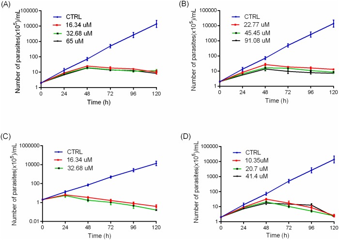
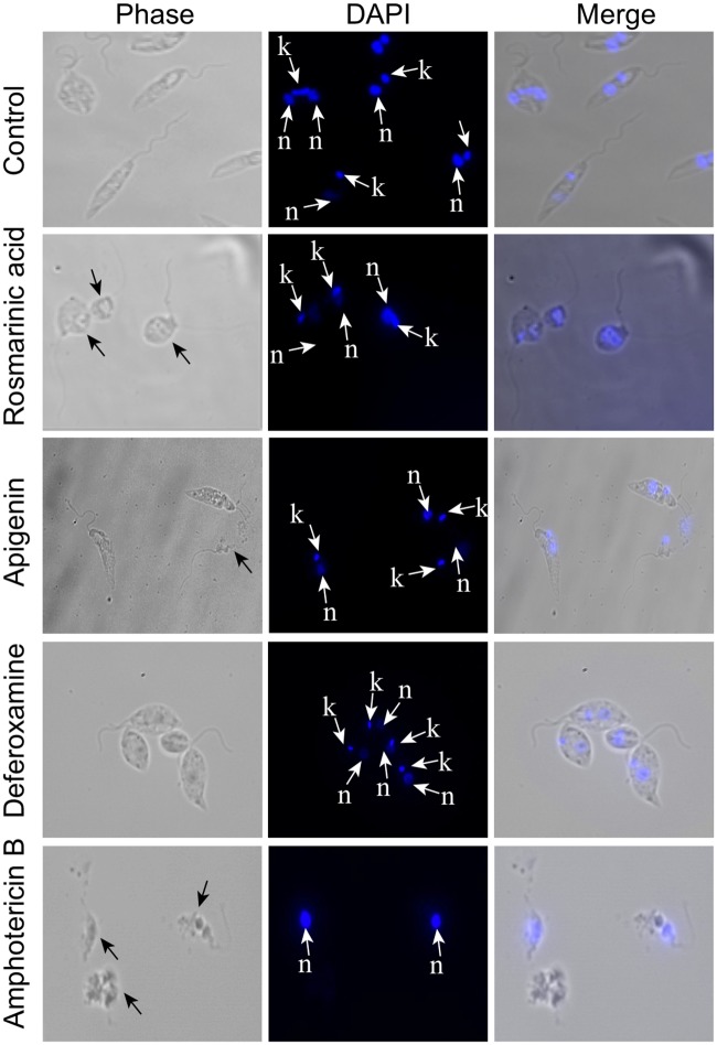
Methodology/principal findings: The dose dependent effect and cytotoxicity of the compounds were determined by the MTT assay. Flow cytometry was used to determine the effect of the compounds on the cell cycle. Parasite morphological analysis was done by microscopy and growth kinetic studies were conducted by culturing cells and counting at 24 hours intervals over 120 hours. The cellular levels of iron in promastigotes treated with compounds was determined by atomic absorption spectroscopy and the effect of compounds on the expression of iron dependent enzymes was investigated using RT-qPCR. The IC50 of the compounds ranged from 16.34 μM to 198 μM compared to amphotericin B and deferoxamine controls. Rosmarinic acid and apigenin were the most effective against the promastigote and the intracellular amastigote forms. Selectivity indexes (SI) of rosmarinic acid and apigenin were 15.03 and 10.45 respectively for promastigotes while the SI of 12.70 and 5.21 respectively was obtained for intracellular amastigotes. Morphologically, 70% of rosmarinic acid treated promastigotes showed rounded morphology similar to the deferoxamine control. About 30% of cells treated with apigenin showed distorted cell membrane. Rosmarinic acid and apigenin induced cell arrest in the G0/G1 phase in promastigotes. Elevated intracellular iron levels were observed in promastigotes when parasites were treated with rosmarinic acid and this correlated with the level of expression of iron dependent genes.

Conclusions/significance: The data suggests that rosmarinic acid exerts its anti-leishmanial effect via iron chelation resulting in variable morphological changes and cell cycle arrest.


Conflict of interest statement

The authors have declared that no competing interests exist.

Figures

Fig 1
Fig 1. Structures of selected phenolic compounds.
Fig 2
Fig 2. Growth kinetics of L. donovani promastigotes.
Cells were treated with different concentrations of (A) rosmarinic acid (B) apigenin (C) amphotericin B and (D) deferoxamine. The growth kinetics curve was determined by counting parasites every 24 hours over a period of 120 hours. CTRL = control. Points represent mean ± SD from three independent experiments.
Fig 3
Fig 3. Effect of phenolic compounds on cell morphology.
Promastigotes were cultivated in the presence or absence of test compounds (apigenin, rosmarinic acid) and control compounds (amphotericin B, deferoxamine) for 24 hours, stained with DAPI and examined by phase contrast microscopy and fluorescence microscopy at x100. Nucleus—n, Kinetoplast—k.
Fig 4
Fig 4. Effects of compounds on the structure of mitochondria.
Promastigotes were cultivated in the presence and absence of rosmarinic acid, apigenin, amphotericin B and deferoxamine for 24 hours and stained with MitoTracker Red CMXROS and DAPI, examined by phase contrast and fluorescence microscopy at x100. Mitochondria—mt, Nucleus—n, Kinetoplast—k.
Fig 5
Fig 5. Cell cycle progression of L. donovani.
Flow cytometry histograms of (A) untreated parasites, (B) apigenin, (C) rosmarinic acid, (D) deferoxamine and (E) amphotericin B after 24 hours incubation. Bar charts (F) are the quantification of cell count in G0/G1, S, G2 and M phases of parasites treated with respective concentrations of the compounds. *P<0.05, **P<0.005 were considered significant compared to the untreated promastigotes using Student’s t test.
Fig 6
Fig 6. Iron content analyses.
(A) Intracellular iron content in promastigotes, (B) iron content in unused and spent culture medium was determined by atomic absorption spectrophotometry. Error bars represents five technical replicates of two independent test. *P <0.05, **P<0.005. Student’s t test and Dunn’s multiple comparison test was used to analyse the data.
Fig 7
Fig 7. Gene expression of iron dependent proteins.
Expression of (A) LIT1, (B) FeSOD and (C) RNR were determined by qRT-PCR. Promastigotes were cultured with the compounds for 24 hours. The data presented is from three independent experiments.
Fig 8
Fig 8. Infectivity index of compounds.
The parasite load in infected macrophages was determined by counting the number of intracellular amastigotes per 100 macrophages. The data represents the mean±SD of three independent experiments in triplicate *P<0.05, **P<0.005, ***P≤0.0005 were considered significant, compared to the untreated cells using student’s t test.

Similar articles

Cited by

References

    1. den Boer M, Argaw D, Jannin J, Alvar J. Leishmaniasis impact and treatment access. Clin Microbiol Infect. 2011;17(10):1471–7. 10.1111/j.1469-0691.2011.03635.x - DOI - PubMed
    1. Akhoundi M, Kuhls K, Cannet A, Votypka J, Marty P, Delaunay P, et al. A Historical Overview of the Classification, Evolution, and Dispersion of Leishmania Parasites and Sandflies. PLoS Negl Trop Dis. 2016;10(3):e0004349 10.1371/journal.pntd.0004349 - DOI - PMC - PubMed
    1. Steverding D. The history of leishmaniasis. Parasit Vectors. 2017;10(1):82 10.1186/s13071-017-2028-5 - DOI - PMC - PubMed
    1. Das VN, Pandey RN, Siddiqui NA, Chapman LA, Kumar V, Pandey K, et al. Longitudinal Study of Transmission in Households with Visceral Leishmaniasis, Asymptomatic Infections and PKDL in Highly Endemic Villages in Bihar, India. PLoS Negl Trop Dis. 2016;10(12):e0005196 10.1371/journal.pntd.0005196 - DOI - PMC - PubMed
    1. Croft SL, Coombs GH. Leishmaniasis--;current chemotherapy and recent advances in the search for novel drugs. Trends Parasitol. 2003;19(11):502–8. - PubMed

Publication types