. 2019 Feb 25;15(2):e1007590.
doi: 10.1371/journal.ppat.1007590. eCollection 2019 Feb.

PML nuclear body-residing proteins sequentially associate with HPV genome after infectious nuclear delivery

Affiliations

PML nuclear body-residing proteins sequentially associate with HPV genome after infectious nuclear delivery

Lucile Guion et al. PLoS Pathog. .

Abstract

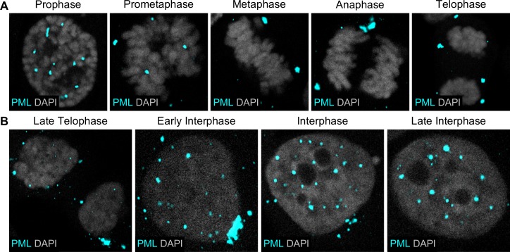
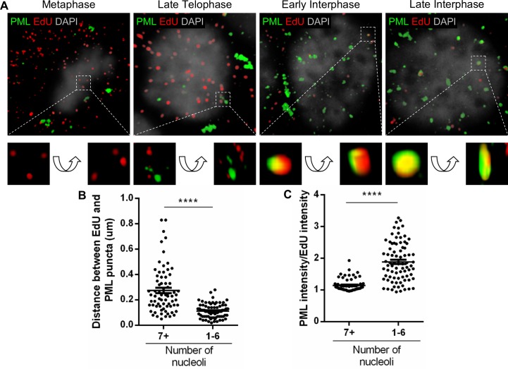
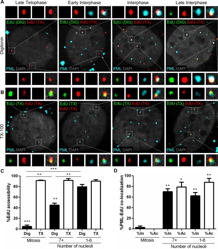
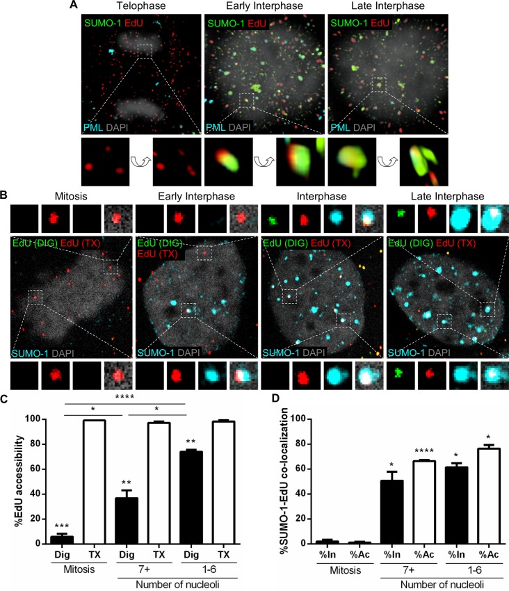
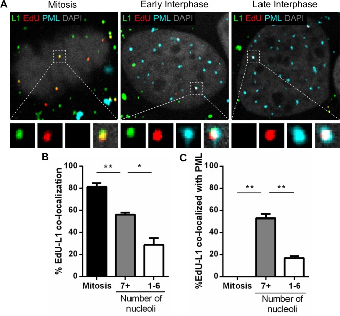
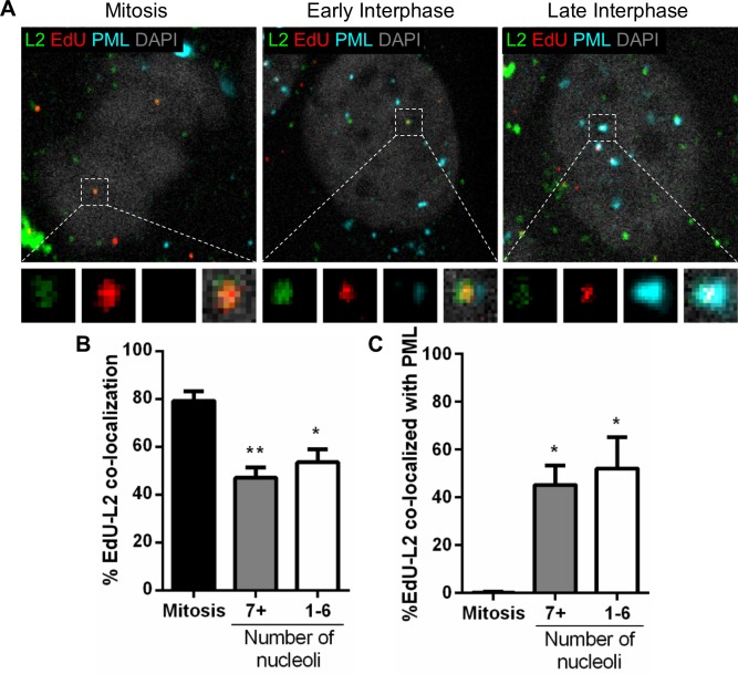
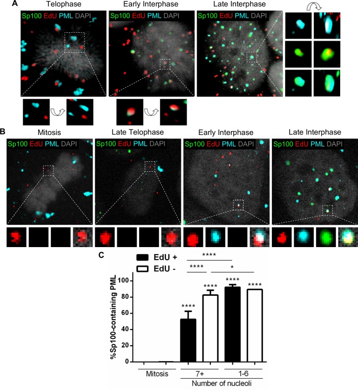
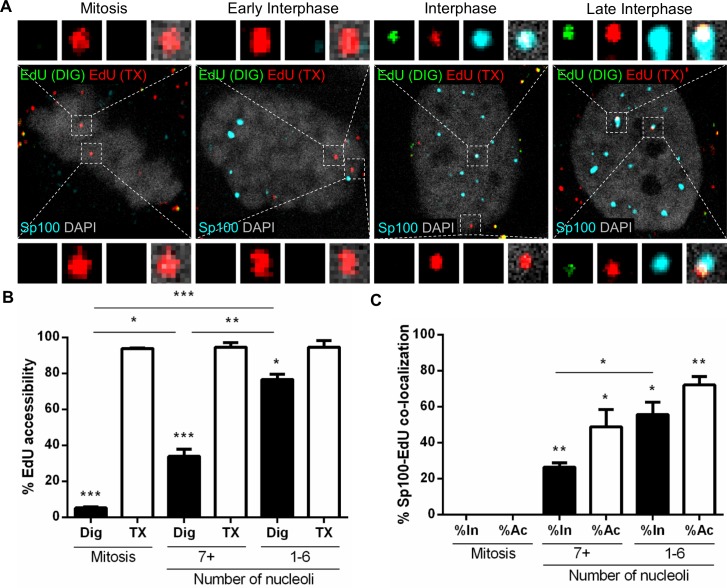
Subnuclear promyelocytic leukemia (PML) nuclear bodies (NBs) are targeted by many DNA viruses after nuclear delivery. PML protein is essential for formation of PML NBs. Sp100 and Small Ubiquitin-Like Modifier (SUMO) are also permanently residing within PML NBs. Often, large DNA viruses disassemble and reorganize PML NBs to counteract their intrinsic antiviral activity and support establishment of infection. However, human papillomavirus (HPV) requires PML protein to retain incoming viral DNA in the nucleus for subsequent efficient transcription. In contrast, Sp100 was identified as a restriction factor for HPV. These findings suggested that PML NBs are important regulators of early stages of the HPV life cycle. Nuclear delivery of incoming HPV DNA requires mitosis. Viral particles are retained within membrane-bound transport vesicles throughout mitosis. The viral genome is released from transport vesicles by an unknown mechanism several hours after nuclear envelope reformation. The minor capsid protein L2 mediates intracellular transport by becoming transmembranous in the endocytic compartment. Herein, we tested our hypothesis that PML protein is recruited to incoming viral genome prior to egress from transport vesicles. High-resolution microscopy revealed that PML protein, SUMO-1, and Sp100 are recruited to incoming viral genomes, rather than viral genomes being targeted to preformed PML NBs. Differential immunofluorescent staining suggested that PML protein and SUMO-1 associated with transport vesicles containing viral particles prior to egress, implying that recruitment is likely mediated by L2 protein. In contrast, Sp100 recruitment to HPV-harboring PML NBs occurred after release of viral genomes from transport vesicles. The delayed recruitment of Sp100 is specific for HPV-associated PML NBs. These data suggest that the virus continuously resides within a protective environment until the transport vesicle breaks down in late G1 phase and imply that HPV might modulate PML NB assembly to achieve establishment of infection and the shift to viral maintenance.


Conflict of interest statement

The authors have declared that no competing interests exist.

Figures

Fig 1
Fig 1. PML protein dynamics is used to approximate time during and after mitosis.
HaCaT cells were grown on glass coverslips, then fixed, permeabilized, incubated with rabbit anti-PML protein antibody (cyan), and mounted with DAPI (white). Confocal images show cells in all stages of mitosis (A) and end of mitosis and stages of interphase (B).
Fig 2
Fig 2. PML protein encompasses incoming viral genomes only after nuclear delivery.
HaCaT cells were infected with EdU-labeled PsVs for 24 h, fixed, permeabilized, and treated with Click-iT reaction buffer with AF555 dye to stain EdU-labeled pseudogenomes (red). Next, cells were incubated with mouse anti-PML protein antibody (green) and mounted in DAPI (white). (A) Images show 3D reconstruction of high resolution z-stacks and close-up images were rotated at a 45° angle on the x, y, z axes in the 3D-rendered images. (B) Distance (μm) between center of EdU and center of PML protein puncta was measured in NIS Elements. (C) PML and EdU mean intensity were measured in NIS Elements, normalized to ROI area, and the ratio was calculated. (B and C) Nucleoli were also counted: 7+ nucleoli corresponds to early interphase, 1–6 to late interphase. Results are shown as average of 2 independent experiments and SEM, with 30–50 cells of each condition (early and late interphase cells) collected in z-stacks spanning the whole nucleus. (B) 7+ nucleoli: 0.275 μm ± 0.021 μm; 1–6 nucleoli: 0.113 μm ± 0.006 μm. (C) 7+ nucleoli: 1.146 ± 0.025; 1–6 nucleoli: 1.888 ± 0.069. p value was determined using Student’s t-test comparing Early to Late Interphase. ****: p < 0.0001.
Fig 3
Fig 3. Viral genome associates with PML protein while still inaccessible to small molecular dyes.
(A and B) HaCaT cells were infected with EdU-labeled PsVs for 24 h, fixed, permeabilized with 0.625 μg/mL digitonin (A) or 0.5% TX-100 (B), and treated with AF555 (green) in Click-iT reaction buffer. Next, the cells were permeabilized again with 0.5% TX-100 and treated with AF647 (red) in Click-iT reaction buffer. Lastly, cells were incubated with rabbit anti-PML protein antibody (cyan) and mounted with DAPI (white). (C and D) Percent accessibility of viral genome (C) was determined by manually counting the number of red only (inaccessible [In]) or red/green (accessible [Ac]) EdU puncta over the total number of EdU puncta associated with condensed chromosomes in mitotic cells or nuclear-localized in interphase cells in digitonin- or TX-100-permeabilized cells. Nucleoli were also counted: 7+ nucleoli corresponds to early interphase, 1–6 to late interphase. Co-localization of PML protein and EdU (D) was quantified by manually counting the number of EdU puncta that co-localized with PML protein signal over the total number of accessible or inaccessible EdU puncta associated with condensed chromosomes in mitotic cells or nuclear-localized in interphase in digitonin-permeabilized cells. Results are shown as average of 3 independent experiments and SEM, with 30–50 cells of each condition (mitotic, early and late interphase cells) collected in z-stacks spanning the whole nucleus. (C) Mitosis: Dig: %Ac = 4.90% ± 2.55%; TX: %Ac = 91.37% ± 0.77%. 7+ nucleoli: Dig: %Ac = 45.05% ± 3.80%; TX: %Ac = 91.62% ± 3.38%. 1–6 nucleoli: Dig: %Ac = 79.55% ± 4.38%; TX: %Ac = 90.35% ± 2.63%. (D) Mitosis: %In at PML = 2.73% ± 2.73%; %Ac at PML = 0% ± 0%. 7+ nucleoli: %In at PML = 70.09% ± 5.43%; %Ac at PML = 78.85% ± 9.05%. 1–6 nucleoli: %In at PML = 62.22% ± 6.19%; %Ac at PML = 76.43% ± 7.57%. p value was determined using Student’s t-test comparing Dig to TX and Dig to Dig (C) or Interphase to Mitosis (D). ***: p < 0.001. **: p < 0.01. *: p < 0.05. ns: p > 0.05.
Fig 4
Fig 4. Viral genome associates with SUMO-1 while still inaccessible to small molecular dyes.
(A) HaCaT cells were infected with EdU-labeled PsVs for 24 h, fixed, permeabilized, and treated with AF555 (red) in Click-iT reaction buffer. Next, cells were incubated with rabbit anti-SUMO-1 antibody (green) and mouse anti-PML protein antibody (cyan) and mounted in DAPI (white). Images show 3D reconstruction of high resolution z-stacks. Close-up images were rotated at a 45° angle on the x, y, z axes in the 3D-rendered images. (B) HaCaT cells were infected with EdU-labeled PsVs for 24 h, fixed, permeabilized with 0.625 μg/mL digitonin and treated with AF555 (green) in Click-iT reaction buffer. Next, the cells were permeabilized again with 0.5% TX-100 and treated with AF647 (red) in Click-iT reaction buffer. Lastly, cells were incubated with rabbit anti-SUMO-1 antibody (cyan) and mounted with DAPI (white). (C and D) Percent accessibility of viral genome (C) was determined by manually counting the number of red only (inaccessible [In]) or red/green (accessible [Ac]) EdU puncta over the total number of EdU puncta associated with condensed chromosomes in mitotic cells or nuclear-localized in interphase cells in digitonin- or TX-100-permeabilized cells. Nucleoli were also counted: 7+ nucleoli corresponds to early interphase, 1–6 to late interphase. Co-localization of SUMO-1 and EdU (D) was quantified by manually counting the number of EdU puncta that co-localized with SUMO-1 signal over the total number of accessible or inaccessible EdU puncta associated with condensed chromosomes in mitotic cells or nuclear-localized in interphase in digitonin-permeabilized cells. Results are shown as average of 3 independent experiments and SEM, with 30–50 cells of each condition (mitotic, early and late interphase cells) collected in z-stacks spanning the whole nucleus. (C) Mitosis: Dig: %Ac = 5.85% ± 2.41%; TX: %Ac = 99.09% ± 0.12%. 7+ nucleoli: Dig: %Ac = 36.78% ± 6.21%; TX: %Ac = 97.22% ± 1.10%. 1–6 nucleoli: Dig: %Ac = 74.00% ± 1.73%; TX: %Ac = 98.30% ± 1.18%. (D) Mitosis: %In at SUMO-1 = 1.99% ± 1.48%; %Ac at SUMO-1 = 1.09% ± 0.63%. 7+ nucleoli: %In at SUMO-1 = 50.73% ± 7.14%; %Ac at SUMO-1 = 66.48% ± 0.77%. 1–6 nucleoli: %In at SUMO-1 = 61.45% ± 3.27%; %Ac at SUMO-1 = 76.47% ± 2.79%. p value was determined using Student’s t-test comparing Interphase to Mitosis. ****: p < 0.0001. ***: p < 0.001. **: p < 0.01. *: p < 0.05. ns: p > 0.05.
Fig 5
Fig 5. PML protein is recruited to the viral genome prior to the loss of L1 protein in late interphase.
(A) HaCaT cells were infected with EdU-labeled PsVs for 24 h, fixed, permeabilized, and treated with AF555 (red) in Click-iT reaction buffer. Next, the cells were incubated with rabbit anti-PML protein antibody (cyan) and mouse 33L1-7 antibody (green) for specific detection of L1 protein and mounted with DAPI (white). (B) Percent co-localization of EdU and L1 was determined by manually counting the number of EdU puncta and L1 puncta (red/green) over the total number of EdU puncta (red only) associated with condensed chromosomes in mitotic cells or nuclear-localized in interphase cells. Nucleoli were also counted: 7+ nucleoli corresponds to early interphase, 1–6 to late interphase. (C) Percent co-localization of EdU with PML protein was determined by manually counting the number of EdU-L1 puncta (red/green) co-localizing with PML protein signal over the total number of EdU puncta (red only) co-localizing with PML protein associated with condensed chromosomes in mitotic cells or nuclear-localized in interphase cells. Results are shown as average of 3 independent experiments and SEM, with 30–50 cells of each condition (mitotic, early and late interphase cells) collected in z-stacks spanning the whole nucleus. (B) Mitosis: %EdU-L1 = 81.48% ± 3.41%. 7+ nucleoli: %EdU-L1 = 56.19% ± 1.72%. 1–6 nucleoli: %EdU-L1 = 29.01% ± 5.87%. (C) Mitosis: %EdU-L1 at PML = 0% ± 0%. 7+ nucleoli: %EdU-L1 at PML = 52.94% ± 3.86%. 1–6 nucleoli: %EdU-L1 at PML = 16.79% ± 1.74%. p value was determined using Student’s t-test comparing Interphase to Mitosis. ****: p < 0.0001. ns: p > 0.05.
Fig 6
Fig 6. L2 protein remains associated with the viral genome in late interphase.
(A) HaCaT cells were infected with EdU-labeled PsVs for 24 h, fixed, permeabilized, and treated with AF555 (red) in Click-iT reaction buffer. Next, the cells were incubated with rabbit anti-PML protein antibody (cyan) and mouse 33L2-1 antibody (green) for specific detection of L2 protein and mounted with DAPI (white). (B) Percent co-localization of EdU and L2 was determined by manually counting the number of EdU puncta and L2 puncta (red/green) over the total number of EdU puncta (red only) associated with condensed chromosomes in mitotic cells or nuclear-localized in interphase cells. Nucleoli were also counted: 7+ nucleoli corresponds to early interphase, 1–6 to late interphase. (C) Percent co-localization of EdU with PML protein was determined by manually counting the number of EdU-L2 puncta (red/green) co-localizing with PML protein signal over the total number of EdU puncta (red only) co-localizing with PML protein associated with condensed chromosomes in mitotic cells or nuclear-localized in interphase cells. Results are shown as average of 3 independent experiments and SEM, with 30–50 cells of each condition (mitotic, early and late interphase cells) collected in z-stacks spanning the whole nucleus. (B) Mitosis: %EdU-L2 = 79.22% ± 4.06%. 7+ nucleoli: %EdU-L2 = 47.11% ± 4.29%. 1–6 nucleoli: %EdU-L2 = 53.59% ± 5.42%. (C) Mitosis: %EdU-L2 at PML = 0.22% ± 0.22%. 7+ nucleoli: %EdU-L2 at PML = 45.20% ± 4.69%. 1–6 nucleoli: %EdU-L2 at PML = 52.11% ± 7.60%. p value was determined using Student’s t-test comparing Interphase to Mitosis. **: p < 0.01. *: p < 0.05.
Fig 7
Fig 7. Sp100 recruitment to the viral genome is delayed compared to PML protein.
(A and B) HaCaT cells were infected with EdU-labeled PsVs for 24 h, fixed, permeabilized, and treated with Click-iT reaction buffer with AF555 dye to stain EdU-labeled pseudogenomes (red). Next, the cells were incubated with mouse anti-PML protein antibody (cyan) and rabbit anti-Sp100 antibody (green) and mounted in DAPI (white). (A) Images show 3D reconstruction of high resolution z-stacks. Close-up images were rotated at a 45° angle on the x, y, z axes in the 3D-rendered images. (B) Representative confocal images of the following quantification. (C) Percent Sp100-containing PML was determined by manually counting the PML protein puncta co-localizing with Sp100 puncta also co-localizing with EdU puncta (EdU+) or not (EdU-) associated with condensed chromosomes in mitotic cells or nuclear-localized in interphase cells. Nucleoli were also counted: 7+ nucleoli corresponds to early interphase, 1–6 to late interphase. Results are shown as average of 3 independent experiments and SEM, with 30–50 cells of each condition (mitotic, early and late interphase cells) collected in z-stacks spanning the whole nucleus. (C) Mitosis: EdU+ = 0% ± 0%; EdU- = 0.13% ± 0.13%. 7+ nucleoli: EdU+ = 52.56% ± 9.95%; EdU- = 82.48% ± 5.83%. 1–6 nucleoli: EdU+ = 92.10% ± 3.19%; EdU- = 89.38% ± 0.08%. p value was determined using Student’s t-test. ****: p < 0.0001. *: p < 0.05. ns: p > 0.05.
Fig 8
Fig 8. Sp100 is recruited after the viral genome becomes accessible to small molecular dyes.
(A) HaCaT cells were infected with EdU-labeled PsVs for 24 h, fixed, permeabilized with 0.625 μg/mL digitonin and treated with AF555 (green) in Click-iT reaction buffer. Next, the cells were permeabilized again with 0.5% TX-100 and treated with AF647 (red) in Click-iT reaction buffer. Lastly, cells were incubated with rabbit anti-Sp100 antibody (cyan) and mounted with DAPI (white). (B and C) Percent accessibility of viral genome (B) was determined by manually counting the number of red only (inaccessible [In]) or red/green (accessible [Ac]) EdU puncta over the total number of EdU puncta associated with condensed chromosomes in mitotic cells or nuclear-localized in interphase cells in digitonin- or TX-100-permeabilized cells. Nucleoli were also counted: 7+ nucleoli corresponds to early interphase, 1–6 to late interphase. Co-localization of Sp100 and EdU (C) was quantified by manually counting the number of EdU puncta that co-localized with Sp100 signal over the total number of accessible or inaccessible EdU puncta associated with condensed chromosomes in mitotic cells or nuclear-localized in interphase in digitonin-permeabilized cells. Results are shown as average of 3 independent experiments and SEM, with 30–50 cells of each condition (mitotic, early and late interphase cells) collected in z-stacks spanning the whole nucleus. (B) Mitosis: Dig: %Ac = 5.22% ± 0.69%; TX: %Ac = 93.87% ± 0.38%. 7+ nucleoli: Dig: %Ac = 33.98% ± 3.89%; TX: %Ac = 94.60% ± 2.57%. 1–6 nucleoli: Dig: %Ac = 76.67% ± 2.86%; TX: %Ac = 94.59% ± 3.64%. (C) Mitosis: %In at Sp100 = 0% ± 0%; %Ac at Sp100 = 0% ± 0%. 7+ nucleoli: %In at Sp100 = 26.49% ± 2.46%; %Ac at Sp100 = 48.95% ± 9.61%. 1–6 nucleoli: %In at Sp100 = 55.79% ± 6.74%; %Ac at Sp100 = 72.22% ± 4.66%. p value was determined using Student’s t-test comparing Interphase to Mitosis. ****: p < 0.0001. ***: p < 0.001. **: p < 0.01. *: p < 0.05. ns: p > 0.05.
Fig 9
Fig 9. Mutations in SUMO motifs on L2 protein render pseudovirions defective for nuclear delivery.
(A and E) HaCaT cells were infected with EdU-labeled WT or mutant PsVs for 24 h, fixed, permeabilized, and treated with AF555 (red) in Click-iT reaction buffer. Next, cells were incubated with rabbit anti-PML protein antibody (cyan) (A) or mouse anti-α-tubulin antibody (white) (E) and mounted with DAPI (white (A), blue (E)). (A) Representative confocal images of EdU and PML localization in interphase cells. (E) Representative confocal images of EdU on microtubules in mitotic cells. (B) Number of EdU in whole cell was determined by manually counting EdU puncta present in z-stacks spanning whole nucleus of interphase cells. WT = 12.99 ± 0.38; K35R = 12.35 ± 0.39; SIM 105-9A = 10.27 ± 0.35; SIM 145-8A = 11.44 ± 0.36; SIM 286 = 9A = 12.83 ± 0.35. (C) Number of EdU in nucleus was determined by manually counting EdU puncta present in z-stacks spanning the whole nucleus of interphase cells. WT = 2.83 ± 0.24; K35R = 0.91 ± 0.10; SIM 105-9A = 0.29 ± 0.43; SIM 145-8A = 0.43 ± 0.06; SIM 286 = 9A = 0.02 ± 0.01. (D) Percent co-localization of EdU and PML protein was determined by manually counting the number of EdU puncta that co-localized with PML protein signal over the total number of EdU puncta present in z-stacks spanning the whole nucleus of interphase cells. WT = 70.93% ± 0.69%; K35R = 68.12% ± 10.58%; SIM 105-9A = 44.48% ± 12.66%; SIM 145-8A = 75.28% ± 1.09%; SIM 286 = 9A = 0% ± 0%. (F) Number of EdU localized on chromosomes was determined by manually counting the number of EdU puncta that co-localized with mitotic chromosomes in z-stacks spanning whole mitotic cells. WT = 11.66 ± 0.92; K35R = 6.73 ± 0.81; SIM 105-9A = 2.92 ± 0.33; SIM 145-8A = 4.47 ± 0.48; SIM 286 = 9A = 0.29 ± 0.10. Each quantification is shown as average of 2 independent experiments and SEM, with 100 cells in each condition and experiment. p value was determined using Student’s t-test comparing mutants to WT. ****: p < 0.0001. **: p < 0.01. ns: p > 0.05.

Similar articles

Cited by

References

    1. Bernardi R, Pandolfi PP. Structure, dynamics and functions of promyelocytic leukaemia nuclear bodies. Nat Rev Mol Cell Biol. 2007;8(12):1006–16. 10.1038/nrm2277 . - DOI - PubMed
    1. Ishov AM, Sotnikov AG, Negorev D, Vladimirova OV, Neff N, Kamitani T, et al. PML is critical for ND10 formation and recruits the PML-interacting protein daxx to this nuclear structure when modified by SUMO-1. J Cell Biol. 1999;147(2):221–34. . - PMC - PubMed
    1. Cuchet D, Sykes A, Nicolas A, Orr A, Murray J, Sirma H, et al. PML isoforms I and II participate in PML-dependent restriction of HSV-1 replication. J Cell Sci. 2011;124(2):280–91. 10.1242/jcs.075390 . - DOI - PMC - PubMed
    1. Zhong S, Müller S, Ronchetti S, Freemont PS, Dejean A, Pandolfi PP. Role of SUMO-1-modified PML in nuclear body formation. Blood. 2000;95(9):2748–52. . - PubMed
    1. Shen TH, Lin HK, Scaglioni PP, Yung TM, Pandolfi PP. The mechanisms of PML-nuclear body formation. Mol Cell. 2006;24(3):331–9. 10.1016/j.molcel.2006.09.013 . - DOI - PMC - PubMed

Publication types