Review
. 2019 May:201:77-86.
doi: 10.1016/j.biomaterials.2019.02.008. Epub 2019 Feb 12.

Designing next generation of photon upconversion: Recent advances in organic triplet-triplet annihilation upconversion nanoparticles

Affiliations
Review

Designing next generation of photon upconversion: Recent advances in organic triplet-triplet annihilation upconversion nanoparticles

Ling Huang et al. Biomaterials. 2019 May.

Abstract

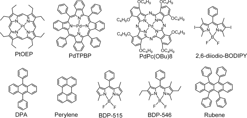
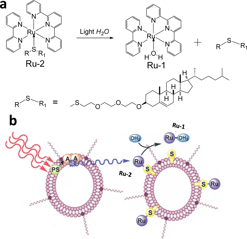
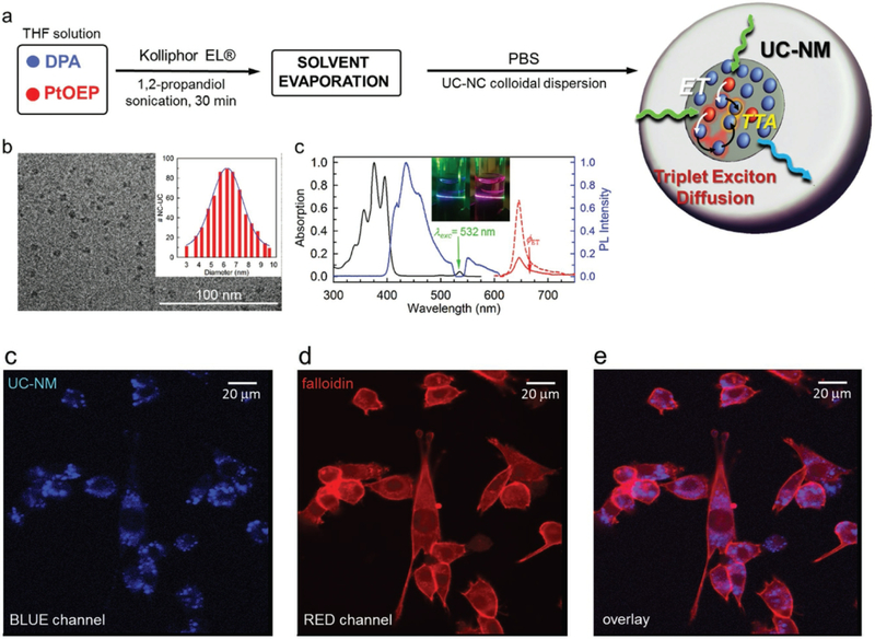
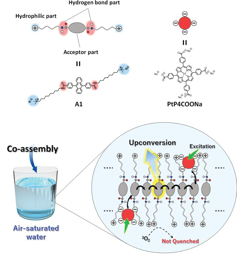
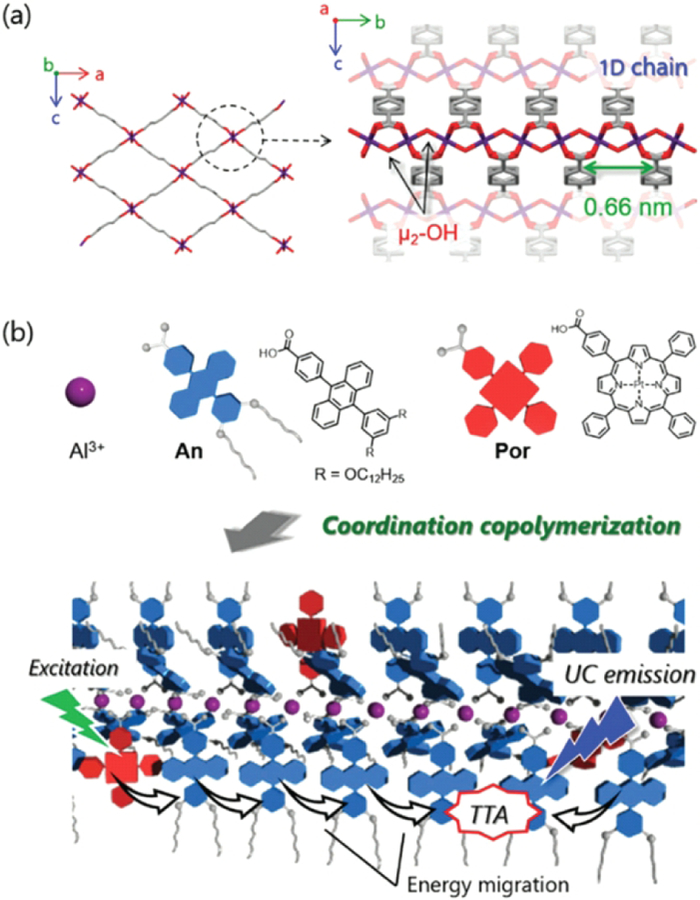
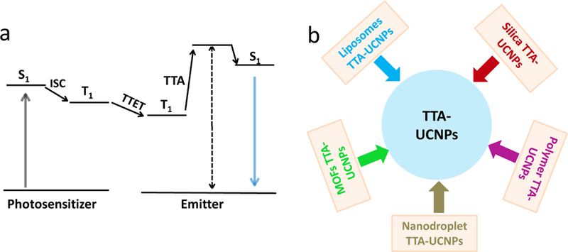
Organic triplet-triplet annihilation upconversion (TTA-UC) nanoparticles have emerged as exciting therapeutic agents and imaging probes in recent years due to their unique chemical and optical properties such as outstanding biocompatibility and low power excitation density. In this review, we focus on the latest breakthroughs in such new version of upconversion nanoparticle, including their design, preparation, and applications. First, we will discuss the key principles and design concept of these organic-based photon upconversion in regard to the methods of selection of the related triplet TTA dye pairs (photosensitizer and emitter). Then, we will discuss the recent approaches s to construct TTA-UCNPs including silica TTA-UCNPs, lipid-coated TTA-UCNPs, polymer encapsulated TTA-UCNPs, nano-droplet TTA-UCNPs and metal-organic frameworks (MOFs) constructed TTA-UCNPs. In addition, the applications of TTA-UCNPs will be discussed. Finally, we will discuss the challenges posed by current TTA-UCNP development.

Keywords: And cancer therapy; Bioimaging; Nanoparticles; Photo-targeting; Triplet-triplet annihilation upconversion.


Figures

Figure 1.
Figure 1.
The commercial photosensitizers (PtOEP, PdTPBP, PdPc (OBu)8, 2,6-diiodio-BPDIPY) and emitters (DPA, perylene, BDP-515, BDP-546, and Rubene).
Figure 2.
Figure 2.
(a) Molecular structures Ru-1 and Ru-2. (b) Schematic illustration the TTA-UC liposomes structure and TTA-UC trigger the Ru-1 releasing process with red light irradiation,λex = 630 nm Reference
Figure 3.
Figure 3.
Upper panel: (a) schematic illustration the preparation of the TTA-UCNPs containing PtOEP and DPA. (b) Cryo-TEM images of TTA-UC nanomicelles (c) UV-vis absorption spectra, TTA-UC emission spectra and phosphorescence emission spectrum of TTA-UC nanomicelles. Bottom panel (c-d) Confocal imaging of TTA-UC nanomicelles with 532 laser irradiation in 3T3 cell, (c) blue channel, (d) red channel and (d) overlay imaging.
Figure 4.
Figure 4.
Schematic illustration of formed the aqueous TEM-UC system process. Top panel: molecular structure of A-1 and PtP4COONa. Bottom panel: the A-1 and PtP4COONa by self-assembly generation the TEM-UC system in air-saturated water. Reference .
Figure 5.
Figure 5.
(a) Schematic illustration of silica coated TTA-UCNPs, and molecular structure of PdOEP, DPA and F-127. (b, c) Fluorescence cell imaging (b, λex = 543 nm, upconversion imaging) and (c, λex = 405 nm, conventional fluorescence image) (d) the overlay of imaging of (b) and (c). (e) In vivo and ex vivo upconversion imaging by silica coated TTA-UCNPs as contrast agents. Reference .
Figure 6.
Figure 6.
Left: a schematic illustration of dual color silica-TTA-UCNPs. Right: mice bright field imaging of TTA-UCNPs and the blue and green upconversion emission for mice breast and colorectal tumor. Reference .
Figure 7.
Figure 7.
(a) Schematic illustration of a glass capillary microfluidic device to synthesize the polymer encapsulated TTA-UCNPs. (b, c) High-speed optical microscopy observed the process preparation the polymer encapsulated TTA-UCNPs, scar bar is 200 μm.
Figure 8.
Figure 8.
Chemical structures of the PIB-PEG; (b) Schematic illustration of a PIB-PEG encapsulated TTA-UCNPs. (c) In vitro upconversion imaging in living A549 lung carcinoma cells under different conditions. Reference .
Figure 9.
Figure 9.
(a) Schematic illustration the PLA-PEG coated TTA-UCNPs. (b) Fluorescence emission spectra of c-RGDfK under different condition (c) NPTTA-c-RGDfK imaging in tumor-bearing mice under different conditions. (d) Quantitative analysis of the fluorescence intensity for TTA-UCNPs under different conditions. (e) Quantitative analysis the fluorescence intensity of tumor site under different conditions, λex = 530 nm, PdOEP (photosensitizer), DPA (emitter) Reference –
Figure 10.
Figure 10.
Left: a schematic illustration of TTA-UC nanocapsules, and molecular structures of PdOEP, PtTPBP, DPA, BDP-G, and BDP-Y. Right: (a) In vivo lymphatic imaging of the living mouse (d) TTA-UC fluorescence spectra nanocapsules. (e) Hematoxylin and eosin (H&E) staining mice lymph node imaging. Reference .
Figure 11.
Figure 11.
Left up: chemical structure of PEA and BDP-F; Left down: The TTA-UC photoactivation of Cou-C from TTA-CS, Bottom inset: a schematic illustration of a TTA-UC-induced prodrug photoactivation process. Right (a) schematics illustration of the photoactivation (c) Hematoxylin and eosin (H&E) staining of tumor tissue sections from different treatment groups (d) Representative digital photos of tumors for the four groups of mice, excitation power intensity is 100 mW/cm2, Reference .
Figure 12.
Figure 12.
Upper panel: (a) crystal structure of TTA-UC MOF. (b) Schematic illustration of PdTCPP energy transfer to emitters. Bottom panel: In vitro and in vivo imaging of TTA-UC MOF. (a) In vitro and (b) in vivo imaging with TTA-UC MOF. (c, d) In vivo lymph node imaging with TTA-UC MOF as an imaging contrast agent in living mice model, λex = 532 nm Reference
Figure 13.
Figure 13.
(a) Crystal structure of MIL-53. (b) Molecular structures of An and Por, and schematic illustration of TTA-UC process in coordination copolymers, λex = 515 nm Reference
Scheme 1.
Scheme 1.
(a) The photophysical process of TTA-UC. (b) The reported strategies for preparation TTA-UCNPs.

References

    1. Chen G, Qiu H, Prasad PN, Chen X. Upconversion nanoparticles: design, nanochemistry, and applications in theranostics, Chem. Rev 114 (2014) 5161–5214. - PMC - PubMed
    1. Zhou J, Liu Q, Feng W, Sun Y, Li F. Upconversion luminescent materials: advances and applications, Chem. Rev 115 (2015) 395–465. - PubMed
    1. Tu L, Liu X, Wu F, Zhang H. Excitation energy migration dynamics in upconversion nanomaterials, Chem. Soc. Rev 44 (2015) 1331–1345. - PubMed
    1. Chen G, Ågren H, Ohulchanskyy TY, Prasad PN. Light upconverting core-shell nanostructures: nanophotonic control for emerging applications, Chem. Soc. Rev 44 (2015) 1680–1713. - PubMed
    2. Liu J, Bu W, Pan L, Shi J. NIR-triggered anticancer drug delivery by upconverting nanoparticles with integrated azobenzene-modified mesoporous silica, Angew. Chem 125 (2013) 4471–4475; - PubMed
    3. Xing H, Bu W, Zhang S, Zheng X , Li M, Chen F, He Q, Zhou L, Peng W, Hua Y, Shi J. Multifunctional nanoprobes for upconversion fluorescence, MR and CT trimodal imaging, Biomaterials 33 (2012) 1079–1089; - PubMed
    4. Liu Y, Meng X, Bu W, Upconversion-based photodynamic cancer therapy. Coordination Chemistry Reviews 379 (2019) 82–98.
    1. Zhou J, Liu Z, Li F. Upconversion nanophosphors for small-animal imaging, Chem. Soc. Rev 41 (2012) 1323–1349. - PubMed

Publication types