Review
. 2019 Dec:59:19-28.
doi: 10.1016/j.sbi.2019.01.006. Epub 2019 Feb 22.

Making and breaking heme

Affiliations
Review

Making and breaking heme

Arianna I Celis et al. Curr Opin Struct Biol. 2019 Dec.

Abstract

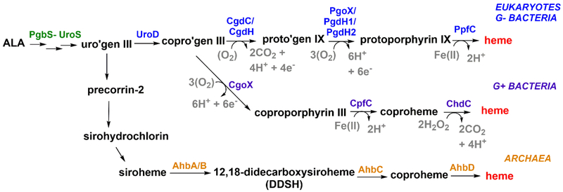
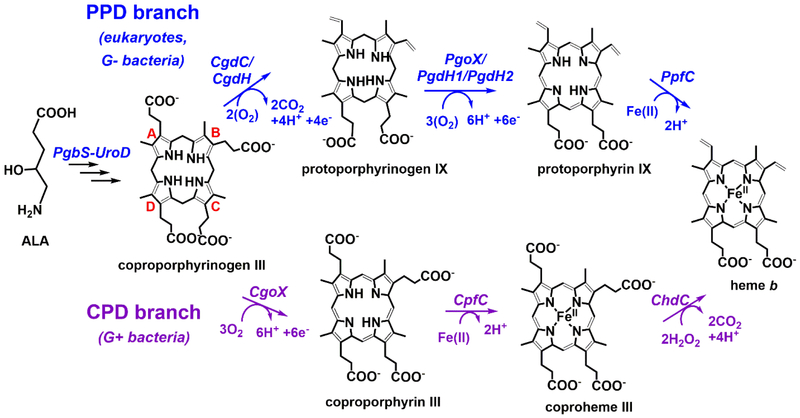
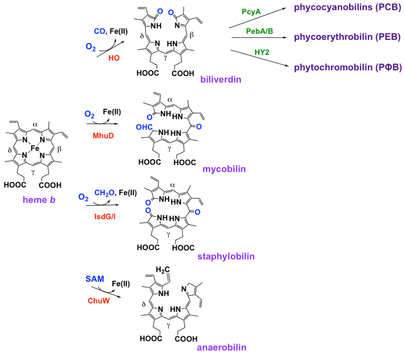
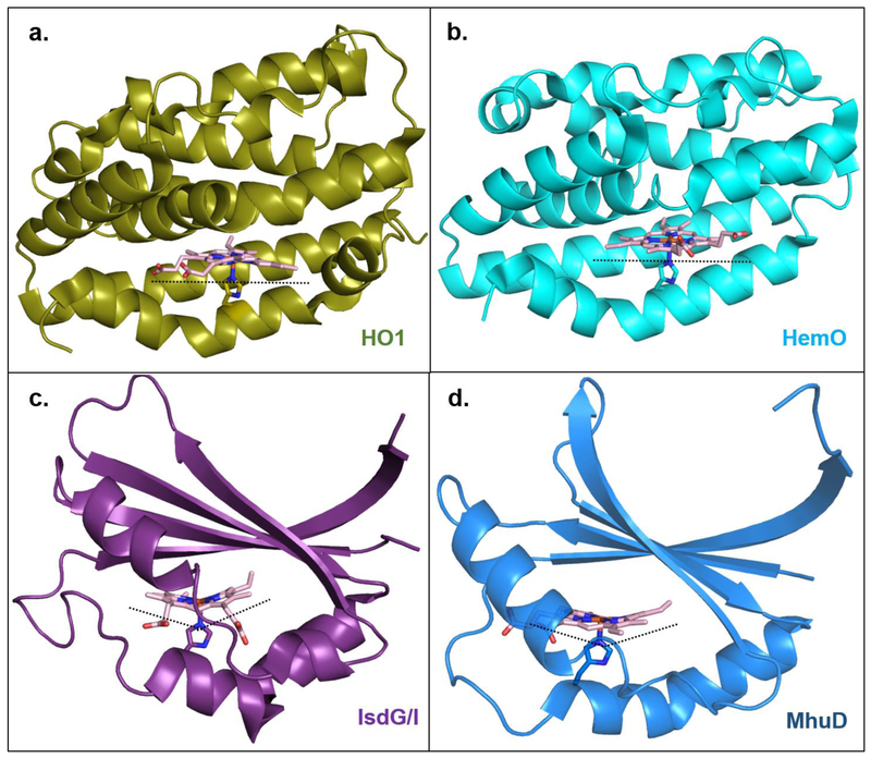
Mechanisms for making and breaking the heme b cofactor (heme) are more diverse than previously expected. Biosynthetic pathways have diverged at least twice along taxonomic lines, reflecting differences in membrane organization and O2 utilization among major groups of organisms. At least three families of heme degradases are now known, again differing in whether and how O2 is used by the organism and possibly the purpose for turning over the tetrapyrrole. Understanding these enzymes and pathways offers a handle for antimicrobial development and for monitoring heme use in organismal and ecological systems.


Figures

Figure 1.
Figure 1.. Multiple biosynthesis pathways leading to heme b.
Eukaryotes and gram-negative bacteria share the canonical pathway (blue). Gram-positive bacteria (purple) and denitrifying/sulfate reducing bacteria and archaea (orange) have distinct pathways that branch off following the coproporphyrinogen III (copro’gen) intermediate. The oxidation of porphyrinogens can use O2 as a 2e-/2H+ acceptor, generating H2O2 as a byproduct (i.e., steps catalyzed by CgdC, PgoX); alternatively, NAD(P)+ can serve as a 2e-/H+ acceptor in the presence of an active site acid (PgdH1, PgdH2). CgdH is a radical SAM-dependent enzyme. The electron acceptors for CgdH and CgoX have not been experimentally verified.
Figure 2.
Figure 2.. The PPD and CPD branches of heme b biosynthesis carry out similar chemical transformations in different orders.
Although some of the enzymes between the branches are homologous, a change in the order in which each is used creates different porphyrin intermediates. The terminal enzyme of the CPD pathway (ChdC) is completely unique. As knowledge about these pathways has evolved, new nomenclature for the enzymes that catalyze each individual step has been developed. For clarity, the most recently proposed nomenclature is used here3.
Figure 3.
Figure 3.. CgdC, CgdH, and ChdC all have an active site gate which closes upon substrate binding.
CgdC ((a); PDBID: 1TLB), CgdH ((b), PDBID: 1OLT) and ChdC ((c), PDBID: 1T0T) are isofunctional enzymes with no structural relationship and highly divergent reaction mechanisms. Interestingly, they all bind substrate at each monomeric site and structurally they share an “active site gate” (shown orange) made up of a partially disordered and mobile alpha helix, which closes in towards substrate upon its binding [4,9,11]. Note: Structures shown here are of individual monomers in their apo-/open- form. CgdC is a dimer, CgdH is a monomer, and ChdC is a pentamer in solution.
Figure 4.
Figure 4.. PgoX and CgoX share similar structural folds but show large differences in their active sites.
PgoX ((a); PDBID: 3NKS) and CgoX ((b); PDBID: 3I6D) share an overall folding pattern, consisting of three domains; (I) FAD-binding (yellow) domain, (II) membrane-binding domain (blue), and (III) substrate-binding domain (pink). Note: about 40% of amino acids in domain II of CgoX could not be crystallographically characterized [22]. PgoX and CgoX both non-covalently bind FAD (lime green) in the same location and orientation. The well-known PgoX inhibitor acifluoren (hot pink), also binds in both enzymes, but is mostly solvent-exposed in the CgoX active site, while it is buried deep inside the active site of PgoX. The different binding location and orientation of acifluoren in CgoX positions it to have a pi-stacking interaction with FAD (inset). This and the significantly larger active site in CgoX support the possibility that CgoX may use NAD(P) as an alternate electron acceptor to O2 to catalyze its reaction. Structures of PgdH1 and PgdH2 are not available to date.
Figure 5.
Figure 5.. CpfC does not have the “active site lip” characteristic of canonical PpfC enzymes, leaving its active site largely solvent exposed.
Juxtaposed structures of (a) H. sapien PfpC bound to protoporphyrin IX (only one monomeric unit shown, pink, PDBID: 2QD1) and (b) B. subtilis CpfC bound to N-methylmesoporphyin (purple with porphyrin shown in teal, PDBID: 1C1H). PpfC has a characteristic “active site lip” (shown in light pink) that moves toward and completely occludes the active site upon substrate binding (surface of active site cavity shown in grey). CpfC enzymes lack this “active site lip” and have an active site that is largely solvent exposed even in the presence of substrate. Accessibility to the porphyrin substrate/product in CpfC may allow for ease in porphyrin transfer to the succeeding enzyme, ChdC.
Figure 6.
Figure 6.. Interaction between yeast frataxin (Yfh1) and PpfC.
Complex formation between Yfh1 and PpfC in yeast involves one PfpC monomer (pink, PDBID: 1LBQ) and one Yfh1 homotrimer (yellow, PDBID: 4EC2). Each Yfh1 subunit binds two Fe(II) molecules (orange spheres, only one per monomer shown). Out of the three Yfh1 monomers, one subunit directly interacts with a PpfC monomer (interacting residues shown in hot pink), a second one is positioned for Fe(II) delivery (residues important for iron transfer shown in purple), and a third does not interact at all [33].
Figure 7.
Figure 7.. Different mechanisms and products of heme degradation.
Organisms have developed various mechanisms to break down heme in order to meet their specific needs. Each reaction is catalyzed by a different enzyme and generates unique products.
Figure 8.
Figure 8.. Multiple enzymes, multiple binding modes, multiple ways to degrade heme.
Canonical heme oxygenase enzymes HO1 and HemO ((a); PDBID: 1N45 and (b); PDBID: 1SK7), respectively) bind heme in a planar orientation. HO1 stereospecifically produces α-biliverdin, while HemO produces β- and γ-biliverdin due to the ~100° rotation of the heme ring in this active site relative to HO1. IsdG-like heme oxygenase enzymes IsdG/I and MhuD ((c); PDBID: 2ZDO and (d); PDBID: 4NL5, respectively) “ruffle” the heme porphyrin which results in cleavage of meso-carbons and products that are different than canonical HO’s (see figure 7).The heme ring in MhuD is rotated ~90° relative to the heme in IsdG/I, also contributing to the different bilin products that are produced.

References

    1. Panek H, O'Brian MR: A whole genome view of prokaryotic haem biosynthesis. Microbiology 2002, 148:2273–2282. - PubMed
    1. Dailey HA, Gerdes S, Dailey TA, Burch JS, Phillips JD: Noncanonical coproporphyrin-dependent bacterial heme biosynthesis pathway that does not use protoporphyrin. Proc Natl Acad Sci U S A 2015, 112:2210–2215. - PMC - PubMed
    1. Bali S, Lawrence AD, Lobo SA, Saraiva LM, Golding BT, Palmer DJ, Howard MJ, Ferguson SJ, Warren MJ: Molecular hijacking of siroheme for the synthesis of heme and d1 heme. Proc Natl Acad Sci U S A 2011, 108:18260–18265.

      • First article to describe a heme biosynthetic pathway for Archaea that differs from the canonical (PPD branch) heme biosynthesis pathway.

    1. Phillips JD, Whitby FG, Warby CA, Labbe P, Yang C, Pflugrath JW, Ferrara JD, Robinson H, Kushner JP, Hill CP: Crystal structure of the oxygen-dependant coproporphyrinogen oxidase (Hem13p) of Saccharomyces cerevisiae. Journal of Biological Chemistry 2004, 279:38960–38968. - PubMed
    1. Dailey HA, Dailey TA, Gerdes S, Jahn D, Jahn M, O'Brian MR, Warren MJ: Prokaryotic Heme biosynthesis: Multiple pathways to a common essential product. Microbiol Mol Biol Rev 2017, 81.

      •• Extensive and thorough review article that describes and compares all heme biosynthesis pathways, and the involved enzymes, known to date; the PPD branch, the CPD branch, and the Ahb pathway branch.

Publication types

LinkOut - more resources