

Radiomics robustness assessment and classification evaluation: A two-stage method demonstrated on multivendor FFDM
- PMID: 30802972
- PMCID: PMC6510593
- DOI: 10.1002/mp.13455
Radiomics robustness assessment and classification evaluation: A two-stage method demonstrated on multivendor FFDM
Abstract
Purpose: Radiomic texture analysis is typically performed on images acquired under specific, homogeneous imaging conditions. These controlled conditions may not be representative of the range of imaging conditions implemented clinically. We aim to develop a two-stage method of radiomic texture analysis that incorporates the reproducibility of individual texture features across imaging conditions to guide the development of texture signatures which are robust across mammography unit vendors.
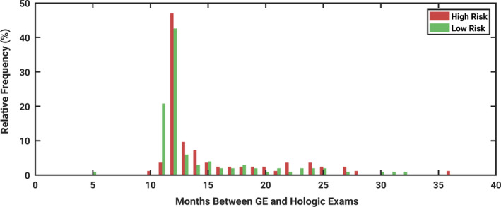
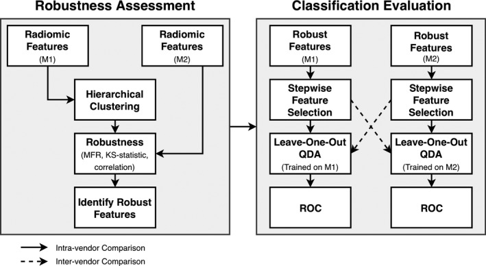
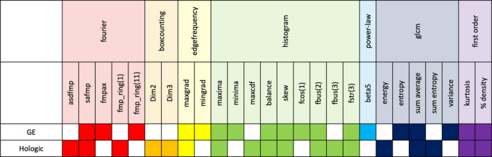
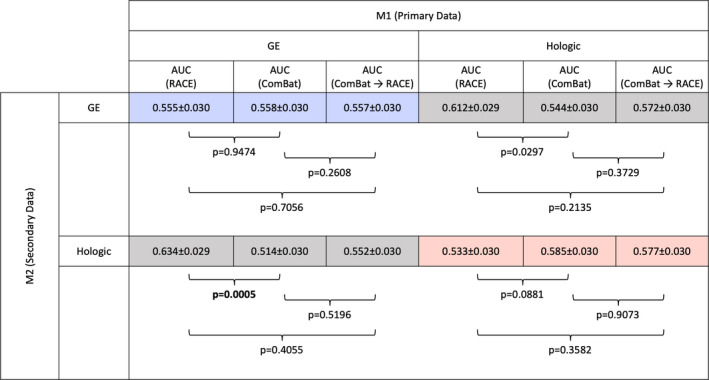
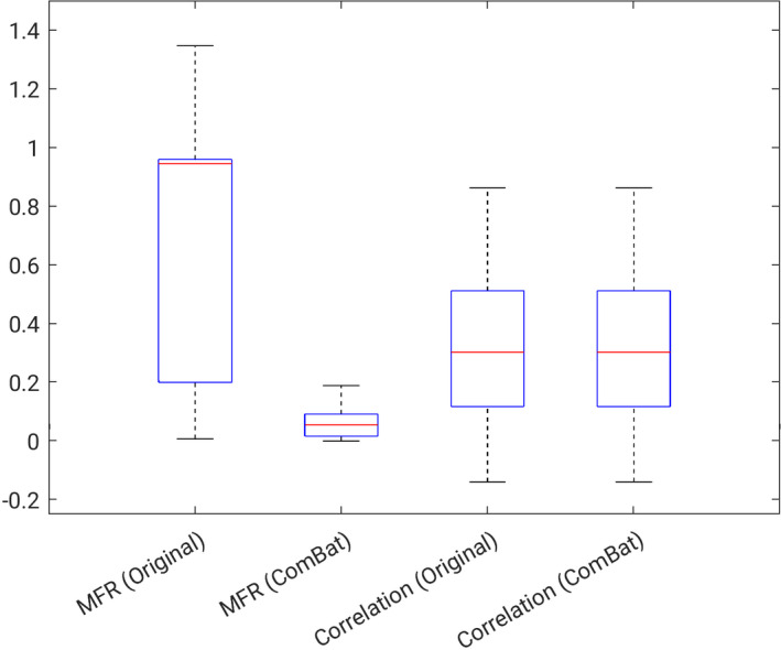
Methods: Full-field digital mammograms were retrospectively collected for women who underwent screening mammography on both a Hologic Lorad Selenia and GE Senographe 2000D system. Radiomic features were calculated on manually placed regions of interest in each image. In stage one (robustness assessment), we identified a set of nonredundant features that were reproducible across the two different vendors. This was achieved through hierarchical clustering and application of robustness metrics. In stage two (classification evaluation), we performed stepwise feature selection and leave-one-out quadratic discriminant analysis (QDA) to construct radiomic signatures. We refer to this two-state method as robustness assessment, classification evaluation (RACE). These radiomic signatures were used to classify the risk of breast cancer through receiver operator characteristic (ROC) analysis, using the area under the ROC curve as a figure of merit in the task of distinguishing between women with and without high-risk factors present. Generalizability was investigated by comparing the classification performance of a feature set on the images from which they were selected (intravendor) to the classification performance on images from the vendor on which it was not selected (intervendor). Intervendor and intravendor performances were also compared to the performance obtained by implementing ComBat, a feature-level harmonization method and to the performance by implementing ComBat followed by RACE.
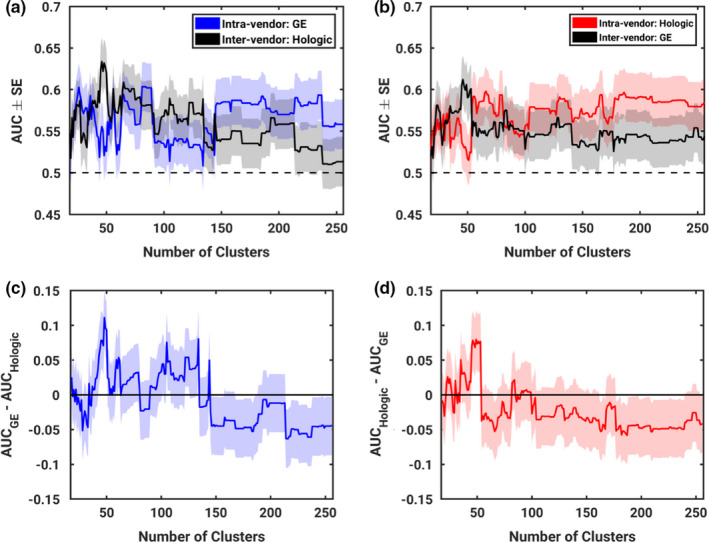
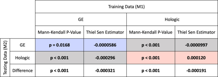
Results: Generalizability, defined as the difference between intervendor and intravendor classification performance, was shown to monotonically decrease as the number of clusters used in stage one increased (Mann-Kendall P < 0.001). Intravendor performance was not shown to be statistically different from ComBat harmonization while intervendor performance was significantly higher than ComBat. No significant difference was observed between either of the single methods and the use of ComBat followed by RACE.
Conclusions: A two-stage method for robust radiomic signature construction is proposed and demonstrated in the task of breast cancer risk assessment. The proposed method was used to assess generalizability of radiomic texture signatures at varying levels of feature robustness criteria. The results suggest that generalizability of feature sets monotonically decreases as reproducibility of features decreases. This trend suggests that considerations of feature robustness in feature selection methodology could improve classifier generalizability in multifarious full-field digital mammography datasets collected on various vendor units. Additionally, harmonization methods such as ComBat may hold utility in classification schemes and should continue to be investigated.
Keywords: breast cancer; radiomics; robustness.
© 2019 American Association of Physicists in Medicine.
Figures
References
-
- National Center for Health Statistics (US) . Health, United States, 2016: With Chartbook on Long‐term Trends in Health [Internet]. Hyattsville (MD): National Center for Health Statistics (US); 2017 [cited 2018 May 29]. Available from: http://www.ncbi.nlm.nih.gov/books/NBK453378/ - PubMed
-
- Tabár L, Vitak B, Chen TH‐H, et al. Swedish two‐county trial: impact of mammographic screening on breast cancer mortality during 3 decades. Radiology. 2011;260:658–663. - PubMed
-
- Saftlas AF, Hoover RN, Brinton LA, et al. Mammographic densities and risk of breast cancer. Cancer. 1991;67:2833–2838. - PubMed
-
- Boyd NF, Byng JW, Jong RA, et al. Quantitative classification of mammographic densities and breast cancer risk: results from the Canadian National Breast Screening Study. J Natl Cancer Inst. 1995;87:670–675. - PubMed
-
- McCormack VA, dos Santos Silva I. Breast density and parenchymal patterns as markers of breast cancer risk: a meta‐analysis. Cancer Epidemiol Biomark Prev Publ Am Assoc Cancer Res Cosponsored Am Soc Prev Oncol. 2006;15:1159–1169. - PubMed
Publication types
MeSH terms
Grants and funding
LinkOut - more resources
Full Text Sources
Medical
