. 2019 Feb 26;3(4):633-643.
doi: 10.1182/bloodadvances.2018026179.

In vitro and in vivo efficacy of an anti-CD203c conjugated antibody (AGS-16C3F) in mouse models of advanced systemic mastocytosis

Affiliations

In vitro and in vivo efficacy of an anti-CD203c conjugated antibody (AGS-16C3F) in mouse models of advanced systemic mastocytosis

Yanyan Zhang et al. Blood Adv. .

Abstract

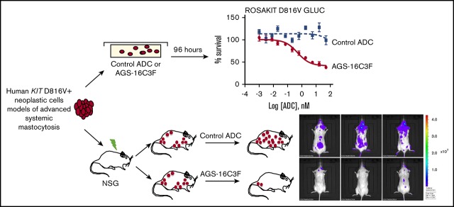
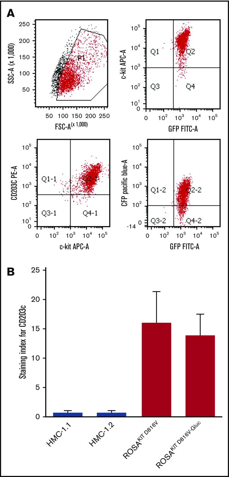
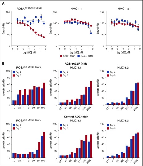
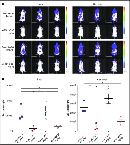
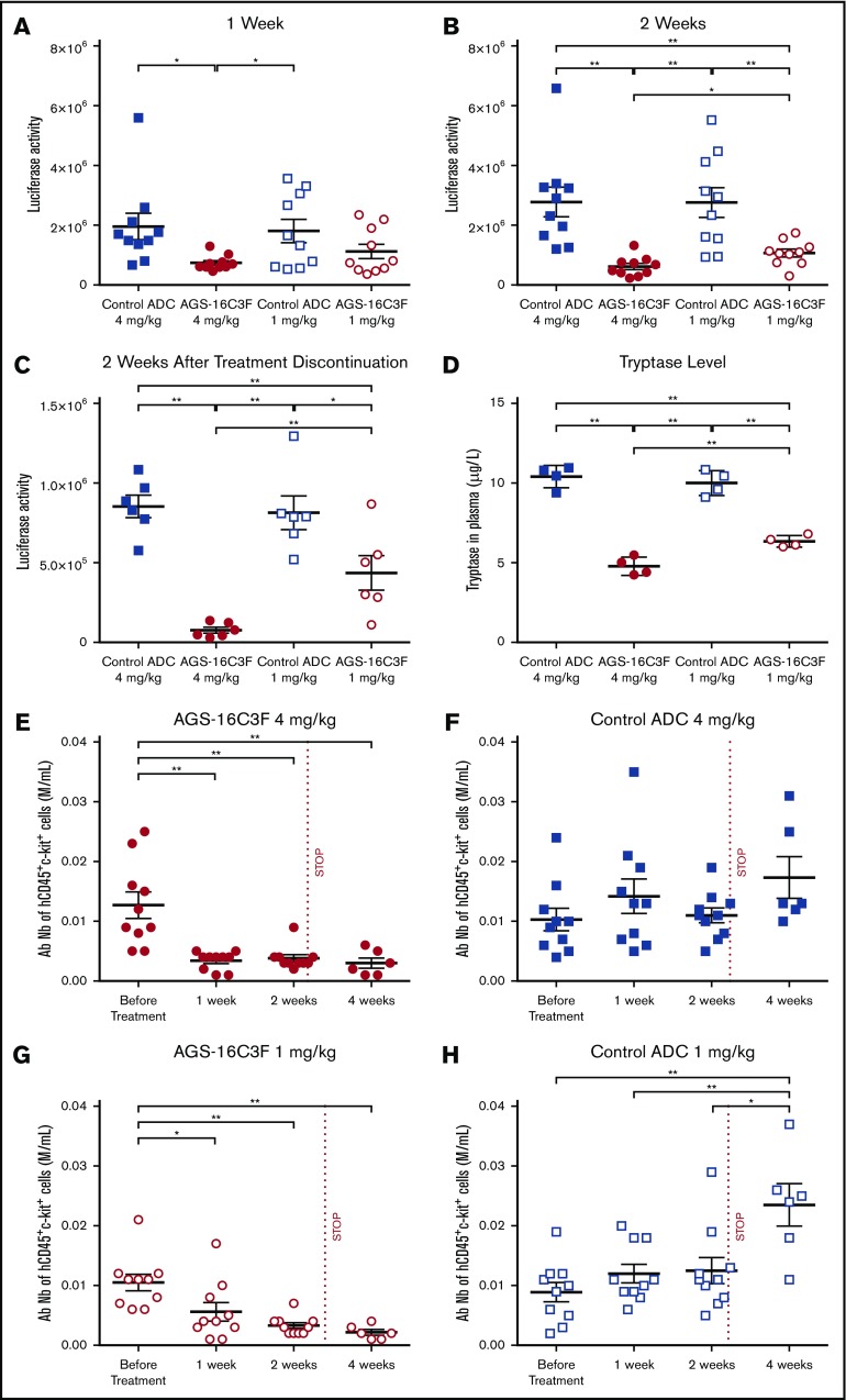
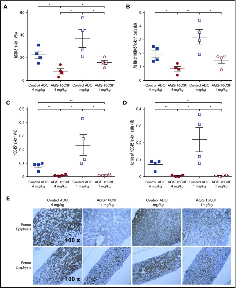
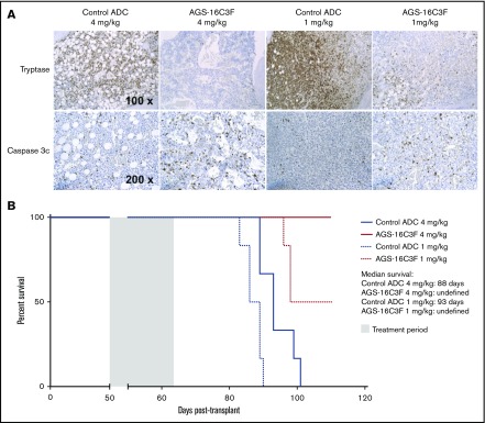
Antibody-drug conjugates (ADCs) are a new class of therapeutics that use antibodies to deliver potent cytotoxic drugs selectively to cancer cells. CD203c, an ecto-nucleotide pyrophosphatase-phosphodiesterase 3, is overexpressed on neoplastic mast cells (MCs) in systemic mastocytosis (SM), thus representing a promising target for antibody-mediated therapy. In this study, we have found that human neoplastic MC lines (ROSAKIT D816V and ROSAKIT D816V-Gluc), which express high levels of CD203c, are highly and specifically sensitive to the antiproliferative effects of an ADC against CD203c (AGS-16C3F). In these cell lines, AGS-16C3F induced cell apoptosis at very low concentrations. To characterize the effects of AGS-16C3F on leukemia progression in vivo, ROSAKIT D816V-Gluc NOD-SCID γ mouse models of advanced SM (AdvSM) were treated with AGS-16C3F or an ADC control for 2 weeks. Whereas AGS-16C3F had no apparent toxicity in xenotransplanted mice, in vivo neoplastic MC burden significantly decreased in both hematopoietic and nonhematopoietic organs. Furthermore, animals treated with AGS-16C3F had prolonged survival compared with the animals treated with control ADC, and AGS-16C3F efficiently prevented disease relapse. In conclusion, these preclinical studies identified CD203c as a novel therapeutic target on neoplastic MCs, and AGS-16C3F as a promising ADC for the treatment of patients with AdvSM.


Conflict of interest statement

Conflict-of-interest disclosure: At the time of the research, H.K.-M. and F.D. were both employees of Agensys, Inc. The remaining authors declare no competing financial interests.

Figures

None
Graphical abstract
Figure 1.
Figure 1.
CD203c expression in human neoplastic mast cell lines. (A) Representative flow cytometric evaluation of CD203c expression on ROSAKIT D816V-Gluc cells. (B) Assessment of surface expression of CD203c by flow cytometry on HMC-1.1, HMC-1.2, ROSAKIT D816V, and ROSAKIT D816V-Gluc cells. Results are expressed as staining index (SI, median fluorescence intensity values of tested Ab relative to its isotype control) and represent the mean ± standard deviation of triplicates. APC-A, allophycocyanin α; GFP, green fluorescent protein; SSC-A, side-scatter area.
Figure 2.
Figure 2.
Antiproliferative and pro-apoptotic effects of AGS-16C3F and cHmLYS-1c3.G2k-mcMMAF on human neoplastic mast cell lines. (A) ROSAKIT D816V-Gluc, HMC-1.1, and HMC-1.2 cells were treated with or without different concentrations of AGS-16C3F or cHmLYS-1c3.G2k-mcMMAF. On day 4 of treatment, viability was evaluated. Results are expressed as the percentage of control and represent the mean ± standard deviation of triplicates. (B) ROSAKIT D816V-Gluc, HMC-1.1, and HMC-1.2 cells were incubated in their medium (control) or with different concentrations of AGS-16C3F or cHmLYS-1c3.G2k-mcMMAF for 4 and 5 days. Results are expressed as the percentage of apoptotic cells, as measured by Annexin V/PI labeling.
Figure 3.
Figure 3.
AGS-16C3F decreased luciferase activity in vivo after 2 weeks of treatment. (A) IVIS showing localization and density of Gluc in treated mice. Units in rainbow color scales are photons per second per centimeter squared per steradian (p/s/cm2/sr). (B) Total relative units per second were calculated for Gluc intensity shown in panel A by reactive oxygen intermediates analysis. Left, back; right, abdomen. Each group comprises 3 mice. Data for each animal are represented; the horizontal bar is the average. *P < .05; **P < .01 assessed by Student t test.
Figure 4.
Figure 4.
Luciferase and tryptase activity are reduced in plasma, and absolute numbers of ROSAKIT D816V-Gluccells are reduced in PB of AGS-16C3F-treated xenotransplanted mice. (A) Luciferase activity after 1 week of treatment, (B) after 2 weeks, and (C) 2 weeks after stopping treatment. (D) Tryptase level measured at 4 days after the last injection of AGS-163CF. Each group comprises 4 mice. (E-H) Absolute neoplastic MC numbers in PB calculated by the equation: total WBC × % of hCD45+/KIT+ cells (fluorescence-activated cell sorter analyses of percentage of hCD45+/KIT+ in PB). Each group comprises 10 mice. Data for each animal are represented; the horizontal bar is the average. *P < .05; **P < .01 assessed by Student t test.
Figure 5.
Figure 5.
Treatment with AGS-16C3F decreased the number of ROSAKIT D816V-Gluccells in hematopoietic organs. Mice were euthanized 4 days after the last injection of AGS-16C3F. (A,C) Fluorescence-activated cell sorter analyses of the percentage of hCD45+/KIT+ in BM (A) and spleen (C). (B,D) Absolute numbers of neoplastic MCs in BM (B) and spleen (D), calculated by: total cell number in 1 femur and in total spleen × % of hCD45+/KIT+ cells in BM and spleen. (E) The femurs from 4 mice were collected and stained immunohistochemically with anti-human CD45 Ab. The sections were counterstained with hematoxylin. *P < .05; **P < .01.
Figure 6.
Figure 6.
Treatment with AGS-16C3F increased apoptosis of ROSAKIT D816V-Gluccells in hematopoietic organs and increased survival of mice. Mice were euthanized 4 days after the last injection of AGS-16C3F. (A) Tryptase staining (upper line) and cleaved caspase 3 (lower line) in femur from control ADC or from AGS-16C3F-treated mice. (B) AGS-16C3F prolonged life span of mice. Survival analyses (n = 6 animals/group). Treatment was performed between days 50 and 64, as indicated in the blue box. Statistical difference: P = .02, between the high and low dose of control-treated mice; P < .001 between AGS-16C3F and high- or low-dose control-treated mice independently (log-rank test).

Similar articles

Cited by

References

    1. Ustun C, Arock M, Kluin-Nelemans HC, et al. . Advanced systemic mastocytosis: from molecular and genetic progress to clinical practice. Haematologica. 2016;101(10):1133-1143. - PubMed
    1. Valent P, Escribano L, Broesby-Olsen S, et al. ; European Competence Network on Mastocytosis. Proposed diagnostic algorithm for patients with suspected mastocytosis: a proposal of the European Competence Network on Mastocytosis. Allergy. 2014;69(10):1267-1274. - PubMed
    1. Valent P, Akin C, Hartmann K, et al. . Advances in the classification and treatment of mastocytosis: current status and outlook toward the future. Cancer Res. 2017;77(6):1261-1270. - PMC - PubMed
    1. Arock M, Akin C, Hermine O, Valent P. Current treatment options in patients with mastocytosis: status in 2015 and future perspectives. Eur J Haematol. 2015;94(6):474-490. - PubMed
    1. Pardanani A. Systemic mastocytosis in adults: 2017 update on diagnosis, risk stratification and management. Am J Hematol. 2016;91(11):1146-1159. - PubMed

Publication types