

Extrapulmonary uterine lymphangioleiomyomatosis (LAM) and dysfunctional uterine bleeding: the first presentation of LAM in a tuberous sclerosis complex patient
- PMID: 30804158
- PMCID: PMC6388898
- DOI: 10.1136/bcr-2018-226358
Extrapulmonary uterine lymphangioleiomyomatosis (LAM) and dysfunctional uterine bleeding: the first presentation of LAM in a tuberous sclerosis complex patient
Abstract
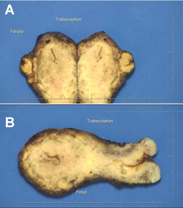
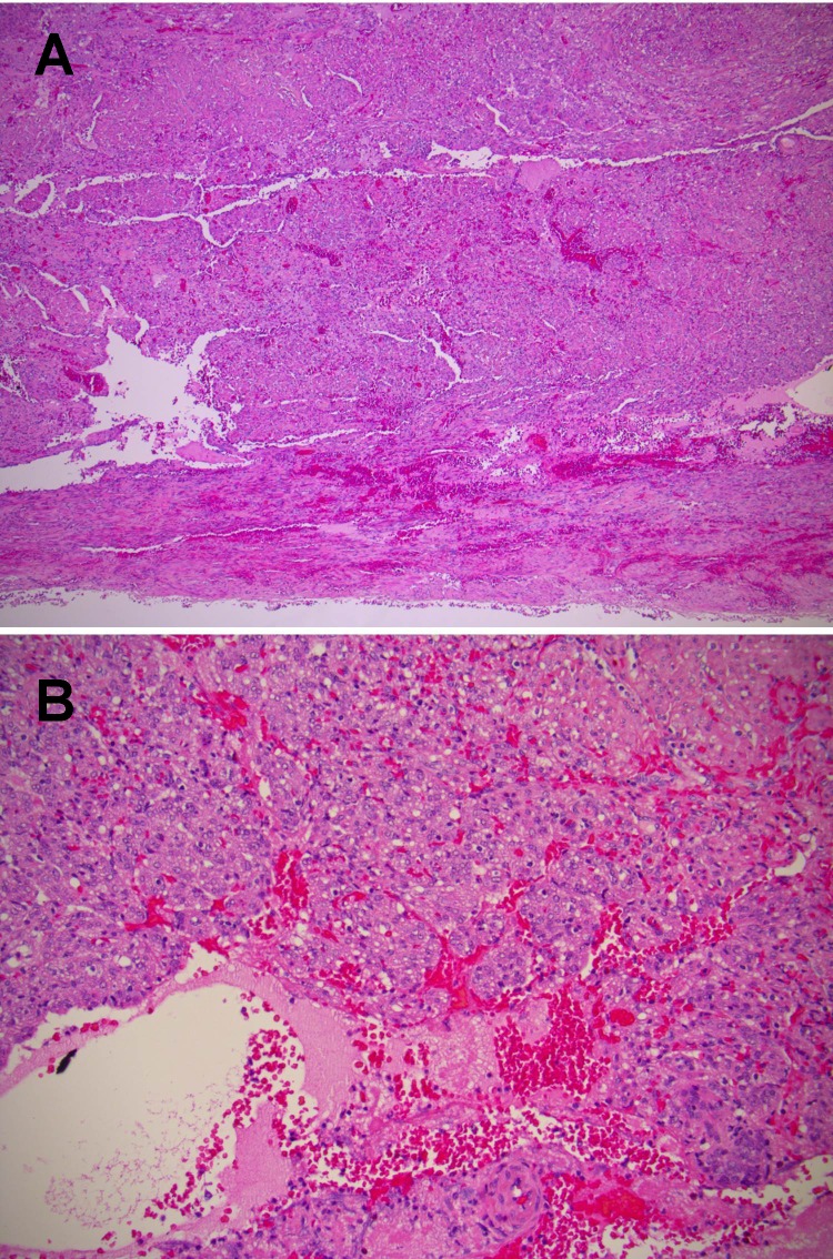
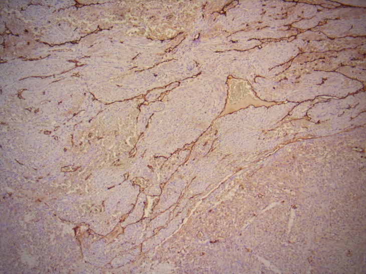
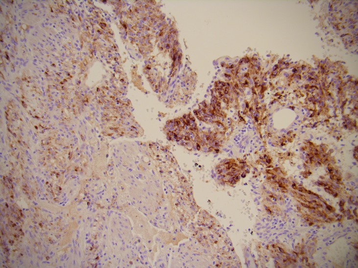
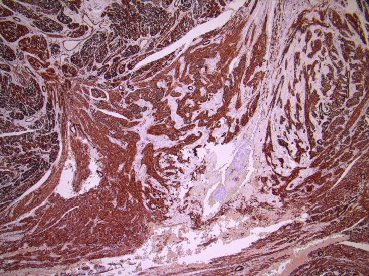
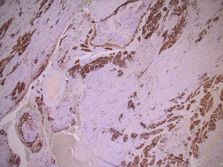
Lymphangioleiomyomatosis (LAM) is a rare disease that typically affects women of childbearing age. It most commonly affects the lungs (P-LAM) but can occasionally occur in extra-pulmonary sites (E-LAM). There is a strong association between LAM and the tuberous sclerosis complex (TSC). We report a case of a 42-year-old female TSC sufferer who presented with dysfunctional uterine bleeding. She was not known to have LAM. An endometrial biopsy revealed a spindled-cell lesion suspicious of leiomyosarcoma, which correlated with cross-sectional imaging. She underwent a hysterectomy that showed a bizarre (symplastic) leiomyomatous endometrial polyp with background uterine LAM. We discuss the clinical and pathological implications of this unusual case of E-LAM and the importance of clinicopathological correlation in TSC sufferers. The association of uterine LAM with TSC is important and LAM should be considered as a differential of dysfunctional uterine bleeding and a benign mimic to uterine leiomyosarcoma in patients with TSC.
Keywords: obstetrics and gynaecology; pathology.
© BMJ Publishing Group Limited 2019. No commercial re-use. See rights and permissions. Published by BMJ.
Conflict of interest statement
Competing interests: None declared.
Figures
Similar articles
-
CT and MR imaging findings of lymphangioleiomyomatosis involving the uterus and pelvic cavity.Korean J Radiol. 2011 Mar-Apr;12(2):261-5. doi: 10.3348/kjr.2011.12.2.261. Epub 2011 Mar 3. Korean J Radiol. 2011. PMID: 21430946 Free PMC article.
-
[Incidental diagnosis of lymphangioleiomyomatosis in gynecological surgery-a case series].Pathologie (Heidelb). 2025 May;46(3):179-184. doi: 10.1007/s00292-025-01414-0. Epub 2025 Feb 7. Pathologie (Heidelb). 2025. PMID: 39918558 Free PMC article. German.
-
[Sporadic Lymphangioleiomyomatosis (sLAM) and Tuberous Sclerosis Complex (TSC) - Pulmonary Manifestations].Pneumologie. 2017 Feb;71(2):86-95. doi: 10.1055/s-0042-111522. Epub 2016 Sep 1. Pneumologie. 2017. PMID: 27585353 Review. German.
-
Lymphangiomyomatosis of the uterus associated with tuberous sclerosis and malignant neoplasia of the female genital tract: a report of two cases.Int J Gynecol Pathol. 1995 Oct;14(4):344-51. doi: 10.1097/00004347-199510000-00010. Int J Gynecol Pathol. 1995. PMID: 8598338
-
Extrapulmonary lymphangioleiomyomatosis and lymphangiomatous cysts in tuberous sclerosis complex.Mayo Clin Proc. 1995 Jul;70(7):641-8. doi: 10.4065/70.7.641. Mayo Clin Proc. 1995. PMID: 7791386 Review.
Cited by
-
Uterine lymphangioleiomyomatosis in a premenopausal woman with tuberous sclerosis: A case report.Case Rep Womens Health. 2024 Sep 12;43:e00650. doi: 10.1016/j.crwh.2024.e00650. eCollection 2024 Oct. Case Rep Womens Health. 2024. PMID: 39314985 Free PMC article.
-
A Case of Uterine Lymphangioleiomyomatosis Complicated by Tuberous Sclerosis Complex.Case Rep Obstet Gynecol. 2022 Dec 13;2022:2893975. doi: 10.1155/2022/2893975. eCollection 2022. Case Rep Obstet Gynecol. 2022. PMID: 36561726 Free PMC article.
-
Sporadic uterine Lymphangioleiomyomatosis (LAM): Report of a unique case arising in the lower uterine segment with short review.Gynecol Oncol Rep. 2021 Jun 17;37:100812. doi: 10.1016/j.gore.2021.100812. eCollection 2021 Aug. Gynecol Oncol Rep. 2021. PMID: 34195331 Free PMC article.
References
-
- Lu HC, Wang J, Tsang YM, et al. . Lymphangioleiomyomatosis initially presenting with abdominal pain: a case report. Clin Imaging 2003;27:166–70. - PubMed
Publication types
MeSH terms
LinkOut - more resources
Full Text Sources
Medical