. 2019 Mar 12;116(11):4983-4988.
doi: 10.1073/pnas.1819156116. Epub 2019 Feb 25.

SHARPIN at the nexus of integrin, immune, and inflammatory signaling in human platelets

Affiliations

SHARPIN at the nexus of integrin, immune, and inflammatory signaling in human platelets

Ana Kasirer-Friede et al. Proc Natl Acad Sci U S A. .

Abstract

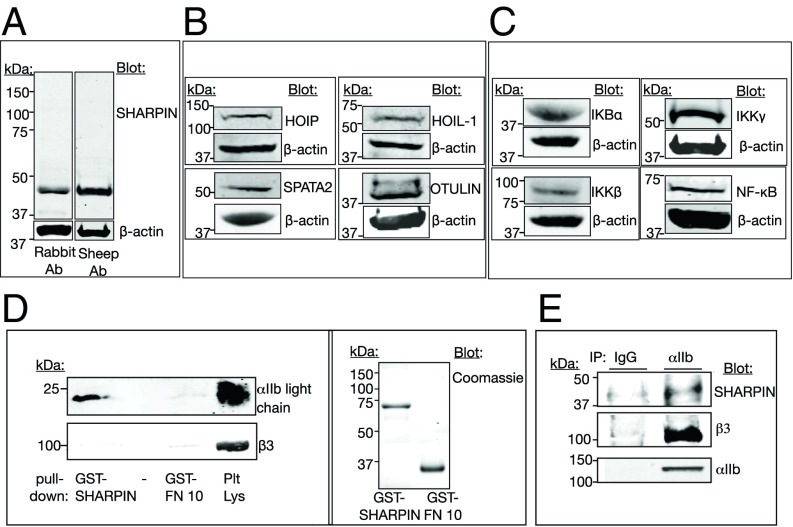
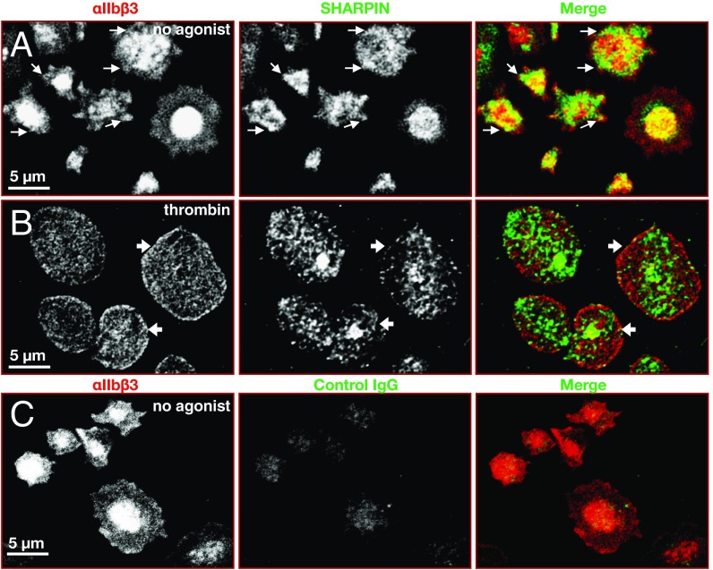
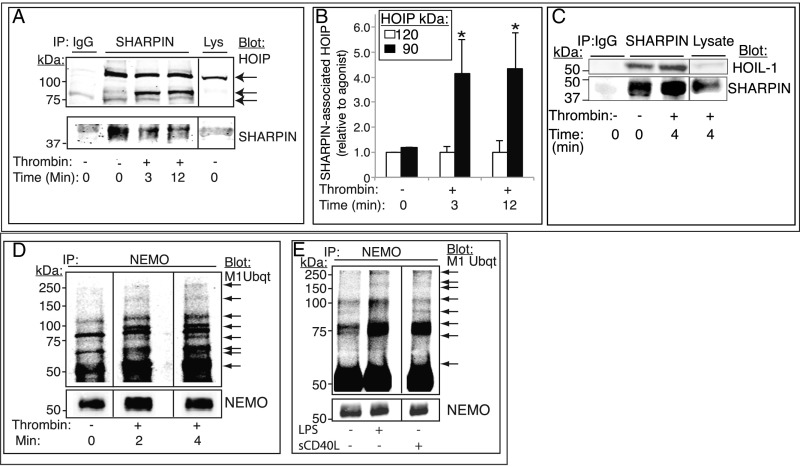
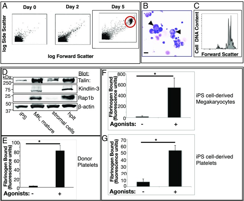
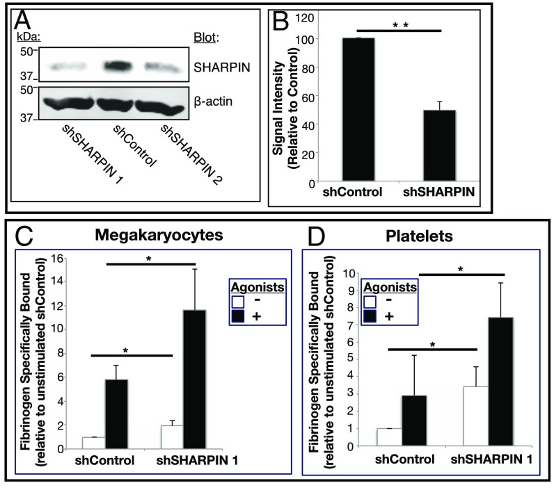
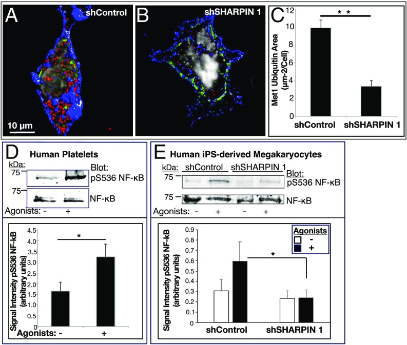
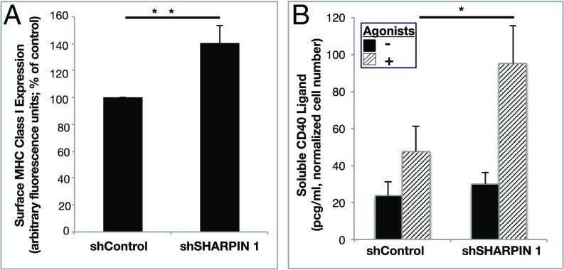
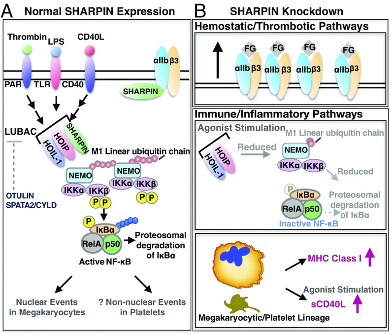
Platelets mediate primary hemostasis, and recent work has emphasized platelet participation in immunity and inflammation. The function of the platelet-specific integrin αIIbβ3 as a fibrinogen receptor in hemostasis is well defined, but the roles of αIIbβ3 or integrin-associated proteins in nonhemostatic platelet functions are poorly understood. Here we show that human platelets express the integrin-associated protein SHARPIN with functional consequences. In leukocytes, SHARPIN interacts with integrin α cytoplasmic tails, and it is also an obligate member of the linear ubiquitin chain assembly complex (LUBAC), which mediates Met1 linear ubiquitination of proteins leading to canonical NF-κB activation. SHARPIN interacted with αIIb in pull-down and coimmunoprecipitation assays. SHARPIN was partially localized, as was αIIbβ3, at platelet edges, and thrombin stimulation induced more central SHARPIN localization. SHARPIN also coimmunoprecipitated from platelets with the two other proteins comprising LUBAC, the E3 ligase HOIP and HOIL-1. Platelet stimulation with thrombin or inflammatory agonists, including lipopolysaccharide or soluble CD40 ligand (sCD40L), induced Met1 linear ubiquitination of the NF-κB pathway protein NEMO and serine-536 phosphorylation of the p65 RelA subunit of NF-κB. In human megakaryocytes and/or platelets derived from induced pluripotent stem (iPS) cells, SHARPIN knockdown caused increased basal and agonist-induced fibrinogen binding to αIIbβ3 as well as reduced Met1 ubiquitination and RelA phosphorylation. Moreover, these SHARPIN knockdown cells exhibited increased surface expression of MHC class I molecules and increased release of sCD40L. These results establish that SHARPIN functions in the human megakaryocyte/platelet lineage through protein interactions at the nexus of integrin and immune/inflammatory signaling.

Keywords: LUBAC; hemostasis; inflammation; integrin; platelet.


Conflict of interest statement

The authors declare no conflict of interest.

Figures

Fig. 1.
Fig. 1.
SHARPIN, LUBAC, and NF-κB pathway proteins are expressed in human platelets. (A) Lysate from unstimulated peripheral blood platelets was run on SDS-polyacrylamide gels, and protein expression was detected on Western blots. (A) SHARPIN was detected with antibodies raised in either rabbit (Left lane) or sheep (Right lane) hosts. (B) Expression of LUBAC members HOIP and HOIL-1 as well as SPATA2 and OTULIN, two proteins involved in Met1 deubiquitination. (C) Expression of NF-κB pathway proteins. Each protein depicted is of the expected molecular size, and β-actin reprobes from the same gels are shown. (D and E) SHARPIN associates with the integrin αIIb subunit. (D) Pulldowns in RIPA buffer using GST-SHARPIN or an irrelevant fusion protein (GST-FN10) as bait were probed with antibodies against integrin αIIb light chain or β3, under reducing gel conditions (Left), and then 5% of each total pulldown sample was used to assess protein loading on gels stained with Coomassie brilliant blue (Right). (E) SHARPIN coimmunoprecipitates with platelet αIIb. Nonidet P-40 lysate from unstimulated platelets was immunoprecipitated with an antibody to αIIb or with a nonspecific IgG and probed for SHARPIN, β3, and αIIb.
Fig. 2.
Fig. 2.
Localization of αIIbβ3 and SHARPIN in human platelets. Shown are confocal super-resolution immunofluorescence images of human platelets adherent to fibrinogen, at 100× magnification. Unstimulated (A) or 0.5 U/mL thrombin-stimulated (B) platelets were fixed and stained with antibodies against αIIbβ3, SHARPIN, or (C) control IgG as indicated. Edge localization of SHARPIN and αIIbβ3 was apparent in unstimulated platelets (arrows), but edge localization of SHARPIN appeared to be reduced in thrombin-stimulated platelets (arrowheads). Experiments were conducted three times, with similar results.
Fig. 3.
Fig. 3.
LUBAC-mediated Met1 ubiquitination of NEMO and NF-κB pathway signaling are triggered in platelets by hemostatic and inflammatory agonists. (AC) SHARPIN association with HOIP (A and B) and HOIL-1 (C) was analyzed in SHARPIN immunoprecipitates from human platelets that had been incubated with vehicle or thrombin (1 U/mL). (A) Western blot analysis with an antibody specific for the C-terminal LDD domain of HOIP revealed a full-length 120-kDa band under all incubation conditions and additional 90-kDa cleavage bands in thrombin-stimulated platelets. (B) Extent of HOIP associated with SHARPIN, determined by densitometry and normalized to SHARPIN intensity (n = 4; mean ± SEM; *P < 0.05). (D and E) Met1 linear ubiquitination of NEMO. Platelets were incubated in the absence or presence of 1 U/mL thrombin for 0–4 min (D) or with either 1 μg/mL LPS or 1 μg/mL sCD40L for 4 min (E). Lysates were immunoprecipitated with an anti-NEMO antibody and probed on Western blots for Met1 ubiquitin. Arrows indicate new or more intense Met1 ubiquitin bands of variable molecular sizes following agonist stimulation. All samples within a panel were run on the same gel, and an 8% aliquot of each immunoprecipitation sample was run separately to determine gel loading. The data shown are representative of at least three separate experiments.
Fig. 4.
Fig. 4.
Characterization of human iPS cell-derived megakaryocytes and platelets. (A) Human iPS cell-derived hematopoietic progenitors were incubated with doxycycline, stem cell factor, and thrombopoietin to induce differentiation to megakaryocytes (21, 46). Note the progressive appearance with time of a population of cells with increased forward and side light scatter characteristic of megakaryocytes (red circle). (B) Cytospins of maturation day 5/6 megakaryocytes stained with Wright Giemsa. (Original magnification 40×; scale bar: 35 μm.) Note the large cells with multilobed nuclei (arrows). (C) Flow cytometry analysis of DNA ploidy using PI in day 6 mature megakaryocytes indicating populations of cells with variably increased ploidy. (D) Western blots of lysates prepared from human iPS cells, megakaryocytes (MK), stromal cells, or peripheral blood platelets (hplt) were probed for known integrin-regulating proteins. (EG) Fibrinogen binding to human donor platelets (E) or to iPS cell-derived megakaryocytes (F) and platelets (G). Cells were incubated for 30 min with vehicle or an agonist mixture of 50 μM ADP, 50 μM epinephrine, and 100 μM PAR1 peptide (SFLLRN) in the presence of FITC-fibrinogen. Specific fibrinogen binding was determined by flow cytometry as described in Materials and Methods (n = 4; mean ± SEM; *P < 0.05).
Fig. 5.
Fig. 5.
shRNA-mediated SHARPIN knockdown in megakaryocytes and platelets leads to increased fibrinogen binding to αIIbβ3. Immature megakaryocytes were transduced with either of two GFP-IRES-shRNA lentiviruses targeting SHARPIN (shSHARPIN) or with a scrambled control shRNA (shControl). (A) Following megakaryocyte maturation, cells were lysed, and Western blots were probed with a SHARPIN antibody or a β-actin antibody as a loading control. (B) Summary of signal intensity of SHARPIN bands from cells, as treated in A (n = 6; mean ± SEM; **P = 0.01). (C and D) Specific Alexa Fluor 647-fibrinogen binding to iPS cell-derived megakaryocytes (C) or platelets (D) incubated in the absence or presence of an agonist mixture (ADP, epinephrine, and PAR1 agonist peptide) as in Fig. 4. Data were collected for GFP-positive live cells and depicted as specific fibrinogen binding relative to unstimulated shControl cells (n = 7; mean ± SEM; *P < 0.05).
Fig. 6.
Fig. 6.
SHARPIN knockdown in megakaryocytes and platelets reduces Met1 ubiquitination and NF-κB pathway signaling. (AC) Reconstructed immunofluorescence images of cells transduced with shControl RNA (A) or shSHARPIN 1 RNA (B). GFP-positive, shRNA expressing megakaryocytes were plated on fibrinogen and stained for Met1 ubiquitin (red), αIIb (blue), and DNA (gray). Images were acquired at 100× magnification. Met1 and αIIb are represented as isosurfaces. (C) Quantification of Met1 ubiquitination was carried out using Volocity image analysis software and is expressed as total Met1 ubiquitin area per cell. The data were collected from 3D projections of 13 optical slices of 0.2 μm each. A minimum of 20 cells were analyzed in each of three separate experiments (mean ± SEM; **P < 0.01). (D and E) Human platelets (D) or shControl- and shSHARPIN-treated megakaryocytes (E) were incubated for 15 min in the absence or presence of an agonist mixture (ADP, epinephrine and PAR1 agonist peptide as in Fig. 4). Cells were lysed and blots were probed with an antibody to phosphoserine-536 of the p65 (RelA) subunit of NF-κB and reprobed for total RelA. Quantification is represented as phosphoserine-536 RelA signal intensity normalized for total RelA NF-κB. (n = 4 in D; n = 8 in E; mean ± SEM; *P < 0.05).
Fig. 7.
Fig. 7.
SHARPIN knockdown affects potential immune/inflammatory functions of megakaryocyte lineage cells. iPS cell-derived immature megakaryocytes were infected with lentivirus expressing either shSHARPIN 1 or shControl shRNA. (A) Following megakaryocyte maturation, surface expression of MHC class I HLA-ABC was determined by flow cytometry (n = 6; mean ± SEM; **P < 0.01). (B) Mature megakaryocytes were incubated for 20 min with vehicle or agonist mixture as in Fig. 4, and sCD40L was determined in cell supernatants by ELISA. Results are normalized for cell count (n = 5; mean ± SEM; *P < 0.05).
Fig. 8.
Fig. 8.
Schematic of protein associations and potential functions of SHARPIN in human megakaryocytes and platelets. (A) Normal SHARPIN expression. SHARPIN associates with αIIbβ3, likely predominantly in resting platelets. In addition, HOIP and HOIL-1 are expressed in platelets and, together with SHARPIN, form LUBAC. Also expressed are OTULIN and SPATA2, involved in Met1 deubiquitination. Stimulation of platelets through receptors for thrombin, LPS, or sCD40L activates LUBAC to add Met1 linear ubiquitin chains (red circles) to NEMO, provoking transautophosphorylation of IKKβ and phosphorylation of IκBα. In nucleated cells, Lys-48-ubiquitination (blue circles) results in proteasomal degradation of IκBα, which frees NF-κB for nuclear translocation. Activation of LUBAC and NF-κB pathway components presumably induces nongenomic responses in anucleate platelets (48, 49). (B) Effects of SHARPIN knockdown. (Upper) Reduction of SHARPIN levels associated with αIIb may prime αIIbβ3 for talin-mediated activation and fibrinogen binding. (Middle) Reduction in SHARPIN levels may also destabilize LUBAC, thereby reducing Met1 linear ubiquitination and NF-κB activation. (Lower) SHARPIN reduction in the megakaryocyte/platelet lineage also leads to an increase in surface expression of MHC class I molecules and increased release of sCD40L, potentially contributing to platelet function in immunity and inflammation.

Similar articles

Cited by

References

    1. Rondina MT, Weyrich AS, Zimmerman GA. Platelets as cellular effectors of inflammation in vascular diseases. Circ Res. 2013;112:1506–1519. - PMC - PubMed
    1. Morse EM, Brahme NN, Calderwood DA. Integrin cytoplasmic tail interactions. Biochemistry. 2014;53:810–820. - PMC - PubMed
    1. Bennett JS. Regulation of integrins in platelets. Biopolymers. 2015;104:323–333. - PubMed
    1. Rantala JK, et al. SHARPIN is an endogenous inhibitor of β1-integrin activation. Nat Cell Biol. 2011;13:1315–1324. - PMC - PubMed
    1. Pouwels J, et al. SHARPIN regulates uropod detachment in migrating lymphocytes. Cell Rep. 2013;5:619–628. - PMC - PubMed

Publication types