. 2019 May 10;294(19):7546-7555.
doi: 10.1074/jbc.RA118.007052. Epub 2019 Feb 25.

Structural and functional consequences of age-related isomerization in α-crystallins

Affiliations

Structural and functional consequences of age-related isomerization in α-crystallins

Yana A Lyon et al. J Biol Chem. .

Abstract

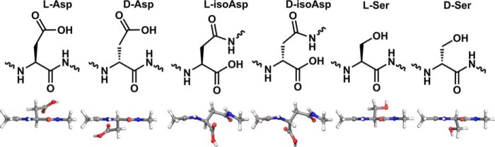
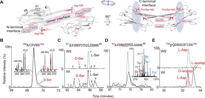
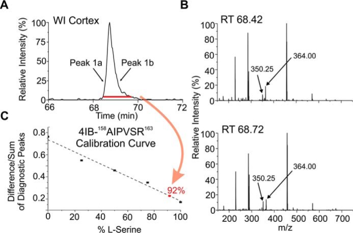
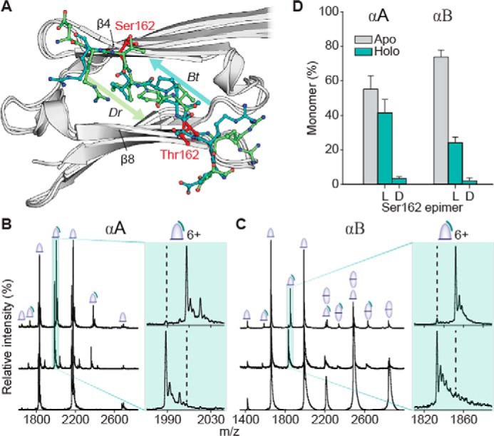
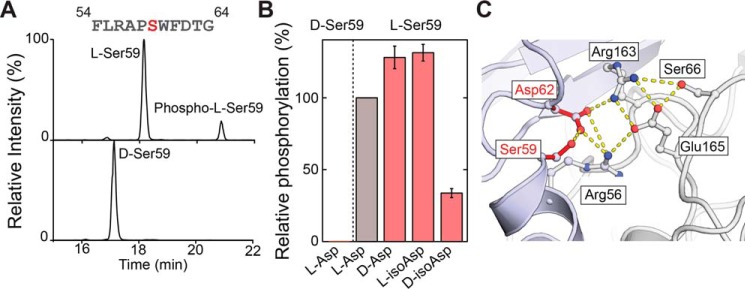
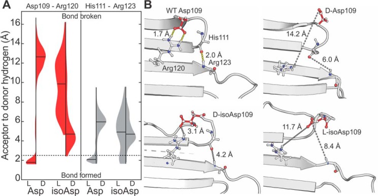
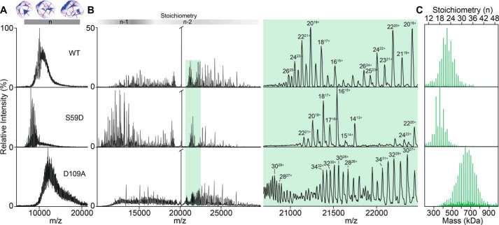
Long-lived proteins are subject to spontaneous degradation and may accumulate a range of modifications over time, including subtle alterations such as side-chain isomerization. Recently, tandem MS has enabled identification and characterization of such peptide isomers, including those differing only in chirality. However, the structural and functional consequences of these perturbations remain largely unexplored. Here, we examined the impact of isomerization of aspartic acid or epimerization of serine at four sites mapping to crucial oligomeric interfaces in human αA- and αB-crystallin, the most abundant chaperone proteins in the eye lens. To characterize the effect of isomerization on quaternary assembly, we utilized synthetic peptide mimics, enzyme assays, molecular dynamics calculations, and native MS experiments. The oligomerization of recombinant forms of αA- and αB-crystallin that mimic isomerized residues deviated from native behavior in all cases. Isomerization also perturbs recognition of peptide substrates, either enhancing or inhibiting kinase activity. Specifically, epimerization of serine (αASer-162) dramatically weakened inter-subunit binding. Furthermore, phosphorylation of αBSer-59, known to play an important regulatory role in oligomerization, was severely inhibited by serine epimerization and altered by isomerization of nearby αBAsp-62. Similarly, isomerization of αBAsp-109 disrupted a vital salt bridge with αBArg-120, a contact that when broken has previously been shown to yield aberrant oligomerization and aggregation in several disease-associated variants. Our results illustrate how isomerization of amino acid residues, which may seem to be only a minor structural perturbation, can disrupt native structural interactions with profound consequences for protein assembly and activity.

Keywords: aging; chaperone; epimer; mass spectrometry (MS); molecular dynamics; protein chemical modification; protein phosphorylation; protein self-assembly; protein structure; radical.


Conflict of interest statement

The authors declare that they have no conflicts of interest with the contents of this article

Figures

Figure 1.
Figure 1.
Corresponding chemical and three-dimensional structures of the isomers and epimers examined herein. 3D structures are shown from above, with the backbone perpendicular to the plane of the page.
Figure 2.
Figure 2.
A, two views of the partial crystal structure of α-crystallin (αB, PDB 4M5S). The structure of αB is used for illustration purposes because αB and αA intermix freely and share high structural similarity. Blue-shaded regions indicate crucial oligomeric interfaces. Pink ribbons denote the isomer-containing peptides, with specific isomerization sites labeled in red. The small cartoon in the middle represents the assembly, with each half-ellipse representing a monomer, the central line indicating the dimer interface, and the peripheral lines representing bound C-terminal peptides. Extracted ion chromatograms: B, αA158AIPVSR163 from the WI (black trace) and WS (gray trace) fractions of the cortex. Insets, RDD mass spectra from the leading and trailing edge of each peak. C, αB, 57APSWFDTGLSEMR69 from the nucleus, revealing abundant isomerization in the WI fraction. D, phosphorylated 57APsWFDTGLSEMR69 detected in the WS cortex (gray trace) and WI cortex (black trace), revealing far less isomerization (where s = phosphoserine). Inset, MS/MS pinpoints the site of phosphorylation to Ser-59. E, αB, 108pQDEHGFISR116 from the cortex (where pQ = pyroglutamate). The abundance of l-isoAsp is much higher in the WI than WS fraction.
Figure 3.
Figure 3.
A, selected-ion chromatogram for 4IB-AIPVSR in the WI cortex digest of the 72-year-old lens. B, RDD spectra from the leading (peak 1a) and trailing (peak 1b) edges of the corresponding LC peak. C, calibration curve is then used to quantify the amount of d-Ser that co-elutes in the LC chromatogram. The curve is generated by making standard solutions that contain known amounts of both isomers and taking the difference over the sum of the two peaks that have the largest differences in the fragmentation spectra. For this peptide, the–29I–NH3 losses from the precursor ion and the –H2O loss from the precursor ion were chosen as the diagnostic peaks. The percent d-Ser/l-Ser in the digest is then determined by averaging the RDD spectra for the entire peak in A (indicated by the red bar). This value maps to the red point in C, 92% l-Ser and 8% d-Ser.
Figure 4.
Figure 4.
Competition experiments reveal a strong preference for l-over d-Ser-162 binding to both αA and αB. A, aligned crystal structures of the α-crystallin domain (gray) with C-terminal peptide bound in two alternate orientations. Arrows indicate orientation (N → C) of bound peptides (green and teal). Equivalent isomerization sites (Ser-162 in αA or Thr-162 in αB) are shown in red. B, native mass spectra of αA core alone (bottom) and mixed with 4:1 ERAIPVSRE and GERAIPVSREG (middle, S = d-Ser). As seen in the magnification of 6+ peak, bottom spectrum, the binding of the lighter mass l-epimer is preferred. The upper trace corresponds to the reverse experiment, i.e. ERAIPVSRE and GERAIPVSREG. Dashed lines guide the eye to expected positions of d-epimer–bound peaks. C, equivalent experiments using the core of αB yield similar results. D, relative average fractions of free versus bound monomer cores from competition experiments, using all 5+, 6+, and 7+ charge states for quantitation. Error bars represent 95% confidence intervals. Crystal structures are as follows: bovine, Bt, in teal, PDB 3L1F, and zebrafish, Dr, in green, PDB 3N3E.
Figure 5.
Figure 5.
A, extracted ion chromatograms following incubation of FLRAPSWFDTG-NH2 and FLAPSWFDTG-NH2 (S = d-Ser) with MAPKAPK-2 reveal that d-Ser is not a viable phosphorylation substrate. B, relative degree of phosphorylation for Asp and Ser isomers of FLRAPSWFDTG-NH2. C, salt-bridge model (PDB 2YGD) of an N-terminal oligomeric interface involving Ser-59 and Asp-62. Hydrogen bonds are shown using dashed yellow lines.
Figure 6.
Figure 6.
A, distance distributions between Asp-109 and Arg-120 (left, red) and His-111 and Arg-123 (right, gray) from MD simulations. Violin plots are shown for each isomer of Asp-109; means are marked with black lines, with the kernel densities normalized to have the same maximum heights. Lengths <2.5 Å can be considered to correspond to bond formation (neglecting consideration of the bond angles), whereas those longer represent absence of the bond (boundary demarcated by dashed line). In all isomers other than l-Asp, the hydrogen-bond donor and acceptors are located too far apart for bond formation the vast majority of the time. B, selected frames from MD simulations highlighting breakage of hydrogen bonds profiled in A and resultant interface destabilization. Yellow dashes indicate H-bonds; short gray dashes show concomitant distances following isomerization of Asp-109; long gray dashes mark the antiparallel dimer interface. His-111 and Arg-123 side chains have not been shown, for clarity.
Figure 7.
Figure 7.
A, native MS of intact oligomeric assemblies of WT, S59D (phosphomimic), and D109A (isoAsp-mimic) αB, which are not directly assignable due to overlap of a multitude of charge states and stoichiometries. B, native MS with CID of the ions from A. Shading above shows regions where oligomers (n-mers) have lost one (n-1) or two (n-2) subunits. Detailed view of the region highlighted in green shows that CID resolves the charge states of the oligomers. C, reconstructed oligomeric distributions. Data were charge deconvolved and then corrected to account for stripped subunits.

References

    1. Truscott R. J. W., Schey K. L., and Friedrich M. G. (2016) Old proteins in man: a field in its infancy. Trends Biochem. Sci. 41, 654–664 10.1016/j.tibs.2016.06.004 - DOI - PMC - PubMed
    1. Catterall J. B., Barr D., Bolognesi M., Zura R. D., and Kraus V. B. (2009) Post-translational aging of proteins in osteoarthritic cartilage and synovial fluid as measured by isomerized aspartate. Arthritis Res. Ther. 11, R55 10.1186/ar2675 - DOI - PMC - PubMed
    1. Truscott R. J., and Friedrich M. G. (2016) The etiology of human age-related cataract. proteins don't last forever. Biochim. Biophys. Acta 1860, 192–198 10.1016/j.bbagen.2015.08.016 - DOI - PMC - PubMed
    1. Hooi M. Y., and Truscott R. J. (2011) Racemisation and human cataract. d-Ser, d-Asp/Asn and d-Thr are higher in the lifelong proteins of cataract lenses than in age-matched normal lenses. Age (Dordr.) 33, 131–141 10.1007/s11357-010-9171-7 - DOI - PMC - PubMed
    1. Geiger T., and Clarke S. (1987) Deamidation, isomerization, and racemization at asparaginyl and aspartyl residues in peptides. Succinimide-linked reactions that contribute to protein degradation. J. Biol. Chem. 262, 785–794 - PubMed

Publication types