. 2019 Apr 1;33(7-8):436-451.
doi: 10.1101/gad.322495.118. Epub 2019 Feb 25.

Synergistic lethality between BRCA1 and H3K9me2 loss reflects satellite derepression

Affiliations

Synergistic lethality between BRCA1 and H3K9me2 loss reflects satellite derepression

Jan Padeken et al. Genes Dev. .

Abstract

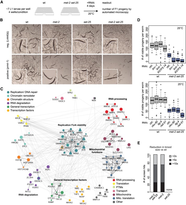
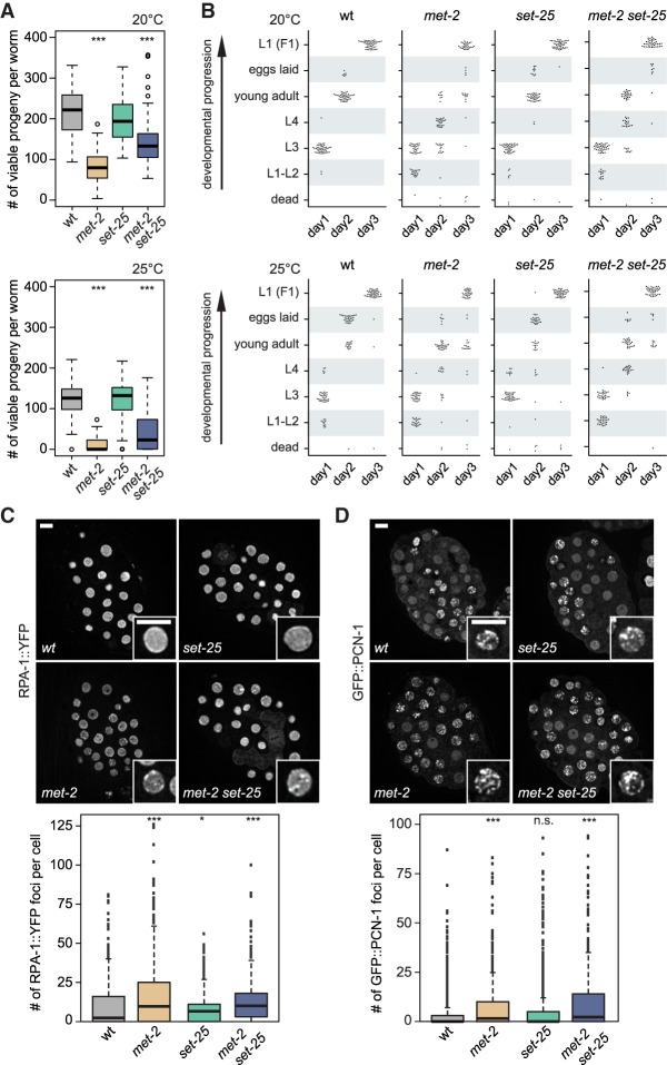
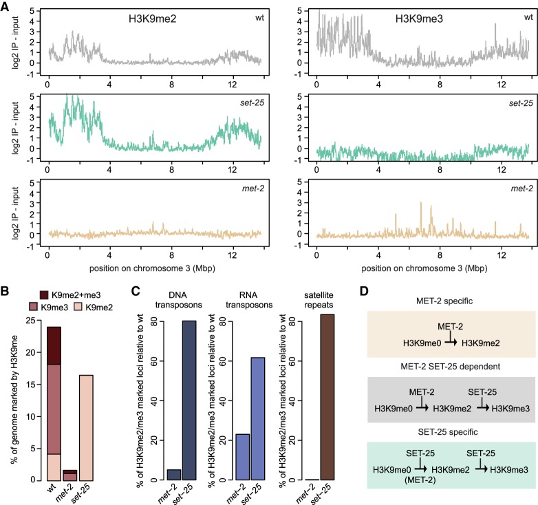
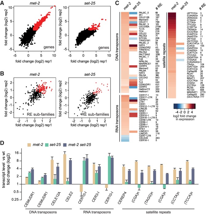
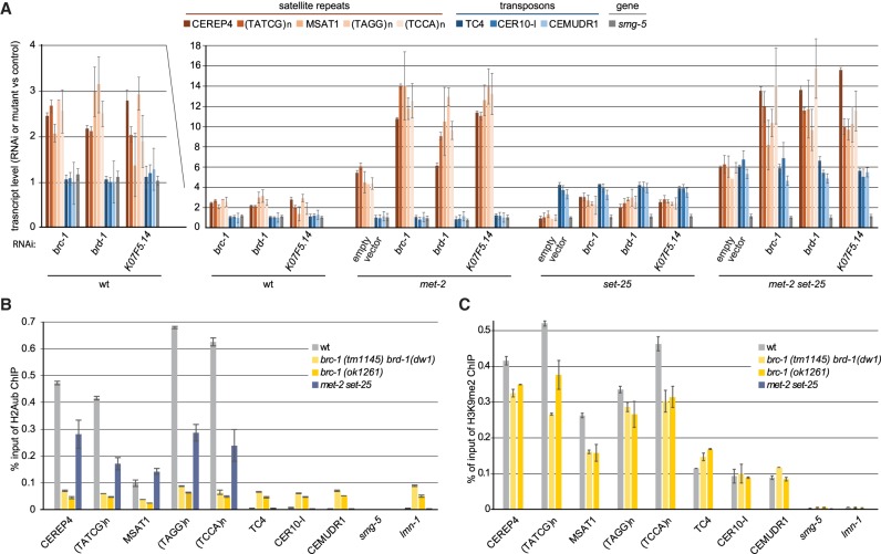
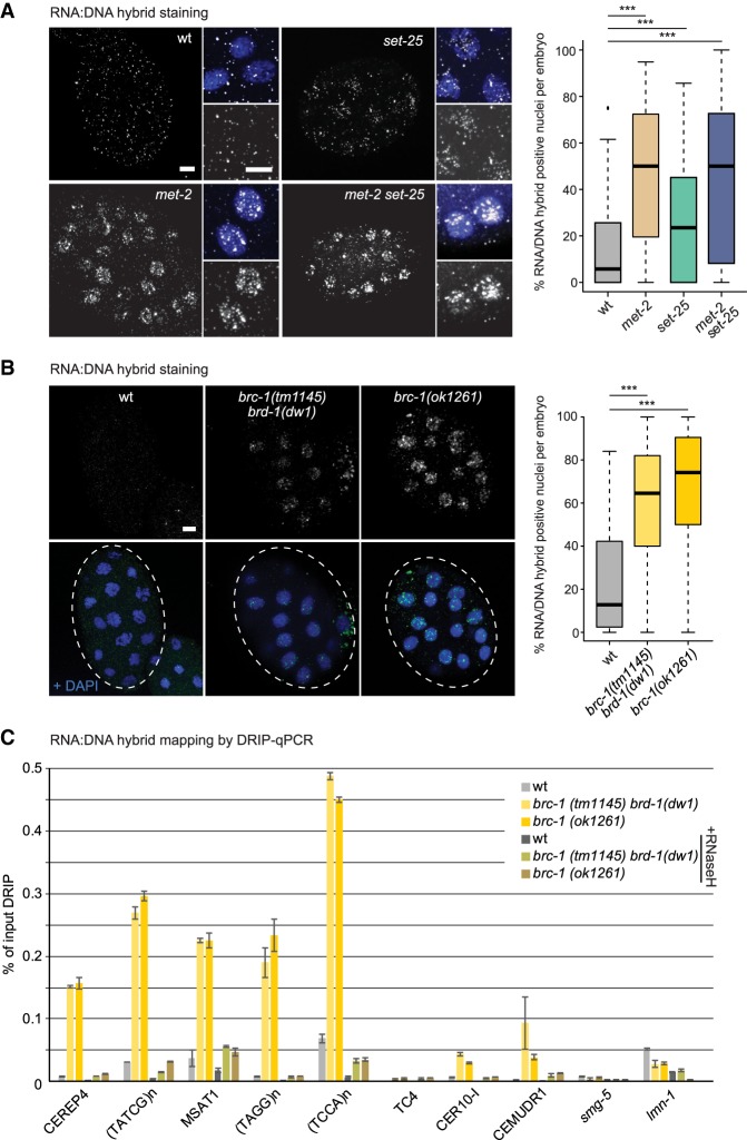
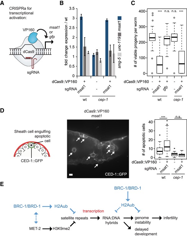
Caenorhabditis elegans has two histone H3 Lys9 methyltransferases, MET-2 (SETDB1 homolog) and SET-25 (G9a/SUV39H1 related). In worms, we found simple repeat sequences primarily marked by H3K9me2, while transposable elements and silent tissue-specific genes bear H3K9me3. RNA sequencing (RNA-seq) in histone methyltransferase (HMT) mutants shows that MET-2-mediated H3K9me2 is necessary for satellite repeat repression, while SET-25 silences a subset of transposable elements and tissue-specific genes through H3K9me3. A genome-wide synthetic lethality screen showed that RNA processing, nuclear RNA degradation, the BRCA1/BARD1 complex, and factors mediating replication stress survival are necessary for germline viability in worms lacking MET-2 but not SET-25. Unlike set-25 mutants, met-2-null worms accumulated satellite repeat transcripts, which form RNA:DNA hybrids on repetitive sequences, additively with the loss of BRCA1 or BARD1. BRCA1/BARD1-mediated H2A ubiquitination and MET-2 deposited H3K9me2 on satellite repeats are partially interdependent, suggesting both that the loss of silencing generates BRCA-recruiting DNA damage and that BRCA1 recruitment by damage helps silence repeats. The artificial induction of MSAT1 transcripts can itself trigger damage-induced germline lethality in a wild-type background, arguing that the synthetic sterility upon BRCA1/BARD1 and H3K9me2 loss is directly linked to the DNA damage provoked by unscheduled satellite repeat transcription.

Keywords: BRCA1 complex; DNA repeats; RNA:DNA hybrids; genome instability; heterochromatin; histone H3K9 methylation; satellite repeats; transcriptional silencing.


Figures

Figure 1.
Figure 1.
Synthetic sterility arises in met-2-deficient worms upon loss of RNA-processing/degradation and DNA damage response genes. (A) Synthetic lethality screen scheme: L1 larvae of wild-type and mutant strains were fed with RNAi-expressing bacteria at 20°C, and both adult viability and number of number of F1 progeny were assessed after 4 d. (B) Representative pictures of worms of indicated genotypes on day 4 after empty vector (L4440Δ) control or prmt-1 (a positive hit) RNAi. (C) Human homologs of RNAi-positive hits mapped onto STRING interactome (Szklarczyk et al. 2014) to illustrate general functions (color-coded) and genetic and physical interaction. Icon size reflects brood size reduction (from >2× to >10×). We include factors in the DNA replication and repair category that were tested secondarily: BRCA1, BARD1, RAD51, ATR. Visualization used visJS2jupyter (Rosenthal et al. 2018). (D) Synthetic sterility demonstrated for met-2 set-25 mutants and depletion of the indicated genes by RNAi after one generation at 25°C. N = 3; n = 25 for each condition. Box plots show median, boxes show 50%, and whiskers show 90% of all data points. (E) All 109 RNAi hits that were synthetic-sterile with met-2 set-25 mutants were tested on two met-2 single mutants (n4256 shown; ok2307 same result, not shown) and the set-25 (n5021) mutant. Number of hits and strength of synergistic effect are plotted. N = 3.
Figure 2.
Figure 2.
MET-2 is essential for organismal fertility and proper developmental timing. (A) Single mutants met-2 and set-25 were tested for fertility at 20°C and 25°C (N = 3; n = 25) and compared with the met-2 set-25 double mutant. Complete viable progeny of singled worms were scored after two generations. (***) P < 0.0001, two-sided Wilcoxon signed-rank test. (B) Developmental timing was analyzed by following singled worms over 3 d of development at 20°C and 25°C of the indicated genotypes. Each dot represents a single worm. N = 3; n = 50. (C,D, top) Representative images of early embryos from adults of indicated genotypes, expressing either RPA-1::YFP (C), grown at 25°C or GFP::PCN-1, grown at 20°C (D). Bars, 5 µm. (Bottom) Quantification shows the number of foci per nucleus for the indicated genotypes. (C) N = 3; n = 25 embryos. (D) N = 2; n = 25 embryos. Box plots show median, boxes 50%, and whiskers 90%, with all other data points shown. (*) P = 0.031, (***) P < 0.0001, (n.s.) P > 0.0, two-sided Wilcoxon signed-rank test.
Figure 3.
Figure 3.
Three pathways for MET-2 and SET-25 targeting define distinct HMT dependence. (A) H3K9me2 and H3K9me3 ChIP-seq was performed on early embryos at 20°C in wild-type (wt), set-25, and met-2 strains. The enrichment of one experiment over input along a typical autosome, chromsome III, is shown (N = 2), demonstrating retention of H3K9me2 in the set-25 mutant and the more general depletion of both marks in met-2. (B) Genome fraction in percentage bearing either H3K9me2, H3K9me3, or both H3K9me2 and me3 marks in wild-type, met-2 (SET-25 dependent), and set-25 (MET-2 dependent) embryos (H3K9me3: log2 immunoprecipitation-input >2, H3K9me2: log2 immunoprecipitation-input >1). (C) Percentage of remaining H3K9me2- or me3-positive loci in the indicated mutants relative to wild type, sorted by repeat classes: DNA-transposons, RNA-transposons, and satellite repeats. (D) Scheme of three targeting pathways for H3K9me. ChIP-seq tracks are shown and color-coded in Supplemental Figure S3G.
Figure 4.
Figure 4.
MET-2 specifically represses satellites, while SET-25 is needed for selected RNA and DNA transposon repression. (A) Scatter plot showing gene expression as log2 fold change over wild type in early embryos of indicated mutants. Two replicas shown. N = 3. (B) Scatter plot showing repeat subfamily expression as log2 fold change over wild type as in A. (C) Heat map showing transcribed repeat subfamilies in met-2 and/or set-25 worms, separated into three major repeat classes (DNA transposons, RNA transposons, simple repeats). (# RE) Copy number of annotated repeats per subfamily. (D) Quantitative PCR (qPCR) confirmation of the most highly expressed single REs demonstrating differential dependence on MET-2 or SET-25 in early embryos. N = 3. RNA5 (CELE2) overlaps with the intron of the gene C48D1.9, and (TAGG)n with the intron of mib-1; neither gene is derepressed by loss of H3K9me.
Figure 5.
Figure 5.
Loss of BRC-1 derepresses satellite repeats, but not RNA transposons, in a strongly additive manner with loss of MET-2. (A) A qPCR analysis of the expression of MET-2-dependent satellite repeats (tan to brown), SET-25-dependent transposons (blue), and the H3K9me-independent smg-5 gene (gray) at the young adult stage of indicated genetic background (wild type, set-25, met-2, and met-2 set-25). L1 larvae were treated with RNAi against brca-1 and partners brd-1 and K07F5.14. Expression is normalized to wild type grown on empty vector-expressing bacteria. Mean and SEM. N = 3. (B) H2Aub ChIP-qPCR and (C) H3K9me2 ChIP-qPCR in early embryos of indicated genotypes on the sequences analyzed in B. Mean and SEM. N = 3. Note that brc-1 (tm1145) carries a second mutation that fully eliminates BRD-1 expression [brd-1 (dw11)] (Janisiw et al. 2018).
Figure 6.
Figure 6.
Loss of MET-2 and BRC-1 complex components provokes RNA:DNA hybrid accumulation on satellite repeats. (A) Representative images taken under identical conditions of early embryos of indicated genotypes grown at 20°C, stained for RNA:DNA hybrids (antibody S9.6, HB-8730, American Type Culture Collection). Enlargements at right of each quarter shows the RNA:DNA hybrid signal alone (top panel) and overlay with DAPI (bottom panel). Bar, 5 µm. At right, the percentage of cells with RNA:DNA nuclear staining per embryo is plotted for indicated genotypes. N = 3; n = 20 embryos. Box plots are as in Figure 2. (***) P < 0.0001, two-sided Wilcoxon signed-rank test. (B) RNA:DNA hybrid immunofluorescence using the S9.6 antibody in wild-type, brc-1, and brc-1 brd-1 mutant embryos, as in A. Similar quantitation for RNA:DNA hybrids is at the right. N = 3; n = 20. (C) RNA:DNA hybrid accumulation on indicated sequences scored by DRIP-qPCR with antibody S9.6. Mutants are as in A and B. qPCR monitors satellite repeats expressed in the brc-1 mutant as well as unaffected transposable elements (CEMUDR1, TC4, and CER10-I) and a single-copy gene (smg-5). RNase H-treated samples quantify unspecific DRIP signal. Mean and SEM; N = 2.
Figure 7.
Figure 7.
Aberrant expression of a satellite repeat can cause loss of fertility. (A) Scheme showing the targeting of catalytically dead Cas9 (dCas9) to msat1 or gfp genes, and activation by 10 copies of the VP16 transactivating domain. (B) qPCR showing the selective induction of the MSAT1 satellite transcripts targeted by dCas9::VP160 as fold change over worms lacking the dCas9::VP160 fusion protein in L3–L4 larvae of wild-type and cep-1 mutant backgrounds (mean and SD.; N = 2). Expression of smg-5 and unc-119 genes serve as negative controls. (C) The total number of viable progeny of MSAT1 overexpressing wild-type and cep-1 mutant strains at 20°C. N = 3; n = 25. Strains lacking the dCas9::VP160 construct and a strain expressing dCas9::VP160 targeted to a GFP reporter transgene are negative controls. Box plots are as in Figure 1 with two-sided Wilcoxon signed-rank test (***) P < 0.0001; (n.s.) P > 0.05. (D) Representative image of a gonad showing apoptotic cells (arrows) expressing the CED-1::GFP (plasma membrane apoptotic marker) in the MSAT1 overexpression strain. Bar, 5 µm. Plotted is the quantitation of apoptosis (number of cells fully engulfed by CED-1::GFP per gonad arm) in wild type and cep-1 mutant (lacking p53 homolog) as a function of MSAT1 overexpression. N = 3; n = 60. (***) P < 0.0001; (n.s.) P > 0.05, two-sided Wilcoxon signed-rank test. (E) Schematic description of how MET-2-mediated H3K9me2 and BRC-1-mediated H2Aub work together to prevent RNA:DNA hybrid accumulation and associated phenotypes.

Similar articles

Cited by

References

    1. Adamo A, Montemauri P, Silva N, Ward JD, Boulton SJ, La Volpe A. 2008. BRC-1 acts in the inter-sister pathway of meiotic double-strand break repair. EMBO Rep 9: 287–292. 10.1038/sj.embor.7401167 - DOI - PMC - PubMed
    1. Alexiadis V, Ballestas ME, Sanchez C, Winokur S, Vedanarayanan V, Warren M, Ehrlich M. 2007. RNAPol-ChIP analysis of transcription from FSHD-linked tandem repeats and satellite DNA. Biochim Biophys Acta 1769: 29–40. 10.1016/j.bbaexp.2006.11.006 - DOI - PMC - PubMed
    1. Avgustinova A, Symeonidi A, Castellanos A, Urdiroz-Urricelqui U, Solé-Boldo L, Martín M, Pérez-Rodríguez I, Prats N, Lehner B, Supek F, et al. 2018. Loss of G9a preserves mutation patterns but increases chromatin accessibility, genomic instability and aggressiveness in skin tumours. Nat Cell Biol 20: 1400–1409. 10.1038/s41556-018-0233-x - DOI - PubMed
    1. Ayrapetov MK, Gursoy-Yuzugullu O, Xu C, Xu Y, Price BD. 2014. DNA double-strand breaks promote methylation of histone H3 on lysine 9 and transient formation of repressive chromatin. Proc Natl Acad Sci 111: 9169–9174. 10.1073/pnas.1403565111 - DOI - PMC - PubMed
    1. Bhatia V, Barroso SI, García-Rubio ML, Tumini E, Herrera-Moyano E, Aguilera A. 2014. BRCA2 prevents R-loop accumulation and associates with TREX-2 mRNA export factor PCID2. Nature 511: 362–365. 10.1038/nature13374 - DOI - PubMed

Publication types

MeSH terms