. 2019 Mar 26;39(3):BSR20181108.
doi: 10.1042/BSR20181108. Print 2019 Mar 29.

miR-206 inhibits osteogenic differentiation of bone marrow mesenchymal stem cells by targetting glutaminase

Affiliations

miR-206 inhibits osteogenic differentiation of bone marrow mesenchymal stem cells by targetting glutaminase

Ying Chen et al. Biosci Rep. .

Abstract

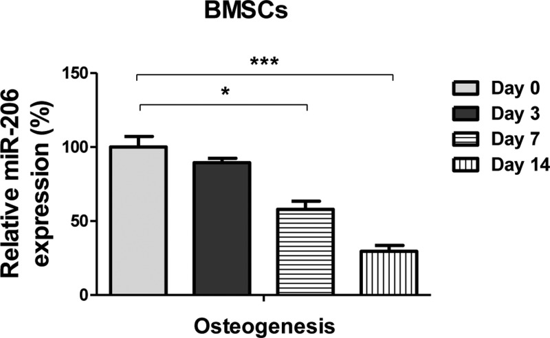
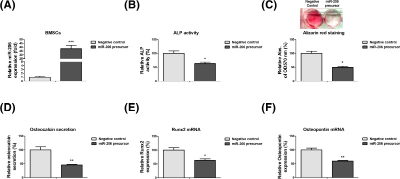
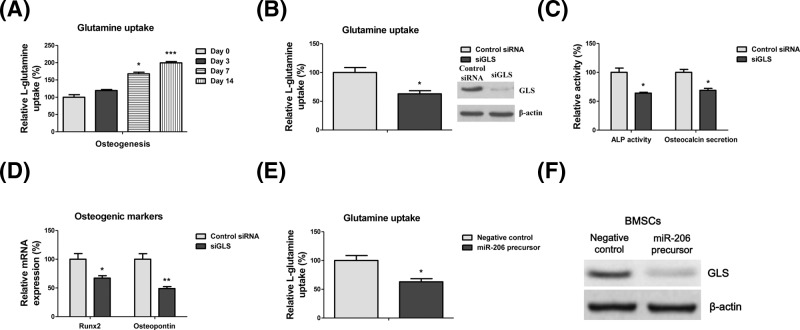
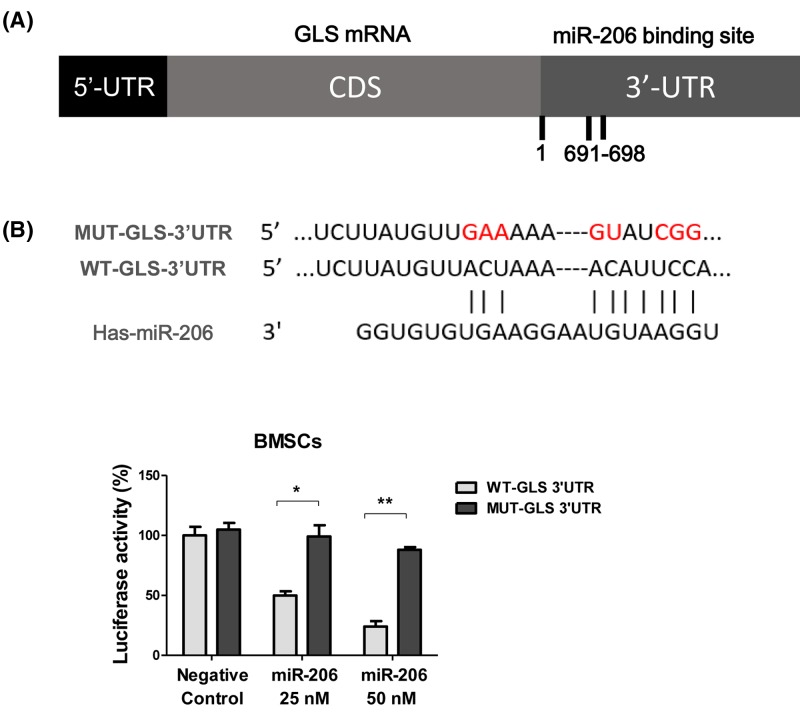
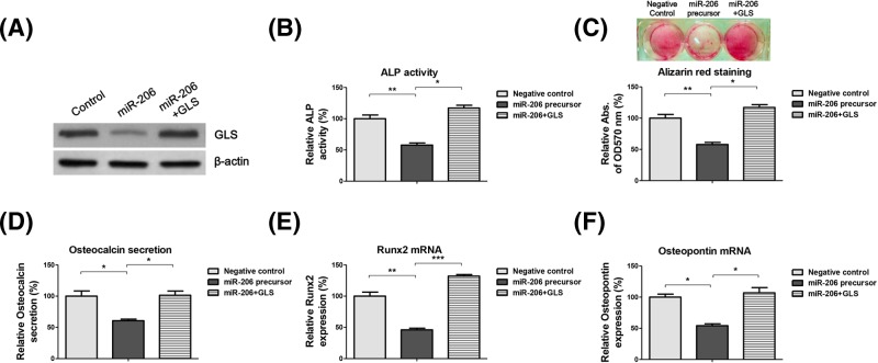
Osteoblast-mediated bone formation is a complex process involving various pathways and regulatory factors, including cytokines, growth factors, and hormones. Investigating the regulatory mechanisms behind osteoblast differentiation is important for bone regeneration therapy. miRNAs are known as important regulators, not only in a variety of cellular processes, but also in the pathogenesis of bone diseases. In the present study, we investigated the potential roles of miR-206 during osteoblast differentiation. We report that miR-206 expression was significantly down-regulated in human bone marrow mesenchymal stem cells (BMSCs) at days 7 and 14 during osteogenic induction. Furthermore, miR-206 overexpressing BMSCs showed attenuated alkaline phosphatase (ALP) activity, Alizarin Red staining, and osteocalcin secretion. The mRNA levels of osteogenic markers, Runx2 and Osteopontin (OPN), were significantly down-regulated in miR-206 overexpressing BMSCs. We observed that significantly increased glutamine uptake at days 7 and 14 during the osteogenic induction and inhibition of glutamine metabolism by knocking down glutaminase (GLS)-suppressed osteogenic differentiation of BMSCs. Here, we discover that miR-206 could directly bind to the 3'-UTR region of GLS mRNA, resulting in suppressed GLS expression and glutamine metabolism. Finally, restoration of GLS in miR-206 overexpressing BMSCs led to recovery of glutamine metabolism and osteogenic differentiation. In summary, these results reveal a new insight into the mechanisms of the miR-206-mediated osteogenesis through regulating glutamine metabolism. Our study may contribute to the development of therapeutic agents against bone diseases.

Keywords: bone marrow mesenchymal stem cells; glutaminase (GLS); microRNA-206; osteogenic differentiation.


Conflict of interest statement

The authors declare that there are no competing interests associated with the manuscript.

Figures

Figure 1
Figure 1. miR-206 is down-regulated in BMSCs during osteogenic differentiation
qRT-PCR was performed to analyze the expressions of miR-206 in BMSCs under osteogenic induction conditions at days 0, 3, 7, or 14. Data are shown as the mean ± S.D. *P<0.05; ***P<0.001. Abbreviation: qRT-PCR, quantitative reverse transcription PCR.
Figure 2
Figure 2. miR-23a/b inhibits the osteogenic differentiation of BMSCs
(A) BMSCs were transfected with native control or miR-206 precursor for 48 h. Expressions of miR-206 were measured by qRT-PCR. RNU6 was the internal control. (B) BMSCs were transfected with native control or miR-206 precursor for 48 h, the relative ALP activity. (C) Alizarin red staining and (D) Osteocalcin secretion were analyzed. (E) BMSCs were transfected with native control or miR-206 precursor for 48 h. The mRNA expressions of Runx2 and (F) OPN were detected by qRT-PCR. GAPDH was the internal control. Data are shown as the mean ± S.D. *P<0.05; **P<0.01; ***P<0.001. Abbreviation: qRT-PCR, quantitative reverse transcription PCR.
Figure 3
Figure 3. Glutamine metabolism is essential for osteogenic differentiation of BMSCs
(A) BMSCs were treated with osteogenic induction condition for 0, 3, 7, or 14 days, the relative glutamine uptakes were measured. (B) BMSCs were transfected with control siRNA or siGLS for 48 h. The glutamine uptake (C) ALP activity and Osteocalcin secretion were measured. (D) BMSCs were transfected with control siRNA or siGLS for 48 h. The mRNA expressions of Runx2 and OPN were detected by qRT-PCR. GAPDH was the internal control. (E) BMSCs were transfected with negative control or miR-206 precursor for 48 h. The relative glutamine uptake and (F) GLS protein expression were measured. β-actin was the internal control. Data are shown as the mean ± S.D. *P<0.05; ***P<0.001
Figure 4
Figure 4. miR-206 directly binds to the 3′-UTR region of GLS
(A) The predicted miR-206 binding sites on 3′-UTR region of GLS. (B) The mutant construct of the binding site (upper). BMSCs were co-transfected with control miR or miR-206 at 25 or 50 nM and wild-type or mutant GLS 3′-UTR for 48 h. The relative luciferase activities were measured (lower). Data are shown as the mean ± S.D. *P<0.05; **P<0.01.
Figure 5
Figure 5. Restoration of GLS recovers glutamine metabolism and osteogenic differentiation of BMSCs
(A) BMSCs were transfected with control vector or miR-206 precursor or miR-206 plus GLS for 48 h. The expressions of GLS were detected by Western blot. β-actin was the internal control. (B) BMSCs were transfected with control vector or miR-206 precursor or miR-206 plus GLS for 48 h. The relative ALP activity, (C) Alizarin Red staining, and (D) Osteocalcin secretion were analyzed. (E) BMSCs were transfected with control vector or miR-206 precursor or miR-206 plus GLS for 48 h. The mRNA expressions of Runx2 and (F) OPN were detected by qRT-PCR. GAPDH was the internal control. Data are shown as the mean ± S.D. *P<0.05; **P<0.01; ***P<0.001. Abbreviation: qRT-PCR, quantitative reverse transcription PCR.

Similar articles

Cited by

References

    1. Rupaimoole R. and Slack F.J. (2017) MicroRNA therapeutics: towards a new era for the management of cancer and other diseases. Nat. Rev. Drug Discov. 16, 203–222 10.1038/nrd.2016.246 - DOI - PubMed
    1. Paul P., Chakraborty A., Sarkar D., Langthasa M., Rahman M., Bari M. et al. . (2018) Interplay between miRNAs and human diseases. J. Cell. Physiol. 233, 2007–2018 10.1002/jcp.25854 - DOI - PubMed
    1. Park J., Wada S., Ushida T. and Akimoto T. (2015) The microRNA-23a has limited roles in bone formation and homeostasis in vivo. Physiol. Res. 64, 711–719 - PubMed
    1. Chen Z., Zhang Y., Liang C., Chen L., Zhang G. and Qian A. (2017) Mechanosensitive miRNAs and bone formation. Int. J. Mol. Sci. 18, pii: E1684, 10.3390/ijms18081684 - DOI - PMC - PubMed
    1. Huiskes R., Ruimerman R., van Lenthe G.H. and Janssen J.D. (2000) Effects of mechanical forces on maintenance and adaptation of form in trabecular bone. Nature 405, 704–706 10.1038/35015116 - DOI - PubMed