Early treadmill exercise increases macrophage migration inhibitory factor expression after cerebral ischemia/reperfusion
- PMID: 30804254
- PMCID: PMC6425847
- DOI: 10.4103/1673-5374.251330
Early treadmill exercise increases macrophage migration inhibitory factor expression after cerebral ischemia/reperfusion
Abstract
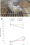
The neuroprotective function of macrophage migration inhibitory factor (MIF) in ischemic stroke was rarely evaluated. This study aimed to investigate the effects of early treadmill exercise on recovery from ischemic stroke and to determine whether these effects are associated with the expression levels of MIF and brain-derived neurotrophic factor (BDNF) in the ischemic area. A total of 40 male Sprague-Dawley rats were randomly assigned to the ischemia and exercise group [middle cerebral artery occlusion (MCAO)-Ex, n = 10), ischemia and sedentary group (MCAO-St, n = 10), sham-surgery and exercise group (Sham-Ex, n = 10), or sham-surgery and sedentary group (Sham-St, n = 10). The MCAO-Ex and MCAO-St groups were subjected to MCAO for 60 minutes, whereas the Sham-Ex and Sham-St groups were subjected to an identical operation without MCAO. Rats in the MCAO-Ex and Sham-Ex groups then ran on a treadmill for 30 minutes once a day for 5 consecutive days. After reperfusion, the hanging time tested by the wire hang test was longer and the relative fractional anisotropy determined by MRI was higher in the peri-infarct region of the MCAO-Ex group compared with the MCAO-St group. The expression levels of MIF and BDNF in the peri-infarct region were upregulated in the MCAO-Ex group. Increased MIF and BDNF levels were positively correlated with relative fractional anisotropy changes in the peri-infarct region. There was no significant difference in the levels of MIF and BDNF in the peri-infarct region between the Sham-Ex and Sham-St groups. Our study demonstrated that early exercise (initiated 48 hours after the MCAO) could improve motor and neuronal recovery after ischemic stroke. Furthermore, the increased levels of MIF and BDNF in the peri-infarct region (penumbra) may be one of the mechanisms of enhanced neurological function recovery. All experiments were approved by the Institutional Animal Care and Use Committee in Asan Medical Center in South Korea (2016-12-126).
Keywords: brain-derived neurotrophic factor; early exercise; ischemic stroke; macrophage migration inhibitory factor; motor recovery; neural regeneration.
Conflict of interest statement
None
Figures


References
-
- Adams HP, Jr, Brott TG, Crowell RM, Furlan AJ, Gomez CR, Grotta J, Helgason CM, Marler JR, Woolson RF, Zivin JA. Guidelines for the management of patients with acute ischemic stroke. A statement for healthcare professionals from a special writing group of the Stroke Council, American Heart Association. Circulation. 1994;90:1588–1601. - PubMed
-
- Assaf Y, Pasternak O. Diffusion tensor imaging (DTI)-based white matter mapping in brain research: a review. J Mol Neurosci. 2008;34:51–61. - PubMed
-
- Bernhagen J, Calandra T, Mitchell RA, Martin SB, Tracey KJ, Voelter W, Manogue KR, Cerami A, Bucala R. MIF is a pituitary-derived cytokine that potentiates lethal endotoxaemia. Nature. 1993;365:756–759. - PubMed
-
- Bernhardt J, Dewey H, Thrift A, Collier J, Connan G. A very early rehabilitation trial for stroke (AVERT): phase II safety and feasibility. Stroke. 2008;39:390–396. - PubMed
LinkOut - more resources
Full Text Sources
Molecular Biology Databases
Miscellaneous

