

Effect of exogenous spastin combined with polyethylene glycol on sciatic nerve injury
- PMID: 30804259
- PMCID: PMC6425831
- DOI: 10.4103/1673-5374.251336
Effect of exogenous spastin combined with polyethylene glycol on sciatic nerve injury
Abstract
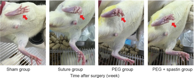
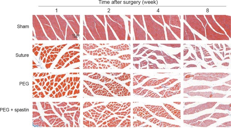
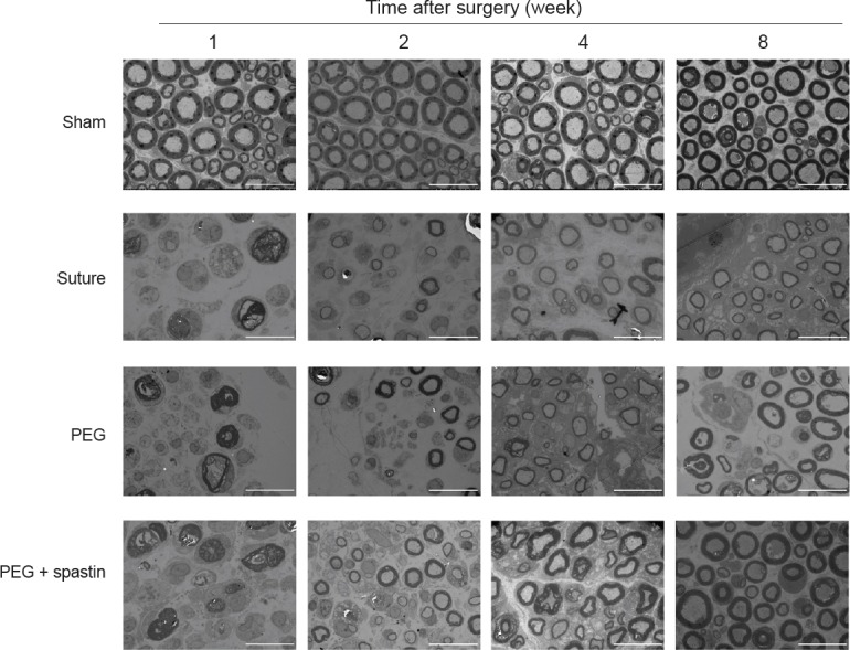
Polyethylene glycol can connect the distal and proximal ends of an injured nerve at the cellular level through axonal fusion to avoid Wallerian degeneration of the injured distal nerve and promote peripheral nerve regeneration. However, this method can only prevent Wallerian degeneration in 10% of axons because the cytoskeleton is not repaired in a timely fashion. Reconstruction of the cytoskeletal trunk and microtubule network has been suggested to be the key for improving the efficiency of axonal fusion. As a microtubule-severing protein, spastin has been used to enhance cytoskeletal reconstruction. Therefore, we hypothesized that spastin combined with polyethylene glycol can more effectively promote peripheral nerve regeneration. A total of 120 male Sprague-Dawley rats were randomly divided into sham, suture, polyethylene glycol, and polyethylene glycol + spastin groups. In suture group rats, only traditional nerve anastomosis of the end-to-end suture was performed after transection of the sciatic nerve. In polyethylene glycol and polyethylene glycol + spastin groups, 50 μL of polyethylene glycol or 25 μL of polyethylene glycol + 25 μL of spastin, respectively, were injected immediately under the epineurium of the distal suture. Sensory fiber regeneration distance, which was used to assess early nerve regeneration at 1 week after surgery, was shortest in the suture group, followed by polyethylene glycol group and greatest in the polyethylene glycol + spastin group. Behavioral assessment of motor function recovery in rats showed that limb function was restored in polyethylene glycol and polyethylene glycol + spastin groups at 8 weeks after surgery. At 1, 2, 4 and 8 weeks after surgery, sciatic functional index values and percentages of gastrocnemius muscle wet weight were highest in the sham group, followed by polyethylene glycol + spastin and polyethylene glycol groups, and lowest in the suture group. Masson staining was utilized to assess the morphology of muscle tissue. Morphological changes in skeletal muscle were detectable in suture, polyethylene glycol, and polyethylene glycol + spastin groups at 1, 2, 4, and 8 weeks after surgery. Among them, muscular atrophy of the suture group was most serious, followed by polyethylene glycol and polyethylene glycol + spastin groups. Ultrastructure of distal sciatic nerve tissue, as detected by transmission electron microscopy, showed a pattern of initial destruction, subsequent disintegration, and gradual repair in suture, polyethylene glycol, and polyethylene glycol + spastin groups at 1, 2, 4, and 8 weeks after surgery. As time proceeded, axonal ultrastructure gradually recovered. Indeed, the polyethylene glycol + spastin group was similar to the sham group at 8 weeks after surgery. Our findings indicate that the combination of polyethylene glycol and spastin can promote peripheral nerve regeneration. Moreover, the effect of this combination was better than that of polyethylene glycol alone, and both were superior to the traditional neurorrhaphy. This study was approved by the Animal Ethics Committee of the Second Military Medical University, China (approval No. CZ20170216) on March 16, 2017.
Keywords: Masson staining; Wallerian degeneration; axonal fusion; microtubule; nerve regeneration; neural regeneration; peripheral nerve injuries; peripheral nerves; polyethylene glycol; spastin.
Conflict of interest statement
None
Figures
Similar articles
-
Polyethylene glycol (PEG) and other bioactive solutions with neurorrhaphy for rapid and dramatic repair of peripheral nerve lesions by PEG-fusion.J Neurosci Methods. 2019 Feb 15;314:1-12. doi: 10.1016/j.jneumeth.2018.12.015. Epub 2018 Dec 23. J Neurosci Methods. 2019. PMID: 30586569 Free PMC article.
-
Polyethylene glycol has immunoprotective effects on sciatic allografts, but behavioral recovery and graft tolerance require neurorrhaphy and axonal fusion.Neural Regen Res. 2025 Apr 1;20(4):1192-1206. doi: 10.4103/NRR.NRR-D-23-01220. Epub 2024 Apr 3. Neural Regen Res. 2025. PMID: 38989956 Free PMC article.
-
Polyethylene glycol treated allografts not tissue matched nor immunosuppressed rapidly repair sciatic nerve gaps, maintain neuromuscular functions, and restore voluntary behaviors in female rats.J Neurosci Res. 2018 Jul;96(7):1243-1264. doi: 10.1002/jnr.24227. Epub 2018 Apr 16. J Neurosci Res. 2018. PMID: 29659046 Free PMC article.
-
Conundrums and confusions regarding how polyethylene glycol-fusion produces excellent behavioral recovery after peripheral nerve injuries.Neural Regen Res. 2018 Jan;13(1):53-57. doi: 10.4103/1673-5374.224363. Neural Regen Res. 2018. PMID: 29451204 Free PMC article. Review.
-
The curious ability of polyethylene glycol fusion technologies to restore lost behaviors after nerve severance.J Neurosci Res. 2016 Mar;94(3):207-30. doi: 10.1002/jnr.23685. Epub 2015 Nov 3. J Neurosci Res. 2016. PMID: 26525605 Free PMC article. Review.
Cited by
-
Spastin interacts with collapsin response mediator protein 3 to regulate neurite growth and branching.Neural Regen Res. 2021 Dec;16(12):2549-2556. doi: 10.4103/1673-5374.313052. Neural Regen Res. 2021. PMID: 33907047 Free PMC article.
-
Polyethylene Glycol Fusion of Nerve Injuries: Review of the Technique and Clinical Applicability.J Hand Microsurg. 2021 Apr;13(2):49-54. doi: 10.1055/s-0040-1718651. Epub 2020 Dec 10. J Hand Microsurg. 2021. PMID: 33867761 Free PMC article. Review.
-
14-3-3 protein augments the protein stability of phosphorylated spastin and promotes the recovery of spinal cord injury through its agonist intervention.Elife. 2024 Jan 17;12:RP90184. doi: 10.7554/eLife.90184. Elife. 2024. PMID: 38231910 Free PMC article.
-
Brain Derived Neurotrophic Factor and Glial Cell Line-Derived Neurotrophic Factor-Transfected Bone Mesenchymal Stem Cells for the Repair of Periphery Nerve Injury.Front Bioeng Biotechnol. 2020 Jul 30;8:874. doi: 10.3389/fbioe.2020.00874. eCollection 2020. Front Bioeng Biotechnol. 2020. PMID: 32850732 Free PMC article.
-
Ulinastatin Promotes Regeneration of Peripheral Nerves After Sciatic Nerve Injury by Targeting let-7 microRNAs and Enhancing NGF Expression.Drug Des Devel Ther. 2020 Jul 9;14:2695-2705. doi: 10.2147/DDDT.S255158. eCollection 2020. Drug Des Devel Ther. 2020. PMID: 32753848 Free PMC article.
References
-
- Ahkong QF, Desmazes JP, Georgescauld D, Lucy JA. Movements of fluorescent probes in theme chanismof cell fusion induced by poly (ethylene glycol) Cell Sci. 1987;88:389–398. - PubMed
-
- Bain JR, Mackinnon SE, Hunter DA. Functional evaluation of complete sciatic, peroneal, and posterior tibial nerve lesions in the rat. Plast Reconstr Surg. 1989;83:129–138. - PubMed
-
- Bittner GD, Keating CP, Kane JR, Britt JM, Spaeth CS, Fan JD, Zuzek A, Wilcott RW, Thayer WP, Winograd JM, Gonzalez-Lima F, Schallert T. Rapid, effective, and long-lasting behavioral recovery produced by microsutures, methylene blue, and polyethylene glycol after completely cutting rat sciatic nerves. J Neurosci Res. 2012;90:967–980. - PubMed
LinkOut - more resources
Full Text Sources
Research Materials
