. 2019 Feb 21;51(2):1-12.
doi: 10.1038/s12276-019-0211-9.

Downregulation of lysyl oxidase and lysyl oxidase-like protein 2 suppressed the migration and invasion of trophoblasts by activating the TGF-β/collagen pathway in preeclampsia

Affiliations

Downregulation of lysyl oxidase and lysyl oxidase-like protein 2 suppressed the migration and invasion of trophoblasts by activating the TGF-β/collagen pathway in preeclampsia

Xiang-Hong Xu et al. Exp Mol Med. .

Abstract

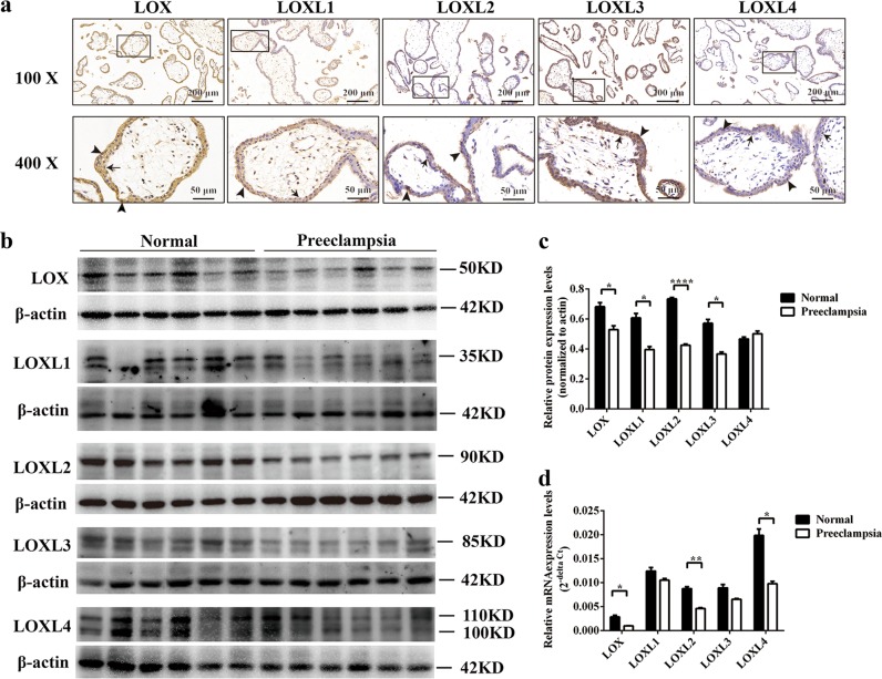
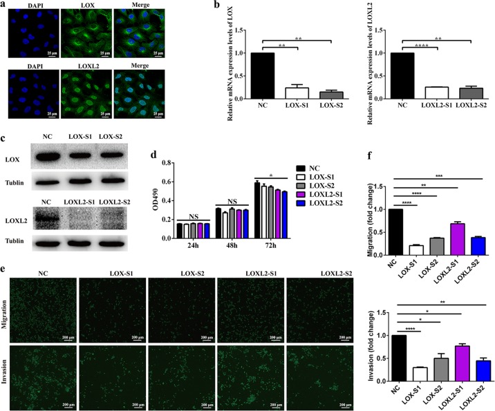
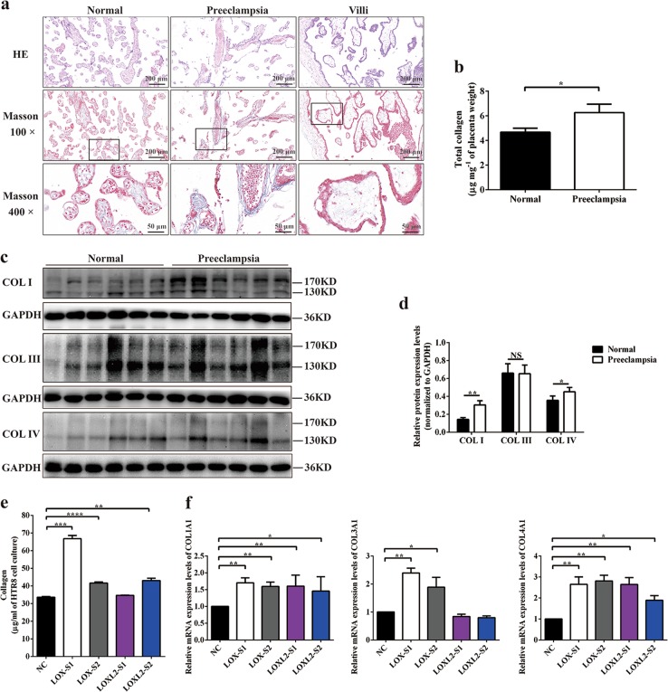
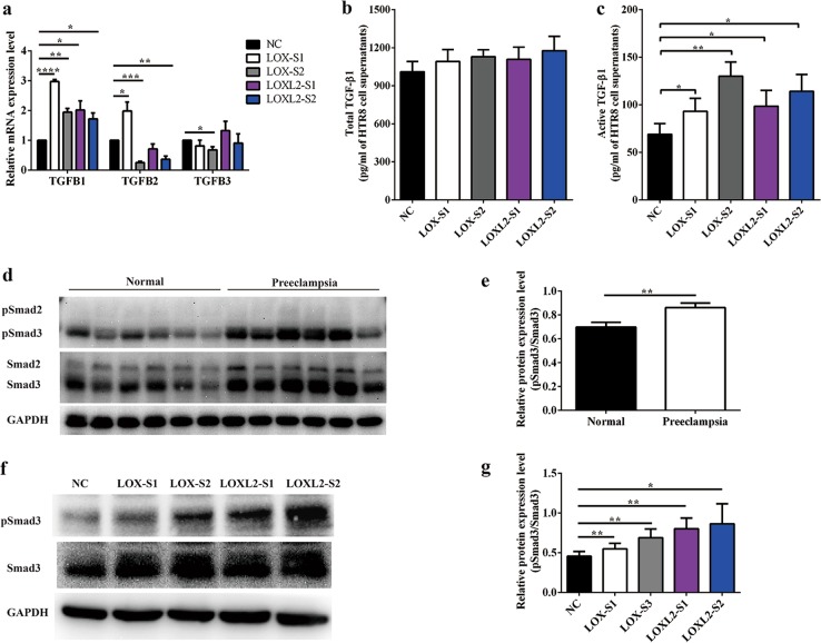
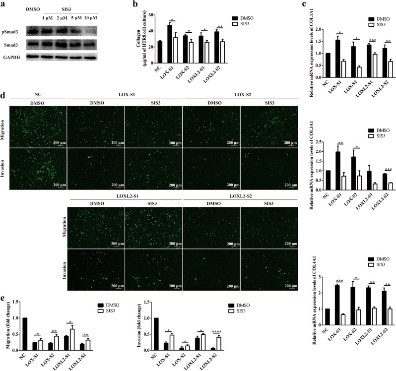
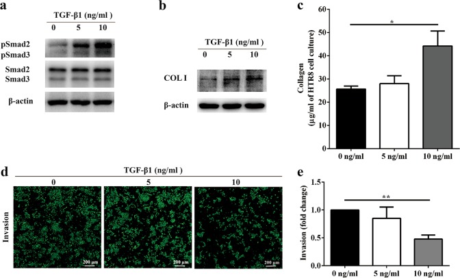
Preeclampsia is a pregnancy-specific disorder that is a major cause of maternal and fetal morbidity and mortality with a prevalence of 6-8% of pregnancies. Although impaired trophoblast invasion in early pregnancy is known to be closely associated with preeclampsia, the underlying mechanisms remain elusive. Here we revealed that lysyl oxidase (LOX) and LOX-like protein 2 (LOXL2) play a critical role in preeclampsia. Our results demonstrated that LOX and LOXL2 expression decreased in preeclamptic placentas. Moreover, knockdown of LOX or LOXL2 suppressed trophoblast cell migration and invasion. Mechanistically, collagen production was induced in LOX- or LOXL2-downregulated trophoblast cells through activation of the TGF-β1/Smad3 pathway. Notably, inhibition of the TGF-β1/Smad3 pathway could rescue the defects caused by LOX or LOXL2 knockdown, thereby underlining the significance of the TGF-β1/Smad3 pathway downstream of LOX and LOXL2 in trophoblast cells. Additionally, induced collagen production and activated TGF-β1/Smad3 were observed in clinical samples from preeclamptic placentas. Collectively, our study suggests that the downregulation of LOX and LOXL2 leading to reduced trophoblast cell migration and invasion through activation of the TGF-β1/Smad3/collagen pathway is relevant to preeclampsia. Thus, we proposed that LOX, LOXL2, and the TGF-β1/Smad3/collagen pathway can serve as potential markers and targets for clinical diagnosis and therapy for preeclampsia.


Conflict of interest statement

The authors declare that they have no conflict of interest.

Figures

Fig. 1
Fig. 1. Decreased expression of LOX and LOXL2 was found in preeclamptic placentas.
a Representative images of expression and localization of LOX family members in first trimester villi by immunohistochemistry analysis. Brownish color represents positive staining of LOX family members. Arrows, trophoblasts; arrowheads, cytotrophoblasts. b Western blot analysis of LOX family members in placentas from normal full-term pregnancies and preeclampsia patients. c Statistical analysis of protein densitometry quantification of western blot analysis (b) by Student’s t-test. Data are presented as the means ± SEM. d mRNA expression of LOX family members was analyzed by quantitative reverse-transcription PCR. Statistical data were analyzed by Student’s t-test. Data are presented as the means ± SEM. *P < 0.05; **P < 0.01; and ****P < 0.0001
Fig. 2
Fig. 2. Knockdown of LOX or LOXL2 decreased trophoblast cell migration and invasion.
a Immunofluorescence staining for LOX and LOXL2 in HTR-8/SVneo cells. Confocal microscopy images were shown. Scale bar = 25 μm. b mRNA expression of LOX and LOXL2 in HTR-8/SVneo cells transfected with LOX siRNA, LOXL2 siRNA, or negative control siRNA was analyzed by quantitative reverse-transcription PCR. c Western blot analysis of LOX and LOXL2 protein expression in HTR-8/SVneo cells transfected with LOX siRNA, LOXL2 siRNA, or negative control siRNA. d Proliferation of HTR-8/SVneo cells lines detected by MTS assays. Statistical analysis of cell proliferation index was performed by one-way analysis of variance. e HTR-8/SVneo cell migration and invasion were determined by Transwell assays. Representative images are shown. f Relative fold changes in cell migration and invasion were calculated. Statistical data were analyzed by Student’s t-test. All data are presented as the means ± SEM of five independent experiments. *P < 0.05; **P < 0.01; ***P < 0.001; ****P < 0.0001; and NS not significant
Fig. 3
Fig. 3. Collagen production was induced in LOX- or LOXL2-downregulated preeclamptic placenta and trophoblast cells.
a Representative hematoxylin-eosin staining and Masson’s trichrome staining of placenta and villi are shown. b Amount of total collagen was determined by measuring the hydroxyproline contents of placental tissues. c Western blot analysis of type I collagen, type III collagen, and type IV collagen in the placenta. d Statistical analysis of protein densitometry quantification of western blot analysis (c) by Student’s t-test. Data are presented as the means ± SEM. e Amount of soluble collagen from cell culture supernatants was quantified using a sircol collagen assay. Data are presented as the means ± SEM of three independent experiments. f Quantification of COL1A1, COL3A1, and COL4A1 mRNA expression levels determined in HTR-8/SVneo cells after siRNA treatment. Data are presented as the means ± SEM of three independent experiments. *P < 0.05; **P < 0.01; ***P < 0.001; ***P < 0.001; and NS not significant
Fig. 4
Fig. 4. TGF-β1/Smad3 signaling was activated in LOX- or LOXL2-downregulated trophoblast cells.
a mRNA expression of TGFB1, TGFB2, and TGFB3 was determined by quantitative reverse-transcription PCR. b, c Total and active transforming growth factor-β1 (TGF-β1) protein levels in culture supernatants from HTR-8/SVneo cells transfected with small interfering RNA (siRNA) were measured by enyme-linked immunosorbent assay. d Western blotting and e quantification analysis of pSmad2/3 and Smad2/3 in placentas from normal pregnant women and preeclampsia patients. f Western blotting and g quantification analysis of pSmad2/3 and Smad2/3 in HTR-8/SVneo cells transfected with siRNA. Statistical data were analyzed by Student’s t-test. Data are presented as the means ± SEM of three independent experiments. *P < 0.05; **P < 0.01; ***P < 0.001; and ****P < 0.0001
Fig. 5
Fig. 5. Inhibiting Smad3 could partially rescue trophoblast cell migration and invasion.
a Western blot analysis of pSmad3 and Smad3 in HTR-8/SVneo cells treated with the Smad3 inhibitor SIS3 (0, 2, 5, or 10 μmol/L) for 24 h. b Amount of soluble collagen in culture supernatants from HTR-8/SVneo cells transfected with small interfering RNA (siRNA) followed by SIS3 treatment (10 μmol/L) was quantified using a sircol collagen assay. c Quantification of COL1A1, COL3A1, and COL4A1 mRNA expression levels was determined in HTR-8/SVneo cells upon siRNA transfection followed by SIS3 treatment (10 μmol/L). d Migration and invasion of HTR-8/SVneo cells upon siRNA transfection followed by SIS3 treatment (10 μmol/L) was determined by Transwell assays. Representative images are shown. e Relative fold changes in cell migration and invasion were calculated. Statistical data were analyzed by Student’s t-test. Data are presented as the means ± SEM of three independent experiments. *P < 0.05; **P < 0.01; ***P < 0.001; and ****P < 0.0001
Fig. 6
Fig. 6. Exogenous transforming growth factor-β1 (TGF-β1) treatment reduced trophoblast cell invasion.
a Western blot analysis of pSmad3 and Smad3 in HTR-8/SVneo cells stimulated by exogenous TGF-β1. b Western blot analysis of type I collagen production in HTR-8/SVneo cells stimulated by exogenous TGF-β1. c Amount of soluble collagen from cell culture supernatants was quantified using a sircol collagen assay. Data are presented as the means ± SEM of three independent experiments. d Invasion of HTR-8/SVneo cells upon TGF-β1 treatment was determined by Transwell assays. Representative images are shown. e Relative fold changes in cell invasion were calculated. Statistical data were analyzed by Student’s t-test. Data are presented as the means ± SEM of three independent experiments. *P < 0.05 and **P < 0.01

Similar articles

Cited by

References

    1. Molnar J, et al. Structural and functional diversity of lysyl oxidase and the LOX-like proteins. Biochim. Biophys. Acta. 2003;1647:220–224. doi: 10.1016/S1570-9639(03)00053-0. - DOI - PubMed
    1. Csiszar K. Lysyl oxidases: a novel multifunctional amine oxidase family. Prog. Nucleic Acid Res. Mol. Biol. 2001;70:1–32. doi: 10.1016/S0079-6603(01)70012-8. - DOI - PubMed
    1. Ma L, et al. Lysyl oxidase 3 is a dual-specificity enzyme involved in STAT3 deacetylation and deacetylimination modulation. Mol. Cell. 2017;65:296–309. doi: 10.1016/j.molcel.2016.12.002. - DOI - PubMed
    1. Cuevas EP, et al. LOXL2 catalytically inactive mutants mediate epithelial-to-mesenchymal transition. Biol. Open. 2014;3:129–137. doi: 10.1242/bio.20146841. - DOI - PMC - PubMed
    1. Vadasz Z, et al. Abnormal deposition of collagen around hepatocytes in Wilson’s disease is associated with hepatocyte specific expression of lysyl oxidase and lysyl oxidase like protein-2. J. Hepatol. 2005;43:499–507. doi: 10.1016/j.jhep.2005.02.052. - DOI - PubMed

Publication types

MeSH terms