. 2019 Feb 21;51(2):1-20.
doi: 10.1038/s12276-019-0209-3.

Extracellular signal-regulated kinase 5 increases radioresistance of lung cancer cells by enhancing the DNA damage response

Affiliations

Extracellular signal-regulated kinase 5 increases radioresistance of lung cancer cells by enhancing the DNA damage response

Weiwei Jiang et al. Exp Mol Med. .

Erratum in

Abstract

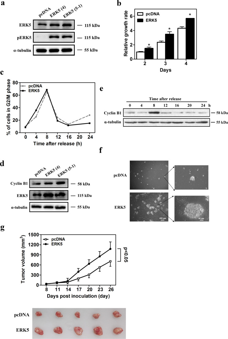
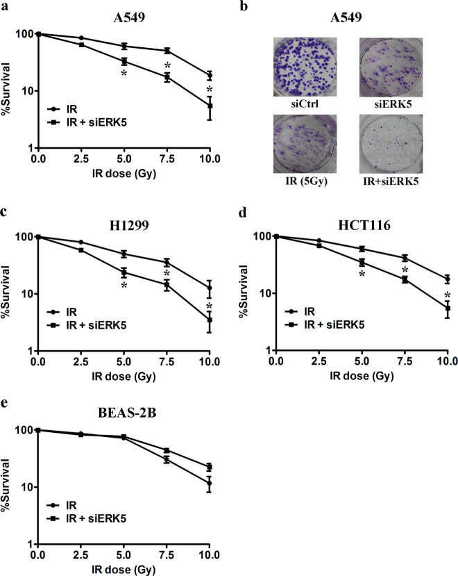
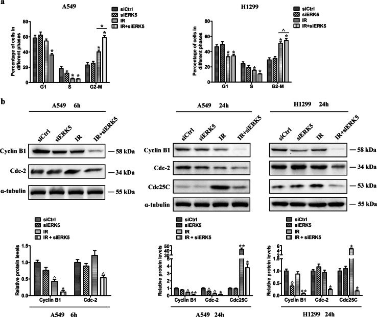
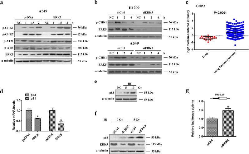
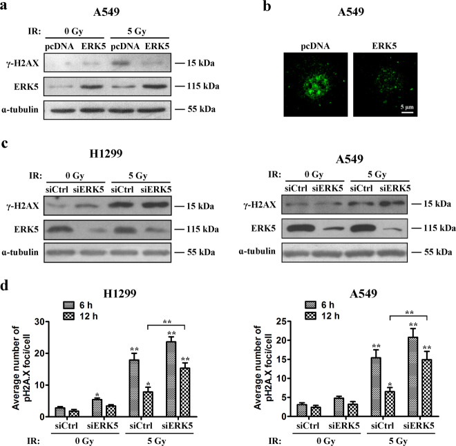
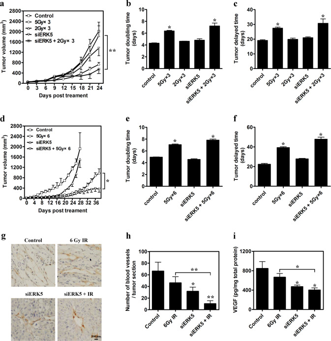
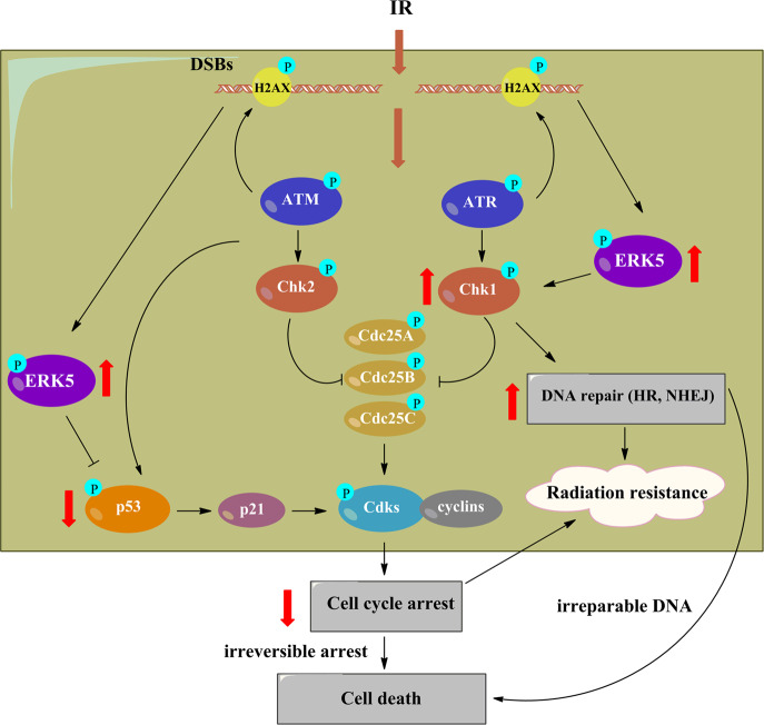
Radiotherapy is a frequent mode of cancer treatment, although the development of radioresistance limits its effectiveness. Extensive investigations indicate the diversity of the mechanisms underlying radioresistance. Here, we aimed to explore the effects of extracellular signal-regulated kinase 5 (ERK5) on lung cancer radioresistance and the associated mechanisms. Our data showed that ERK5 is activated during solid lung cancer development, and ectopic expression of ERK5 promoted cell proliferation and G2/M cell cycle transition. In addition, we found that ERK5 is a potential regulator of radiosensitivity in lung cancer cells. Mechanistic investigations revealed that ERK5 could trigger IR-induced activation of Chk1, which has been implicated in DNA repair and cell cycle arrest in response to DNA double-strand breaks (DSBs). Subsequently, ERK5 knockdown or pharmacological inhibition selectively inhibited colony formation of lung cancer cells and enhanced IR-induced G2/M arrest and apoptosis. In vivo, ERK5 knockdown strongly radiosensitized A549 and LLC tumor xenografts to inhibition, with a higher apoptotic response and reduced tumor neovascularization. Taken together, our data indicate that ERK5 is a novel potential target for the treatment of lung cancer, and its expression might be used as a biomarker to predict radiosensitivity in NSCLC patients.


Conflict of interest statement

The authors declare that they have no conflict of interest.

Figures

Fig. 1
Fig. 1. ERK5 promotes lung cancer cell proliferation and increased the malignant transformation ability of A549 cells.
a The protein expression level of ERK5 and p-ERK5 in A549 cells stably transfected with either empty vector or vector expressing the ERK5 gene. b Stably transfected A549 cells were seeded 2000/well in 96-well plates, and cell growth was detected with MTT assays on days 2, 3, and 4. The data represent the mean ± SD; *p < 0.05 versus control. c Both ERK5-overexpression A549 cells and control cells were synchronized by double thymidine block and harvested at various time points after release. The cells were stained with PI, followed by flow cytometry analysis. d ERK5 overexpression and its effect on upregulation of cyclin B1 were detected via western blot, with ɑ-tubulin as the internal control. e ERK5-overexpression A549 cells were synchronized by double thymidine block and harvested at various time points after release. Then, the expression levels of cyclin B1 were detected via western blotting. f ERK5 overexpression and empty vector control A549 cells were cultured in soft agar and incubated for 3 weeks. The number and shape of colonies were examined. g The same number (2 × 106) of A549 cells that stably expressed ERK5 and empty vector plasmids were injected subcutaneously into the right dorsum of each mouse; tumor growth rates were then compared. The data are presented as the mean ± SD, *p < 0.05. Representative images of excised tumors from each group are also shown (lower panel)
Fig. 2
Fig. 2. ERK5 expression is upregulated by IR stress.
a A549 cells were exposed to different X-ray doses, and the expression levels of ERK5 and p-ERK5 were detected via western blotting at 12 h after exposure. Band intensity was quantified with ImageJ software. The results shown are representative of three different experiments. The data are presented as the mean ± SD; *p < 0.05 and **p < 0.01. b A549 cells were exposed to 10 Gy IR, and the expression levels of ERK5 and p-ERK5 were detected by western blotting at the indicated time after exposure. c A549 cells were exposed to 5 Gy IR, and the expression levels of ERK5, p-ERK5, ERK1/2, and p-ERK1/2 were detected by western blotting at 1.5 and 2 h after exposure. d A549 cells were exposed to 5 Gy IR, and the expression levels of ERK5, p-ERK5, ERK1/2, p-ERK1/2, and Cyclin B1 were detected by western blotting at the indicated time after exposure
Fig. 3
Fig. 3. ERK5 knockdown radiosensitizes lung cancer cells.
Cells were plated at 500 cells per well in a 6-well plate and after 24 h treated with the indicated dose of IR and/or ERK5 siRNA. Cells were then maintained for another 12 days. The formed colonies were fixed and stained. Colonies containing >50 cells were counted, and the colony formation percentage was determined for each cell line with respect to the nontreated controls. Survival curves for advanced human lung adenocarcinoma A549 cells (a) and representative images of stained colonies formed by A549 cells (b), H1299 cells (c), HCT116 cells (d), and transformed nonneoplastic human bronchial epithelial BEAS-2B cells (e). *p < 0.01 compared with the respective control
Fig. 4
Fig. 4. ERK5 inhibits IR-induced apoptosis in lung cancer cells.
a A549 cells stably transfected with either empty vector or vector encoding ERK5 were treated with 5 Gy IR. After 16 h, all cells were harvested for flow cytometry analysis. Annexin V/PI-stained cells were analyzed, and the percentage of apoptotic cells was determined. The experiments were carried out independently in triplicate; representative data are shown. The data are presented as the mean ± SD. *p < 0.05. The Annexin V/PI double-staining profile of A549 cells is also included. b ERK5 was downregulated in H1299 and A549 cells through transfection with ERK5 siRNA. Cells were treated with IR, and cellular apoptosis was detected as in (a). c A549 cells stably transfected with either empty vector or vector encoding ERK5 were irradiated with IR at various doses and further cultured for 48 h. Whole-cell lysates were prepared and subjected to immunoblotting to detect cleaved caspase-8 (c-Casp8), cleaved caspase-3 (c-Casp3), cleaved caspase-9 (c-Casp9), and cleaved PARP (c-PARP) levels to reflect cellular apoptosis. ɑ-Tubulin levels were also detected as the loading control. d ERK5 was downregulated in H1299 and A549 cells after transfection with ERK5 siRNA. Cells were treated with IR, and cellular apoptosis was detected as in (c). The results shown are representative of three different experiments. Densitometric quantification of the immunoblot data in (c, d) is also shown. The data are presented as the mean ± SD; *p < 0.05, **p < 0.01, and ***p < 0.001
Fig. 5
Fig. 5. ERK5 knockdown potentiates IR-induced G2/M arrest.
A549 and H1299 cells were exposed to IR with or without ERK5 siRNA treatment. After the treatment time points, cells were processed for cell cycle analysis using PI staining. a Quantitative data showing the cell cycle distribution in A549 (left) and H1299 cells (right) after treatment with IR (5 Gy) and/or ERK5 siRNA. *p < 0.01 and ^p < 0.05 compared with the respective control or indicated treatment. b Western blots for G2/M cell cycle-related proteins (Cyclin B1, Cdc2, and Cdc25C) at 6 and 24 h. Band intensity was quantified using ImageJ software. The results shown are representative of three different experiments. The data are presented as the mean ± SD. ^p < 0.05, *p < 0.01, and **p < 0.001
Fig. 6
Fig. 6. ERK5 promotes IR-induced phosphorylation of ChK1 in lung cancer cells.
a A549 cells stably transfected with either empty vector or vector expressing ERK5 were treated with IR and further cultured for various time periods. Whole-cell lysates were prepared and subjected to immunoblotting to detect the levels of various checkpoint proteins. b ERK5 was downregulated in H1299 and A549 cells via transfection with ERK5 siRNA. Cells were treated with IR and further cultured for various time periods. Whole-cell lysates were prepared and subjected to immunoblotting to detect phosphorylated Chk1. ɑ-Tubulin was also detected as the loading control. c The expression levels of ERK5 in 226 lung adenocarcinoma tumor tissues and 20 normal tissues were obtained from TCGA. d A549 cells stably transfected with either empty vector or vector encoding ERK5 were harvested for RNA extraction and quantitative real-time PCR analysis using primers specific for human p53, p21, and GAPDH (internal control). The data represent the mean values of triplicate samples. *p < 0.05. e A549 cells were irradiated with IR at various doses, and the protein expression level of p53 was detected via western blotting. f A549 cells were transfected with control vector or ERK5 siRNA and treated with or without IR, and the protein expression level of p53 was detected via western blotting. g A549 cells were transfected with control vector or ERK5 siRNA. Transfected cells were then cotransfected with P53-Luc plasmid and pRL-TK Renilla vector. After 24 h, luciferase expression was measured. p53 luciferase activity was normalized to that of Renilla luciferase activity, and the results are expressed relative to the control values. The results are presented as the means ± SD from at least three independent experiments. *p < 0.05 versus control
Fig. 7
Fig. 7. ERK5 facilitates homologous recombination to repair IR-induced DSBs.
a, b A549 cells stably transfected with either empty vector or vector expressing ERK5 were treated with 5 Gy IR and further cultured for 6 h. Whole-cell lysates were prepared and subjected to immunoblotting to detect ERK5 and phosphorylated H2AX (γH2AX) levels. ɑ-Tubulin levels were also detected as the loading control (a). Immunofluorescent staining for γH2AX was performed on control and ERK5 overexpression A549 cells fixed at 6 h following 5 Gy X-ray irradiation treatment (b). c, d H1299 and A549 cells were transfected with control vector or ERK5 siRNA, treated with or without IR, and further cultured for 6 h. Whole-cell lysates were prepared and subjected to immunoblotting to detect ERK5 and phosphorylated H2AX (γH2AX) levels. ɑ-Tubulin was also detected as the loading control (c). Quantitation of the number of pH2A.X foci in H1299 and A549 cells after 6 and 12 h of treatments are also shown (d). *p < 0.05 and **p < 0.01 compared with the respective control or indicated treatment
Fig. 8
Fig. 8. ERK5 knockdown inhibits LLC tumor growth and increases tumor radiosensitivity.
ac Tumor growth was suppressed in LLC-bearing C57BL/6J mice treated with ERK5 knockdown combined with low-dose IR. When visible tumors were formed, local irradiation treatments (cumulative dose of 6 Gy) were fractionally administered on days 0, 2, and 4, and constructs expressing either Luc shRNA or ERK5 shRNA were injected into the tumor mass on days 1, 3, and 5. The tumor growth inhibitory effects of different treatments were compared (a). Tumor doubling time (b) and tumor delay time (c) are also shown. df Tumor growth was suppressed in LLC-bearing C57BL/6J mice treated with ERK5 knockdown combined with high-dose IR. When visible tumors were formed, local irradiation (total dose of 30 Gy) was fractionally administered on days 0, 2, 4, 6, 8, and 10, and a construct expressing either Luc shRNA or ERK5 shRNA was injected into the tumor mass on days 1, 3, and 5. The tumor growth inhibitory effects of different treatments were compared (d). Tumor doubling time (e) and tumor delay time (f) are also shown. g, h Blood vessel density within tumors was characterized by anti-CD31 immunostaining using an anti-mouse CD31 monoclonal antibody (g) and determined by the average number of vessels in 3 regions of the highest density at ×200 magnification in each section (the assay was repeated in 4 sections per mouse, and 3 mice were tested) (h). i Total protein was extracted from LLC tumors, and the intracellular VEGF level was detected via ELISA using 100 μg total protein per well. The data are presented as the mean ± SD; *p < 0.05 and **p < 0.01 compared with the respective control or indicated treatment
Fig. 9
Fig. 9. ERK5 knockdown enhances radiation-induced tumor growth inhibition of human lung cancer A549 xenografts in athymic nude mice.
Mice were given a s.c. injection of A549 cells (2 × 106) and monitored for tumor growth until the tumor size reached approximately 50 mm3. Then, local irradiation with 6 Gy was fractionally administered on days 0, 2, and 4, and a construct encoding either siCtrl or siERK5 was injected into the tumor mass on days 1, 3, and 5. a The tumor growth inhibitory effects of different treatments were compared. b Tumor weight/mouse at the end of the study. Tumor doubling time (c) and mean body weight per mouse (d) are also shown. e Determination of tumor necrosis after combined treatment with ERK5 siRNA and IR. Tumor necrosis areas are shown by H&E staining and were observed under a light microscope (×100). The viable tumor cells are indicated by a blue arrow. Tumor necrosis was determined with ImageJ software. Two sections/mouse from three mice were prepared. f Determination of tumor apoptosis after combined treatment with ERK5 siRNA and IR. TUNEL assays were used to detect apoptotic cells (original magnification, ×200). Cells positive for TUNEL staining are indicated by a white arrow. The ratio of apoptotic cells to total cells: TUNEL-positive cells were counted in three fields with the highest density of positively stained cells in each section to determine the percentage of apoptotic cells. g, h Mice were given a s.c. injection of A549 cells (2 × 106) and monitored for tumor growth until the tumor size reached approximately 50 mm3. Then, mice were treated with local irradiation (6 Gy, fractionally administered on days 0, 2, and 4), XMD8-92 (25 mg/kg) or a combination of the treatments. The tumor growth inhibitory effects of different treatments were compared (g). Tumor weight/mouse at the end of the study is shown (h). The data are presented as the mean ± SD, *p < 0.05 and **p < 0.01 compared with the respective control or indicated treatment
Fig. 10
Fig. 10
Schematic of the potential mechanisms by which ERK5 inhibits the effect of lung cancer cell radiotherapy

Similar articles

Cited by

References

    1. Siegel RL, Miller KD, Jemal A. Cancer statistics, 2016. CA Cancer J. Clin. 2016;66:7–30. doi: 10.3322/caac.21332. - DOI - PubMed
    1. Koh PK, Faivre-Finn C, Blackhall FH, De Ruysscher D. Targeted agents in non-small cell lung cancer (NSCLC): clinical developments and rationale for the combination with thoracic radiotherapy. Cancer Treat. Rev. 2012;38:626–640. doi: 10.1016/j.ctrv.2011.11.003. - DOI - PubMed
    1. Ismail IH, Nystrom S, Nygren J, Hammarsten O. Activation of ataxia telangiectasia mutated by DNA strand break-inducing agents correlates closely with the number of DNA double strand breaks. J. Biol. Chem. 2005;280:4649–4655. doi: 10.1074/jbc.M411588200. - DOI - PubMed
    1. Minchom A, Aversa C, Lopez J. Dancing with the DNA damage response: next-generation anti-cancer therapeutic strategies. Ther. Adv. Med. Oncol. 2018;10:1758835918786658. doi: 10.1177/1758835918786658. - DOI - PMC - PubMed
    1. Nakamura TM, Du LL, Redon C, Russell P. Histone H2A phosphorylation controls Crb2 recruitment at DNA breaks, maintains checkpoint arrest, and influences DNA repair in fission yeast. Mol. Cell. Biol. 2004;24:6215–6230. doi: 10.1128/MCB.24.14.6215-6230.2004. - DOI - PMC - PubMed

Publication types

MeSH terms