. 2019 Feb 25;10(3):191.
doi: 10.1038/s41419-019-1442-3.

CXCR7 promotes melanoma tumorigenesis via Src kinase signaling

Affiliations

CXCR7 promotes melanoma tumorigenesis via Src kinase signaling

Siran Xu et al. Cell Death Dis. .

Abstract

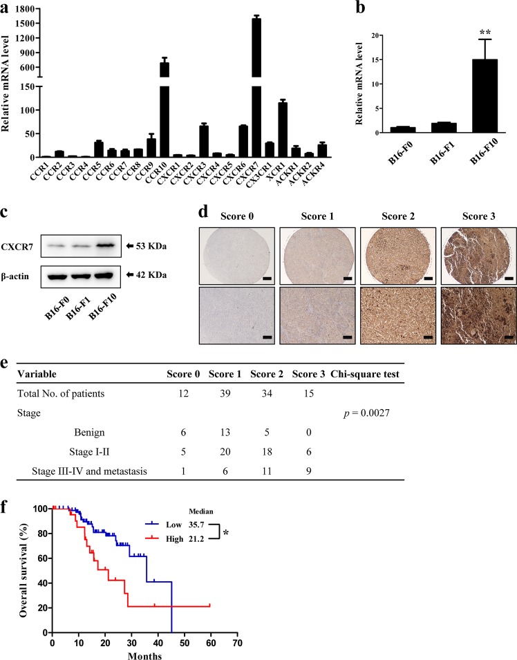
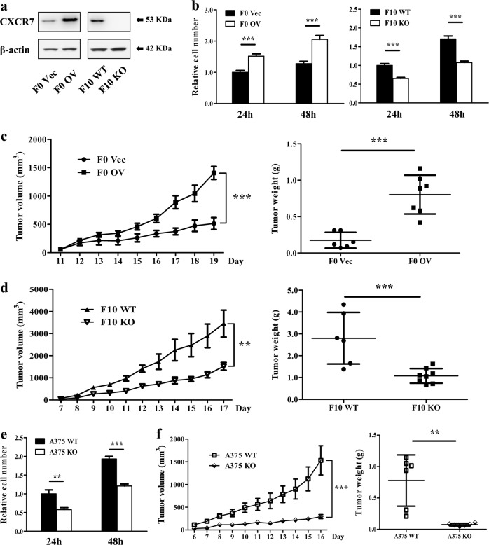
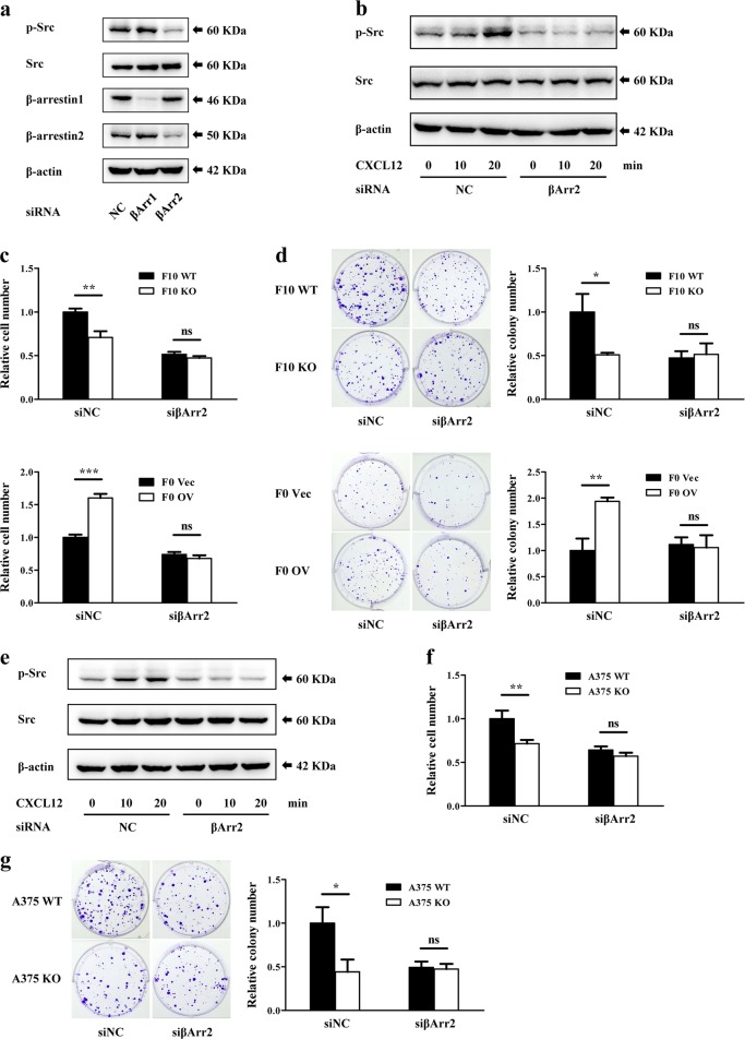
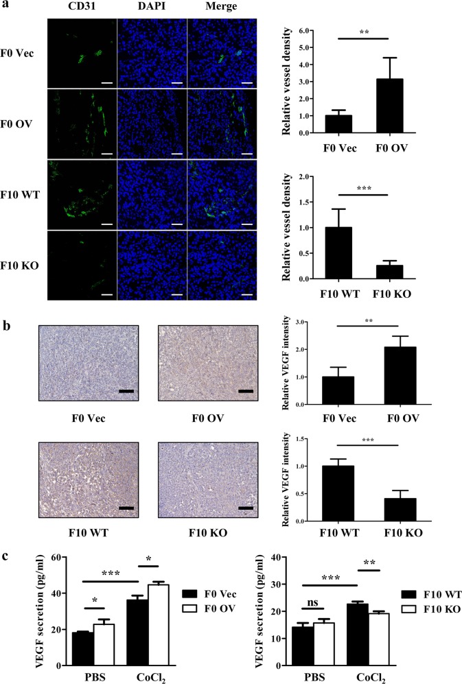
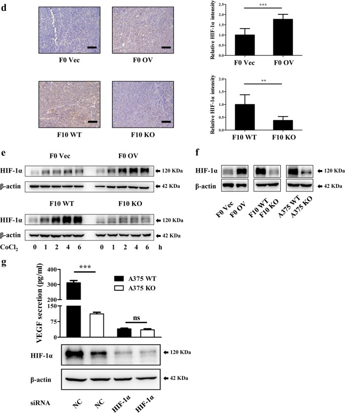
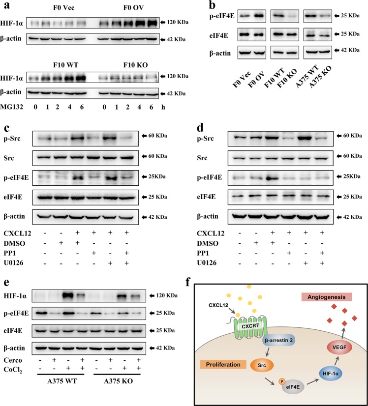
Chemokine receptors have been documented to exert critical functions in melanoma progression. However, current drugs targeting these receptors have limited efficacy in clinical applications, suggesting the urgency to further explore the roles of chemokine receptors in melanoma. Here we found that C-X-C chemokine receptor 7 (CXCR7) was the most highly expressed chemokine receptor in murine melanoma cell lines. In addition, the expression level of CXCR7 was positively correlated with melanoma progression in the clinical samples. High CXCR7 expression was associated with shorter overall survival in melanoma patients. Increased expression of CXCR7 augmented melanoma proliferation in vitro and tumor growth in vivo, whereas knockout of CXCR7 exhibited significant inhibitory effects. Moreover, our data elucidated that CXCR7 activated Src kinase phosphorylation in a β-arrestin2-dependent manner. The administration of the Src kinase inhibitor PP1 or siRNA specific for β-arrestin2 abolished CXCR7-promoted cell proliferation. Importantly, CXCR7 also regulated melanoma angiogenesis and the secretion of vascular endothelial growth factor (VEGF). Subsequent investigations revealed a novel event that the activation of the CXCR7-Src axis stimulated the phosphorylation of eukaryotic translation initiation factor 4E (eIF4E) to accelerate the translation of hypoxia-inducible factor 1α (HIF-1α), which enhanced the secretion of VEGF from melanoma cells. Collectively, our results illuminate the crucial roles of CXCR7 in melanoma tumorigenesis, and indicate the potential of targeting CXCR7 as new therapeutic strategies for melanoma treatment.


Conflict of interest statement

The authors declare that they have no conflict of interest.

Figures

Fig. 1
Fig. 1. The expression of CXCR7 in metastatic melanoma cells and clinical samples.
a The expression levels of chemokine receptors in B16-F10 cells were evaluated by qRT-PCR and normalized to Ccr1 mRNA level. b, c The relative mRNA (b) and protein (c) levels of CXCR7 in B16-F0, B16-F1, and B16-F10 cells. The mRNA levels were normalized to B16-F0 cells. d Representative images of CXCR7 expression in benign, malignant, and metastatic melanoma samples that illustrate scores of 0, 1, 2, and 3. The top images were taken at ×100 original magnification (scale bar = 200 μm) and the bottom images were taken at ×200 original magnification (scale bar = 100 μm). e The correlation of CXCR7 staining scores with tumor stages. The χ2 test was used to assess the correlation between categorical variables. f Overall survival of melanoma patients with high (n = 24) or low (n = 78) CXCR7 expression. The expression cutoff = 3.51 FPKM. Overall survival was analyzed by Kaplan–Meier survival analysis and the log-rank test. The qRT-PCR experiments were independently repeated three times. Data are presented as mean ± SD; *p < 0.05, **p < 0.01 compared with B16-F0 and B16-F1 cells
Fig. 2
Fig. 2. CXCR7 facilitates melanoma cell proliferation in vitro and tumor growth in vivo.
a CXCR7 overexpression and depletion in B16-F0 cells and B16-F10 cells. b The effects of CXCR7 overexpression and depletion on melanoma cell proliferation in vitro. F0 Vec, F0 OV, and F10 WT, F10 KO cells were seeded into 96-well plates and cell proliferations were examined by CCK-8 assays after 24 h and 48 h. The proliferation rates were normalized to F0 Vec cells (left) or F10 WT cells (right) at 24 h. c, d The effects of CXCR7 overexpression (c) and depletion (d) on melanoma tumor volume and tumor weight in vivo (n = 6–8 for each group). e, f The effects of CXCR7 depletion on A375 cell proliferation in vitro (e) and tumor growth in vivo (f; n = 6 for each group). The proliferation rates were normalized to A357 WT cells at 24 h. Proliferation analyses were independently repeated three times. Tumor volumes are shown as mean±SEM, and other data are presented as mean ± SD; **p < 0.01, ***p < 0.001
Fig. 3
Fig. 3. CXCR7 promotes melanoma proliferation through Src activation.
a The effects of CXCR7 modifications on the Src, AKT, and ERK signalings. b CXCR7 expression modulated CXCL12-stimulated Src phosphorylation in the presence of AMD3100. Murine melanoma cell lines were cultured in serum-free medium overnight. After pretreated with AMD3100 (1 μg/ml) for 1 h, the cells were stimulated with recombinant murine CXCL12 (50 ng/ml). The phosphorylation levels of Src were determined by western blot. c PP1 suppressed CXCR7-induced cell proliferation. Melanoma cells were seeded into 96-well plates in the presence of DMSO or PP1 (10 μM). After 48 h, the numbers of cells were examined by CCK-8 assays. The proliferation rates were normalized to F0 Vec cells (top) or F10 WT cells (bottom) without treatment. d Representative images and quantitative results of the clonogenic growth of cells treated with DMSO or PP1 (10 μM). The results were normalized to F0 Vec cells (top) or F10 WT cells (bottom) without treatment. e Serum-starved A375 WT and KO cells were pretreated with AMD3100 (1 μg/ml) for 1 h, and stimulated with recombinant human CXCL12 (100 ng/ml). Cells were harvested for western blot analysis of the phosphorylated Src kinase. f PP1 recapitulated the disrupted cell proliferation induced by loss of CXCR7 in A375 cells. The proliferation rates were normalized to A375 WT cells without treatment. g Clonogenic growth of A375 WT and KO cells with the same treatments described in d. The results were normalized to A375 WT cells without treatment. Proliferation experiments and colony formation assays were independently repeated three times. Data are presented as mean ± SD. **p < 0.01, ***p < 0.001
Fig. 4
Fig. 4. CXCR7 stimulates Src kinase phosphorylation through β-arrestin2.
a The levels of Src phosphorylation in F10 WT cells transfected with siRNA targeting scramble control (NC), β-arrestin1 (βArr1), or β-arrestin2 (βArr2). b, e The effects of β-arrestin2 knockdown on CXCL12-induced Src phosphorylation in melanoma cells. F10 WT (b) and A375 WT (e) cells harboring scramble siRNA or β-arrestin2 siRNA were starved overnight. After pretreated with AMD3100 (1 μg/ml) for 1 h, the cells were exposed to recombinant murine (50 ng/ml) or human (100 ng/ml) CXCL12. The phosphorylation level of Src was detected by western blot. c, f The effects of β-arrestin2 downregulation on melanoma cell proliferation. Indicated cell lines were transfected with scramble siRNA or β-arrestin2 siRNA and seeded into 96-well plates. After 48 h, the cell numbers were determined by CCK-8 assays. The proliferation rates were normalized to F10 WT cells (c, top), F0 Vec cells (c bottom) or A375 WT cells (f) transfected with scramble siRNA. d, g The effects of β-arrestin2 downregulation on melanoma colony formation. Indicated cell lines were transfected with scramble siRNA or β-arrestin2 siRNA and seeded into 6-well plates. After 12 days, the colony numbers were counted. The quantitative results were normalized to F10 WT cells (d, top), F0 Vec cells (d, bottom) or A375 WT cells (g) transfected with scramble siRNA. Proliferation experiments and colony formation assays were independently repeated in triplicate. Data are presented as mean ± SD; *p < 0.05, **p < 0.01, ***p < 0.001; ns not significant
Fig. 5
Fig. 5. CXCR7 contributes to melanoma angiogenesis and promotes VEGF secretion by upregulating HIF-1α expression.
a Immunofluorescent staining of CD31 (green) and DAPI (blue) in tumors derived from murine melanoma cells. The blood vessel densities were calculated by dividing CD31 area by DAPI area and were normalized to F0 Vec tumors (top) or F10 WT tumors (bottom). Left: representative images; scale bar = 50 μm. Right: relative quantitative results (n = 6–8 for each group). b Immunohistochemistry staining of VEGF in tumors derived from mice bearing murine melanoma cells. The staining intensities of VEGF were normalized to F0 Vec tumors (top) or F10 WT tumors (bottom). Left: representative images; scale bar = 100 μm. Right: relative quantitative results of staining intensities (n = 6–8 for each group). c The secretion of VEGF by murine melanoma cells with or without CoCl2 (200 μM) treatment. Cells were pretreated with CoCl2 for 6 h, and then incubated in serum-free medium containing CoCl2 overnight. The conditioned mediums were collected and the levels of VEGF were determined by ELISA. d Immunohistochemistry staining of HIF-1α in tumors derived from mice bearing murine melanoma cells. The staining intensities of HIF-1α were normalized to F0 Vec tumors (top) or F10 WT tumors (bottom). Left: representative images; scale bar = 100 μm. Right: relative quantitative results of staining intensities (n = 6–8 for each group). e HIF-1α expression in murine melanoma cells treated with CoCl2. f HIF-1α expression in melanoma cells under normoxic conditions. g A375 WT and KO cells transfected with siRNA targeting scramble control or HIF-1α were treated by CoCl2. The conditioned mediums and cell lysates were collected to detect VEGF secretion and HIF-1α expression. ELISA assays were independently conducted in triplicate. The quantification of immunofluorescent and immunohistochemistry staining were evaluated in 12 random fields for each tumor. Data are presented as mean ± SD, *p < 0.05, **p < 0.01, ***p < 0.001; ns not significant
Fig. 6
Fig. 6. CXCR7 accelerates HIF-1α translation by facilitating Src-mediated eIF4E phosphorylation.
a HIF-1α expression in murine melanoma cells treated with MG132 (10 μM). b The effects of CXCR7 modifications on eIF4E phosphorylation in melanoma cells. c, d CXCL12 stimulated eIF4E phosphorylation through the Src signaling in melanoma cells. F10 WT (c) and A375 WT (d) cells were starved overnight. After pretreated with AMD3100 (1 μg/ml) combined with PP1 (10 μM) or U0126 (10 μM) for 1 h, the cells were stimulated with recombinant murine (50 ng/ml) or human (100 ng/ml) CXCL12 as indicated. The cell lysates were collected and immunoblotted with the indicated antibodies. e A375 WT and KO cells were pretreated with cercosporamide (cerco, 20 μM) overnight and then incubated with CoCl2 (200 μM) for 4 h. The levels of HIF-1α and phosphorylated eIF4E were examined by western blot. f The proposed model for the functions of CXCR7 in melanoma tumorigenesis. CXCR7 is activated by CXCL12 to support melanoma cell proliferation through β-arrestin2-mediated Src phosphorylation. In addition, the activation of this CXCR7-Src axis stimulates eIF4E phosphorylation to accelerate the translation of HIF-1α, which enhances the secretion of VEGF to facilitate melanoma angiogenesis

Similar articles

Cited by

References

    1. Siegel RL, Miller KD, Jemal A. Cancer statistics, 2018. CA Cancer J. Clin. 2018;68:7–30. doi: 10.3322/caac.21442. - DOI - PubMed
    1. Whiteman DC, Green AC, Olsen CM. The growing burden of invasive melanoma: projections of incidence rates and numbers of new cases in six susceptible populations through 2031. J. Invest. Dermatol. 2016;136:1161–1171. doi: 10.1016/j.jid.2016.01.035. - DOI - PubMed
    1. Luke JJ, Flaherty KT, Ribas A, Long GV. Targeted agents and immunotherapies: optimizing outcomes in melanoma. Nat. Rev. Clin. Oncol. 2017;14:463–482. doi: 10.1038/nrclinonc.2017.43. - DOI - PubMed
    1. Zlotnik A, Yoshie O, Nomiyama H. The chemokine and chemokine receptor superfamilies and their molecular evolution. Genome Biol. 2006;7:243. doi: 10.1186/gb-2006-7-12-243. - DOI - PMC - PubMed
    1. Thelen M, Stein JV. How chemokines invite leukocytes to dance. Nat. Immunol. 2008;9:953–959. doi: 10.1038/ni.f.207. - DOI - PubMed

Publication types

MeSH terms