. 2019 Feb 25;9(1):2711.
doi: 10.1038/s41598-019-39077-6.

Pancreatic acinar differentiation is guided by differential laminin deposition

Affiliations

Pancreatic acinar differentiation is guided by differential laminin deposition

Charlotte Heymans et al. Sci Rep. .

Abstract

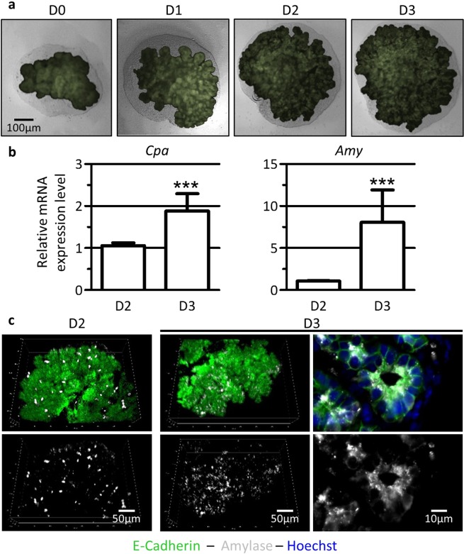
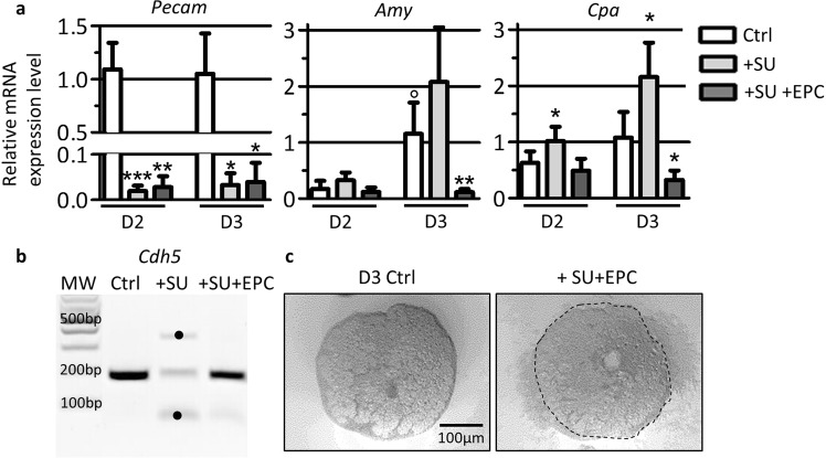
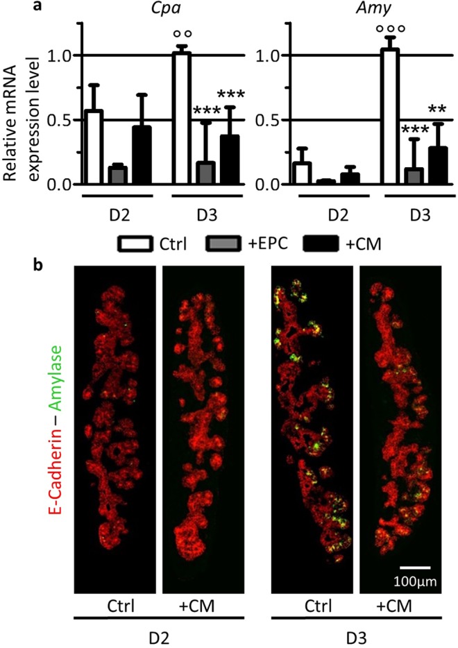
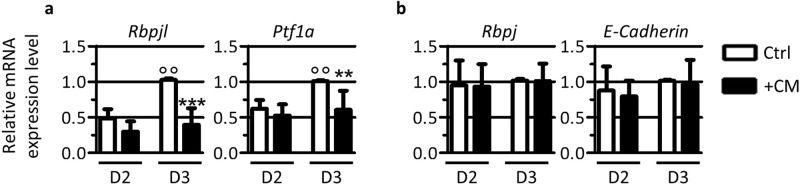
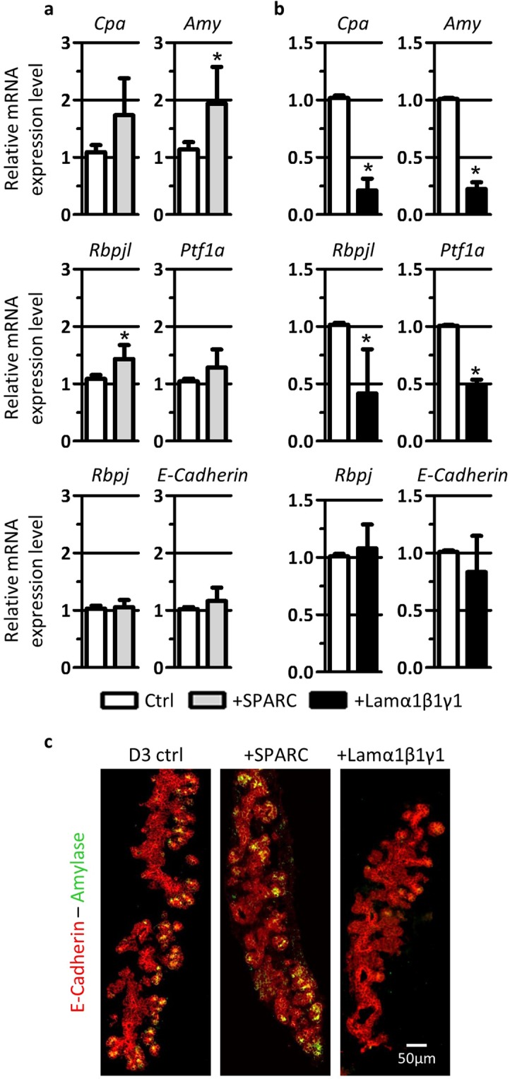
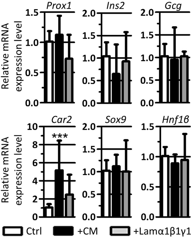
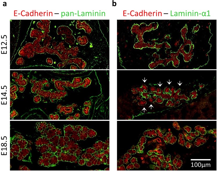
Endothelial cells play multiple roles during pancreas organogenesis. First, they are required to instruct endoderm-derived pancreatic progenitor cells to initiate branching morphogenesis. Later, blood vessels promote β-cell differentiation but also limit acinar development. In this work, we show how endothelial cells might signal to pancreatic progenitors and spatially regulate acinar differentiation. Using an ex vivo culture system of undifferentiated E12.5 pancreata, we demonstrate that embryonic endothelial progenitor cells and their conditioned medium prevent the expression of two members of the pro-acinar transcriptional PTF1L-complex. This effect is not mediated by SPARC, a protein abundantly released in the medium conditioned by endothelial progenitors. On the contrary, heterotrimeric laminin-α1β1γ1, also produced by endothelial progenitor cells, can repress acinar differentiation when used on its own on pancreatic explants. Lastly, we found that laminin-α1 is predominantly found in vivo around the pancreatic trunk cells, as compared to the tip cells, at E14.5. In conclusion, we propose that expression or deposition of laminin-α1β1γ1 around the trunk cells, where blood vessels are predominantly localized, prevent acinar differentiation of these cells. On the contrary, transient decreased expression or deposition of laminin-α1β1γ1 around the tip cells would allow PTF1L-complex formation and acinar differentiation.


Conflict of interest statement

The authors declare no competing interests.

Figures

Figure 1
Figure 1
Pancreatic explants, cultured ex vivo, grow and differentiate. (a) Phase contrast and fluorescence imaging of pancreatic explants dissected at embryonic day (E)12.5 (corresponding to day (D) of culture 0), and cultured up to 3 days on microporous filters (D1-D2-D3). The pancreatic epithelium (green in Pdx1-GFP embryos) develops and progressively invades the surrounding mesenchyme (grey). (b) RT-qPCR analysis of acinar markers Cpa and Amy compared to β-actin, used as housekeeping gene. The expression of acinar markers increases from D2 to D3. (Mann-Whitney: ***p < 0.001). (c) Whole-mount immunofluorescence for Amylase-expressing cells (white) within the pancreatic epithelium (E-Cadherin, green). Nuclei are stained in blue (Hoechst+, right panel). The number of Amylase+ cells (white) increases from D2 to D3.
Figure 2
Figure 2
Endothelial cell ablation stimulates, while addition represses expression of acinar genes. (a) RT-qPCR analysis of endothelial markers Pecam and acinar markers Cpa and Amy reported to β-actin in explants cultured for 2 and 3 days in the presence of control medium (Ctrl), supplemented with SU5416 (+SU) and with EPC (+SU + EPC). Treatment with SU5416 results in loss of Pecam expression in SU-treated explants (+SU). Addition of EPC (+SU + EPC) does not restore Pecam expression. Increased acinar differentiation (Cpa and Amy expression) is observed upon treatment with SU5416 (+SU) for 2 and 3 days of culture. On the contrary, addition of exogenous endothelial cells (+SU + EPC) blocks the induction of acinar differentiation markers. (Mann-Whitney for control explants: °p < 0.05; and for SU- and SU + EPC-treated explants: *p < 0.05; **p < 0.01). (b) PCR for the endothelial marker Cdh5 (174 bp) in control explants, explants treated with SU5416 (+SU) and explants treated with SU5416 and EPC (+SU + EPC) for 3 days. Cdh5 is expressed in control explants, but is absent from SU5416-treated explants (+SU). Addition of EPC on endothelium-deprived explants (+SU + EPC) results in the restoration of Cdh5 amplicon. • = non-specific amplicons. (c) Phase-contrast image of pancreatic explants cultured for 3 days (D3) on filters. Exogenous endothelial cells were added on SU5416-treated pancreatic explants (+SU + EPC) or not (Ctrl). EPC remained clustered around the explants during the culture.
Figure 3
Figure 3
Medium conditioned by EPC limits acinar differentiation. (a) RT-qPCR analysis of acinar markers Cpa and Amy reported to β-actin in explants cultured for 2 and 3 days in the presence of control medium (Ctrl), endothelial cells (+EPC) or of medium conditioned by EPC (+CM). Both EPC and CM limit acinar differentiation. (Mann-Whitney for control explants: °°p < 0.01; °°°p < 0.001; and for EPC- and CM-treated explants: **p < 0.01; ***p < 0.001). (b) Immunofluorescence for Amylase (green) within the pancreatic epithelium (E-Cadherin, red) of control explants (Ctrl) or of explants treated with conditioned medium (+CM) at 2 and 3 days. Fewer Amylase+ cells are observed following culture with CM.
Figure 4
Figure 4
EPC-CM prevents the formation of the acinar PTF1L-complex. (a) RT-qPCR analysis of members of the PTF1L-complex Rbpjl and Ptf1a reported to β-actin in explants cultured for 2 and 3 days with control medium (Ctrl) or EPC-CM (+CM). In control explants, expression of pro-acinar transcription factor Rbpjl and of Ptf1a is induced from 2 to 3 days of culture. EPC-CM blocks the induction of Rbpjl and Ptf1a in developing explants. (b) RT-qPCR analysis of Rbpj and E-Cadherin reported to β-actin in the same culture conditions reveals that CM does not globally affect gene transcription. (Mann-Whitney for control explants: °°p < 0.01; and for EPC-CM-treated explants: **p < 0.01; ***p < 0.001).
Figure 5
Figure 5
SPARC and laminin-α1β1γ1, two proteins abundantly found in EPC-CM, control acinar differentiation. (a) RT-qPCR analysis of acinar and control genes reported to β-actin in pancreatic explants cultured for 3 days in control medium (Ctrl) and in the presence of SPARC (+SPARC). Addition of SPARC favors acinar differentiation as demonstrated by increased levels of acinar markers (Cpa and Amy) and pro-acinar transcription factors (Rbpjl and Ptf1a). Expression level of control genes (Rbpj and E-Cadherin) are not affected. (b) RT-qPCR analysis of acinar and control genes reported to β-actin in pancreatic explants cultured for 3 days in control medium (Ctrl) and in the presence of laminin-α1β1γ1. In the presence of laminin-α1β1γ1, the expression of acinar markers (Cpa and Amy) and pro-acinar transcription factors (Rbpjl and Ptf1a) is reduced as compared to control explants. Expression level of control genes (Rbpjl and E-Cadherin) is not affected. (Mann-Whitney: *p < 0.05). (c) Immunofluorescence for the acinar differentiation marker Amylase (green) and the pancreatic epithelium marker (E-Cadherin, red) in explants at 3 days, as indicated. As compared to control explants showing acinar differentiation (Amylase+ cells), addition of SPARC stimulates, while laminin-α1β1γ1 decreases the Amylase signal.
Figure 6
Figure 6
EPC-CM treatment induces the expression of Carbonic Anhydrase 2 (Car2). RT-qPCR analysis of progenitor, endocrine and ductal markers reported to β-actin in pancreatic explants cultured for 3 days in control medium (Ctrl), EPC-CM (+CM) and in the presence of laminin-α1β1γ1 (+Lamα1β1γ1). EPC-CM and laminin-α1β1γ1 do not affect expression of progenitor (Prox1) and endocrine (Ins2 and Gcg) markers. Conversely, the treatments upregulate the expression of the ductal marker Car2, although the expression of the ductal transcription factors Sox9 and Hnf1β remain stable. (Mann-Whitney: ***p < 0.001).
Figure 7
Figure 7
Differential laminin-α1 deposition during pancreas development in vivo. (a) Immunolabeling for pan-Laminin (green) with the epithelial markers (E-Cadherin, red) in pancreas at E12.5, E14.5 and E18.5. At E12.5, pan-Laminin antibody recognizes an almost continuous structure around the branching pancreatic epithelium. At E14.5 and E18.5, the pan-Laminin signal homogenously surrounds the differentiating pancreatic epithelium. (b) Immunolabeling for laminin-α1 (green) with the epithelial marker (E-Cadherin, red) in developing pancreas (E12.5-E14.5-E18.5). Laminin-α1 antibody recognizes the same structures as pan-Laminin, located around the branching pancreatic epithelium. However, at E14.5, the laminin-α1 signal is much stronger around the trunk cells than around the tip cells (arrows). This is transitory as at E18.5, laminin-α1 is homogenously found around the pancreas.

Similar articles

Cited by

References

    1. Larsen, H. L. & Grapin-Botton, A. The molecular and morphogenetic basis of pancreas organogenesis. Seminars in Cell & Developmental Biology66, 51–68 (2017). - PubMed
    1. Zhou, Q. et al. A Multipotent Progenitor Domain Guides Pancreatic Organogenesis. Developmental Cell13(1), 103–114 (2007). - PubMed
    1. Masui T, Long Q, Beres TM, Magnuson MA, MacDonald RJ. Early pancreatic development requires the vertebrate Suppressor of Hairless (RBPJ) in the PTF1 bHLH complex. Genes Dev. 2007;21:2629–2643. doi: 10.1101/gad.1575207. - DOI - PMC - PubMed
    1. Masui T, et al. Replacement of Rbpj with Rbpjl in the PTF1 complex controls the final maturation of pancreatic acinar cells. Gastroenterology. 2010;139:270–280. doi: 10.1053/j.gastro.2010.04.003. - DOI - PMC - PubMed
    1. Kim SK, Hebrok M, Melton DA. Notochord to endoderm signaling is required for pancreas development. Development. 1997;124:4243–4252. - PubMed

Publication types

MeSH terms