. 2019 Feb 25;9(1):2724.
doi: 10.1038/s41598-019-39368-y.

USP7 Regulates Cytokinesis through FBXO38 and KIF20B

Affiliations

USP7 Regulates Cytokinesis through FBXO38 and KIF20B

Anna Georges et al. Sci Rep. .

Abstract

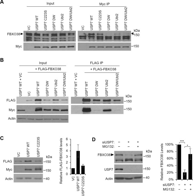
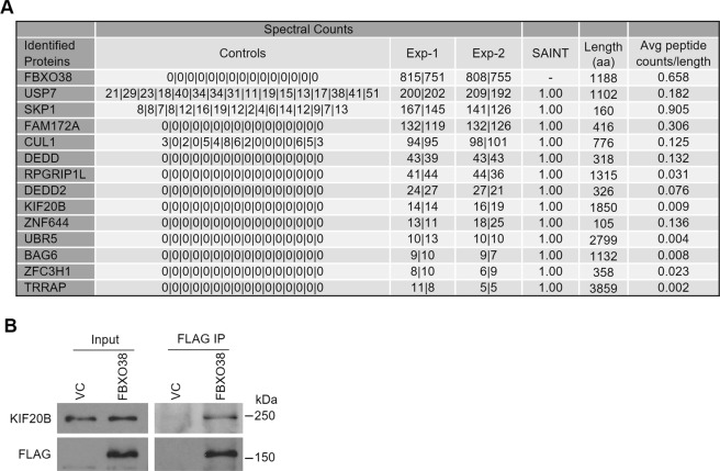
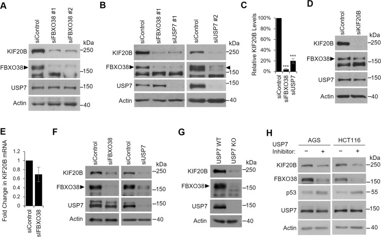
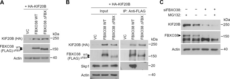
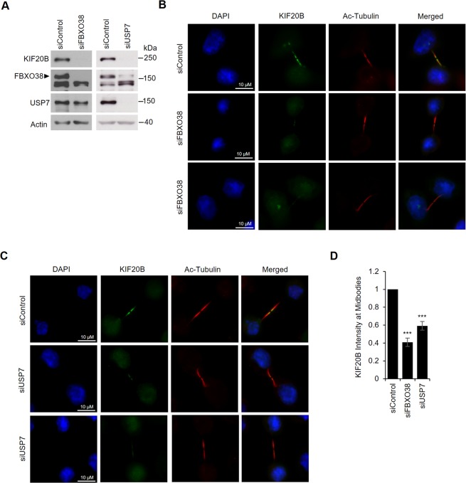
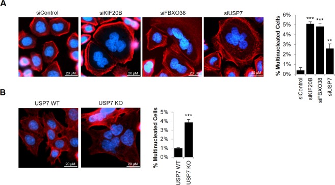
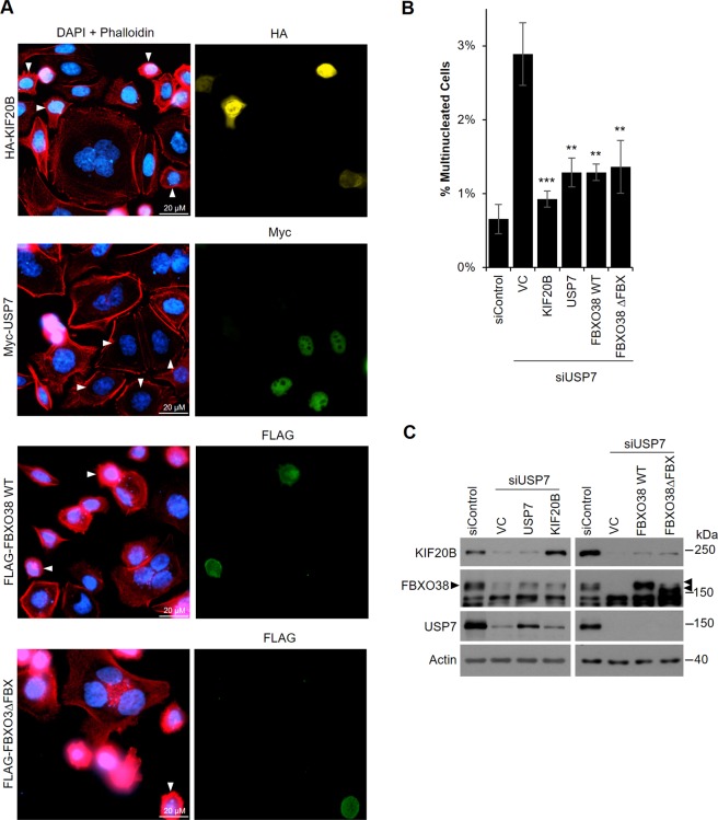
The ubiquitin specific protease 7 (USP7 or HAUSP) is known to regulate a variety of cellular processes by binding and deubiquitylating specific target proteins. To gain a more comprehensive understanding of its interactions and functions, we used affinity purification coupled to mass spectrometry to profile USP7 interactions. This revealed a novel interaction with FBXO38, a poorly characterized F-box protein. We showed that USP7 stabilizes FBXO38 dependent on its catalytic activity by protecting FBXO38 from proteasomal degradation. We used a BioID approach to profile the protein interactions (and putative functions) of FBXO38, revealing an interaction with KIF20B, a Kinesin-6 protein required for efficient cytokinesis. FBXO38 was shown to function independently from an SCF complex to stabilize KIF20B. Consequently, depletion of either FBXO38 or USP7 led to dramatic decreases in KIF20B levels and KIF20B at the midbody, which were manifested in cytokinetic defects. Furthermore, cytokinetic defects associated with USP7 silencing were rescued by restoring FBXO38 or KIF20B. The results indicate a novel mechanism of regulating cytokinesis through USP7 and FBXO38.


Conflict of interest statement

The authors declare no competing interests.

Figures

Figure 1
Figure 1
USP7 interacts with and stabilizes FBXO38. (A) AGS cells were transfected with a plasmid expressing myc-tagged USP7 WT, the USP7 catalytic mutant (C223S), the USP7 NTD pocket mutant (DW), the USP7 Ubl2 pocket mutant (Ubl2), the USP7 double pocket mutant (DW/Ubl2) or an empty vector control (VC). Myc-USP7 constructs were immunoprecipitated with anti-myc antibody and recovered proteins were analyzed by Western blotting using antibodies against myc and endogenous FBXO38. The band corresponding to FBXO38 is indicated with arrow heads. (B) HEK293T cells were transfected with a plasmid expressing myc-tagged USP7 WT or co-transfected with plasmids expressing FLAG-tagged FBXO38 and each of the indicated myc-tagged USP7 plasmids. FBXO38 was immunoprecipitated using anti-FLAG M2 resin and recovered proteins were analyzed by Western blotting using antibodies against FLAG and myc. (C) HEK293T cells were co-transfected with plasmids expressing FLAG-tagged FBXO38 and either myc-USP7 WT, C223S or an empty vector control (VC). Cells were harvested 48 h post transfection and cell lysates were analyzed by Western blotting using antibodies against FLAG, Myc and actin. Quantification of the FLAG-FBXO38 bands (normalized to actin) from two independent experiments is shown on the right. (D) AGS cells were transfected with an siRNA targeting USP7 (+) or a negative control siRNA (−) followed by treatment with the MG132 proteasome inhibitor (+) or DMSO as a negative control (−) for 12 hours. Cell lysates were analyzed by Western blotting using antibodies against FBXO38, USP7 and actin. Quantification of the FBXO38 bands (normalized to actin) from three independent USP7 silencing experiments are shown on the right. ***P < 0.001.
Figure 2
Figure 2
Identification of KIF20B as an interactor of FBXO38. (A) Expression of FLAGBirA*–FBXO38 or FLAGBirA* negative control from integrated cassettes in 293 T-REx cells was induced with tetracycline in the presence of 50 µM biotin for 24 hrs. Bioitinylated proteins were purified on Streptavidin Sepharose beads and recovered proteins were identified by LC-MS/MS. Total spectral counts for two biological replicates (Exp-1 and -2) with two technical replicates each (separated by) are shown for high-confidence FBXO38 interactors (based on Bayesian False Discovery Rate (BFDR) <0.01). Spectral counts for cells expressing FLAGBirA* tag alone (Control) are shown for 16 experiments. The length of each protein (amino acid numbers) and spectral counts per length are also shown for each protein. (B) AGS cells were transfected with a plasmid expressing FLAG-tagged FBXO38 (FBXO38) or an empty vector control (VC) for 48 hours. FBXO38 was immunoprecipitated using anti-FLAG M2 resin and recovered proteins were analyzed by Western blotting using antibodies against FLAG and KIF20B, respectively.
Figure 3
Figure 3
Depletion of FBXO38 or USP7 decreases KIF20B levels. AGS cells were transfected with two different siRNAs targeting FBXO38 (A) or USP7 (B) or with negative control siRNA (siControl) and whole cell lysates were analyzed by Western blotting using the indicated antibodies. Bands corresponding to FBXO38 are indicated with arrow heads. In AGS cells, an additional nonspecific band is also detected by this antibody running just below FBXO38. (C) KIF20B protein bands were quantified in three independent FBXO38 and USP7 silencing experiments in AGS cells and normalized to actin. The average values are shown relative the silencing control. ***P < 0.001. (D) AGS cells were transfected with an siRNA targeting KIF20B, and whole cell lysates were analyzed by Western blotting as in (A). (E) KIF20B mRNA levels were determined by RT-qPCR in AGS cells transfected with siRNA targeting FBXO38 or with negative control siRNA. The experiment was performed in triplicate and values normalized to actin. Average values and standard deviations are shown relative to the silencing control. (F) HCT116 cells were transfected with siRNA targeting FBXO38 or USP7 or with negative control siRNA (siControl) and whole cell lysates were analyzed by Western blotting as in (A). (G) Whole cell lysates of HCT116 USP7 KO or the WT parental cell line (USP7 WT) were analyzed by Western blotting as in (A). (H) AGS or HCT116 cells were treated with 5 µM of compound 4 (USP7 inhibitor) or an inactive enantiomer and harvested after 24 hours. Whole cell lysates were analyzed by Western blotting using the indicated antibodies.
Figure 4
Figure 4
FBXO38 interacts with and stabilizes KIF20B by an SCF-independent mechanism. (A) AGS cells were cotransfected with plasmids expressing HA-tagged KIF20B and FLAG-tagged FBXO38 (WT), the FBXO38 F-box deletion mutant (∆FBX) or an empty vector control (VC). Cell lysates were analyzed by Western blotting using antibodies against FLAG and HA. (B) 293 T cells were cotransfected as in (A), followed by coimmunoprecipitation using anti-FLAG M2 resin. Recovered proteins were analyzed by Western blotting using antibodies against FLAG, HA and Skp1 and myc. Bands corresponding to FBXO38 WT or ∆FBX are indicated with arrow heads. (C) AGS cells were transfected with an siRNA targeting FBXO38 (+) or a negative control siRNA (−) followed by 12 hr treatment with the MG132 proteasome inhibitor (+) or DMSO (−). Cell lysates were analyzed by Western blotting using antibodies against FBXO38, KIF20B and actin.
Figure 5
Figure 5
FBXO38 and USP7 silencing reduces KIF20B levels at the midbodies. AGS cells transfected with an siRNA targeting FBXO38, USP7 or a negative control siRNA were either lysed and analyzed by Western blotting using the indicated antibodies to confirm FBXO38 and USP7 silencing (A) or fixed and stained with DAPI and antibodies against KIF20B and acetylated tubulin (B,C). Representative fluorescence microscopy images of KIF20B localization at the midbodies after FBXO38 silencing (B) or USP7 silencing (C) are shown. (D) The fluorescence intensity of KIF20B at the midbodies in B and C was quantified for 30 midbodies in three independent experiments and values were normalized to the intensity of acetylated tubulin at the midbodies. Average values with standard deviations were plotted for FBXO38 and USP7 silencing shown relative to the silencing control. ***P < 0.001.
Figure 6
Figure 6
FBXO38 or USP7 depletion results in cytokinetic defects. (A) AGS cells were transfected with siRNA targeting KIF20B, FBXO38 or USP7 or with negative control siRNA (siControl). Cells were then fixed, stained with Phalloidin and DAPI, then imaged by fluorescence microscopy. The number of multinucleated cells in ~1500 cells were counted. Average values and standard deviation from three independent experiments are shown in the bar graph on the right. P values relative to siControl are indicated (* = 0.01 < P < 0.05; **= 0.001 < P < 0.01; ***P < 0.001). (B) HCT116 USP7 WT and USP7 KO cells were fixed and stained as in (A) and the number of multinucleated cells were determined. Average values and standard deviation from three independent experiments are shown in the bar graph on the right.
Figure 7
Figure 7
Overexpression of USP7, KIF20B and FBXO38 rescues the cytokinetic defect caused by USP7 silencing. (A) AGS cells were transfected with siRNA targeting USP7 followed by transfection with plasmids expressing HA-KIF20B, myc-USP7, FLAG-FBXO38 WT, FLAG-FBXO38∆FBX or an empty vector control (VC). Alternatively, cells were transfected with negative control siRNA (siControl) followed by transfection with the VC. Cells were then fixed, stained with DAPI and Phalloidin and with antibodies against either HA, myc or FLAG as indicated, then imaged by fluorescence microscopy. Cells expressing the tagged protein are indicated with white arrow heads in the left panel. The number of multinucleated cells (out of ~1500 cells/sample) in the silencing control or in USP7 silenced cells expressing the indicated tagged proteins or containing the VC were counted and plotted in (B). Average values from three independent experiments are shown along with standard deviations. P values are indicated for siUSP7 samples relative to siUSP7 with VC (* = 0.01 < P < 0.05; ** = 0.001 < P < 0.01; ***P < 0.001). (C) Whole cell lysates of cells treated in (A) were analyzed by Western Blotting using the indicated antibodies.

References

    1. Pfoh R, Lacdao IK, Saridakis V. Deubiquitinases and the new therapeutic opportunities offered to cancer. Endocr Relat Cancer. 2015;22:T35–54. doi: 10.1530/ERC-14-0516. - DOI - PMC - PubMed
    1. Frappier L, Verrijzer CP. Gene expression control by protein deubiquitinases. Curr Opin Genet Dev. 2011;21:207–213. doi: 10.1016/j.gde.2011.02.005. - DOI - PubMed
    1. Nicholson B, Suresh Kumar KG. The multifaceted roles of USP7: new therapeutic opportunities. Cell Biochem Biophys. 2011;60:61–68. doi: 10.1007/s12013-011-9185-5. - DOI - PubMed
    1. Kim RQ, Sixma TK. Regulation of USP7: A High Incidence of E3 Complexes. J Mol Biol. 2017;429:3395–3408. doi: 10.1016/j.jmb.2017.05.028. - DOI - PubMed
    1. Song MS, et al. The deubiquitinylation and localization of PTEN are regulated by a HAUSP-PML network. Nature. 2008;455:813–817. doi: 10.1038/nature07290. - DOI - PMC - PubMed

Publication types

Grants and funding