. 2019 Mar;21(3):311-318.
doi: 10.1038/s41556-019-0282-9. Epub 2019 Feb 25.

H4K20me0 recognition by BRCA1-BARD1 directs homologous recombination to sister chromatids

Affiliations

H4K20me0 recognition by BRCA1-BARD1 directs homologous recombination to sister chromatids

Kyosuke Nakamura et al. Nat Cell Biol. 2019 Mar.

Abstract

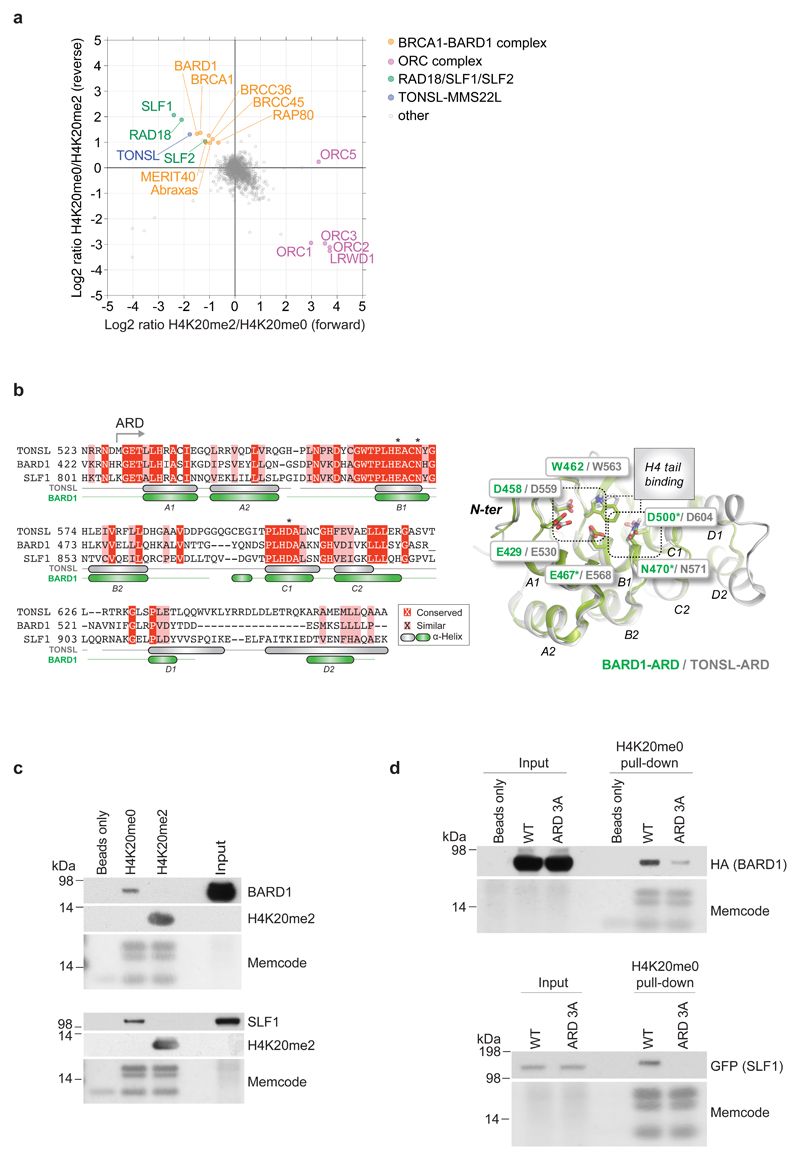
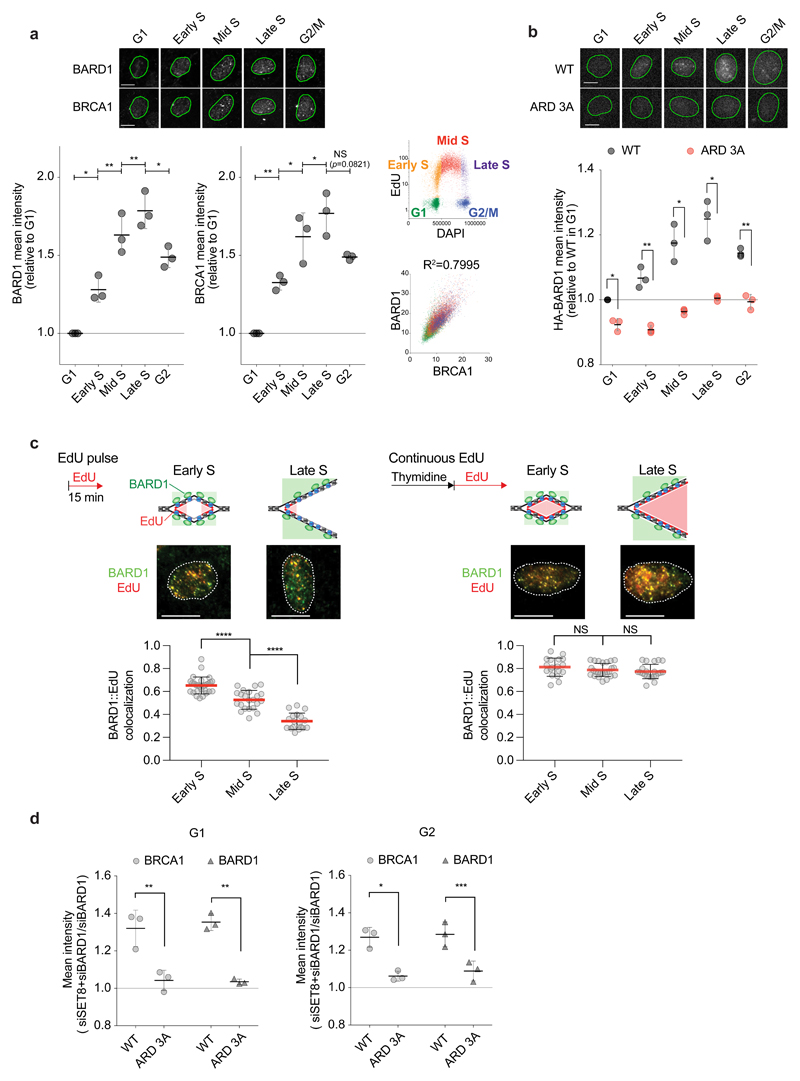
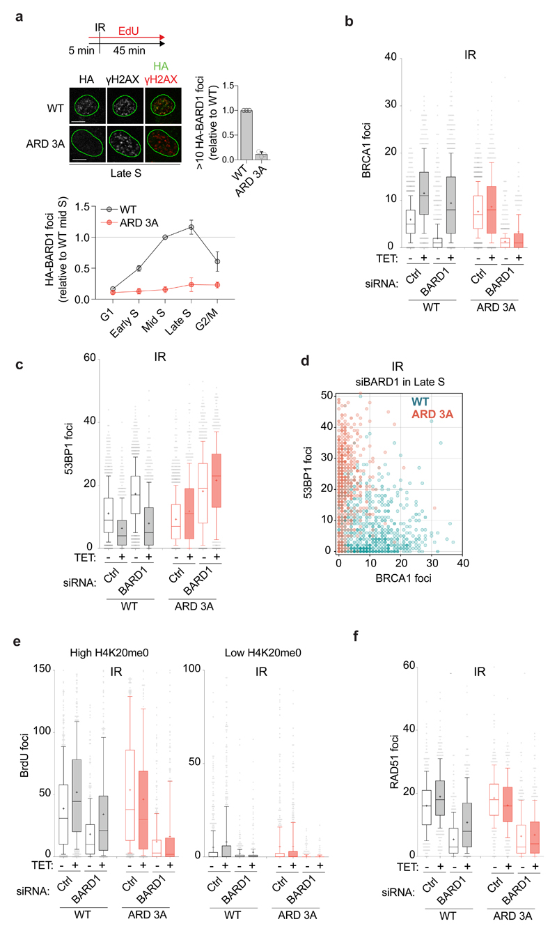
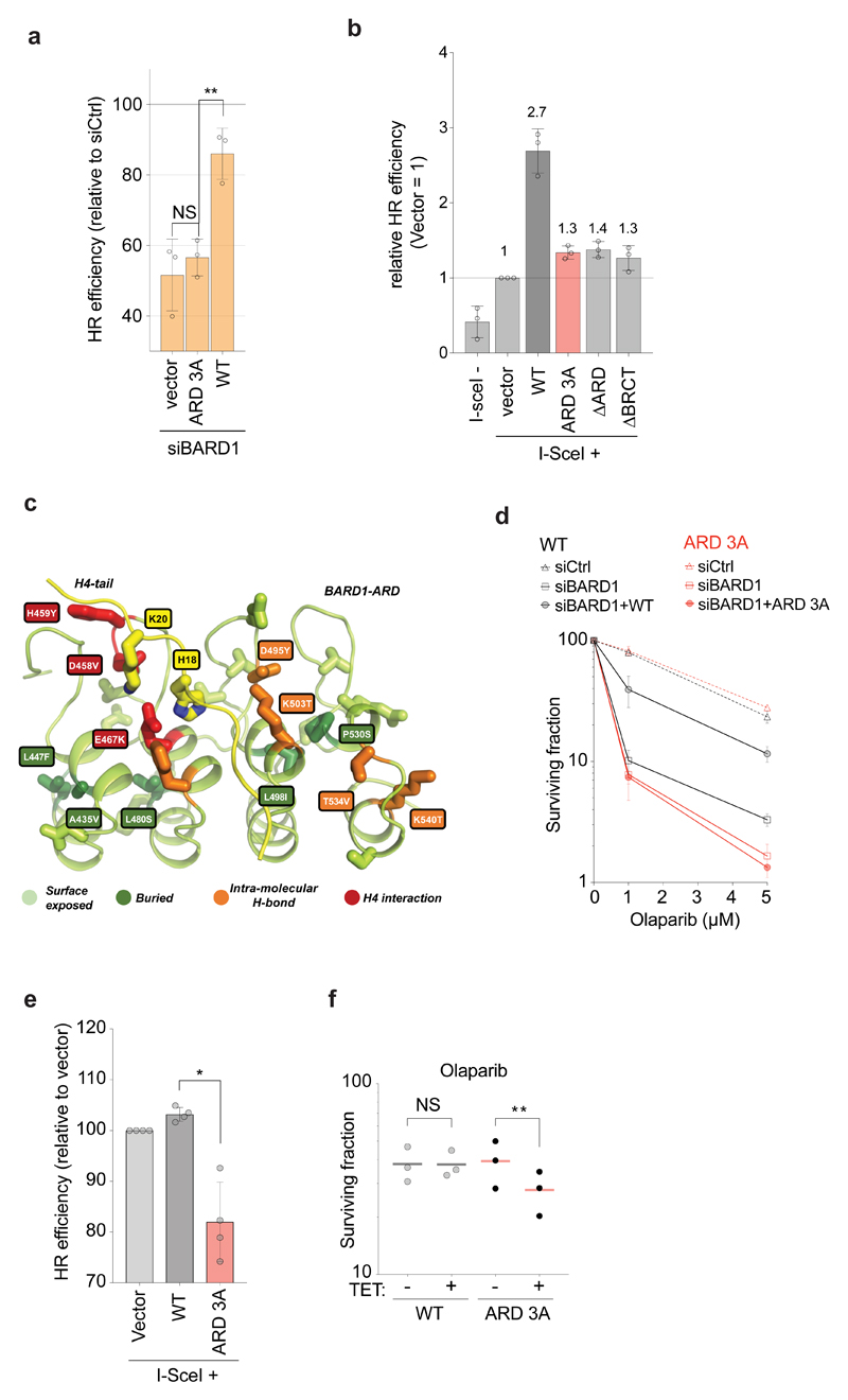
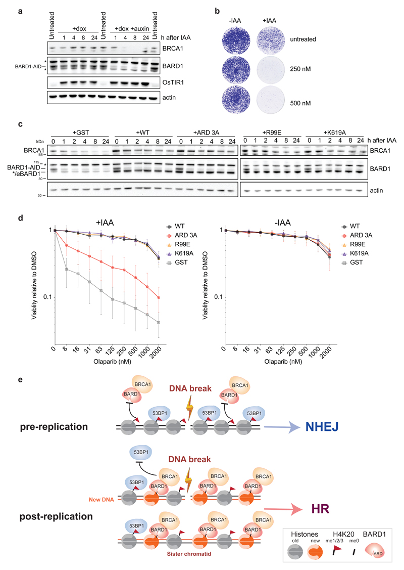
Genotoxic DNA double-strand breaks (DSBs) can be repaired by error-free homologous recombination (HR) or mutagenic non-homologous end-joining1. HR supresses tumorigenesis1, but is restricted to the S and G2 phases of the cell cycle when a sister chromatid is present2. Breast cancer type 1 susceptibility protein (BRCA1) promotes HR by antagonizing the anti-resection factor TP53-binding protein 1(53BP1) (refs. 2-5), but it remains unknown how BRCA1 function is limited to the S and G2 phases. We show that BRCA1 recruitment requires recognition of histone H4 unmethylated at lysine 20 (H4K20me0), linking DSB repair pathway choice directly to sister chromatid availability. We identify the ankyrin repeat domain of BRCA1-associated RING domain protein 1 (BARD1)-the obligate BRCA1 binding partner3-as a reader of H4K20me0 present on new histones in post-replicative chromatin6. BARD1 ankyrin repeat domain mutations disabling H4K20me0 recognition abrogate accumulation of BRCA1 at DSBs, causing aberrant build-up of 53BP1, and allowing anti-resection activity to prevail in S and G2. Consequently, BARD1 recognition of H4K20me0 is required for HR and resistance to poly (ADP-ribose) polymerase inhibitors. Collectively, this reveals that BRCA1-BARD1 monitors the replicative state of the genome to oppose 53BP1 function, routing only DSBs within sister chromatids to HR.


Conflict of interest statement

Competing financial interests

G.S. and A.G. are inventors on a filed patent application covering the therapeutic targeting of ARD interactions with H4K20me0 for cancer therapy. A.G. and T.F. are co-founders of Ankrin Therapeutics.

Figures

Figure 1
Figure 1. BARD1 and SLF1 recognize nucleosomes carrying H4K20me0 via their ARDs.
a, Pull-downs from HeLa nuclear extracts with differentially modified biotinylated recombinant di-nucleosomes analysed by SILAC-based mass spectrometry. b, Alignment of the TONSL ARD (aa. 528-626), BARD1 ARD (aa. 427-546) and SLF1 ARD (aa. 806-903) (left). Overlay of the BARD1 ARD structure with the TONSL ARD from the TONSL-ARD-MCM2-HBD-H3-H4 structure (right). Asterisks indicate the amino acids mutated in ARD 3A in d. c, Pull-downs with modified biotinylated di-nucleosomes in HeLa nuclear extracts. Representative of 3 biological replicates. d, Pull-downs with biotinylated di-nucleosomes in nuclear extracts from HeLa cells expressing either WT or ARD 3A mutant forms of BARD1 or SLF1. Representative of 3 (BARD1) and 2 (SLF1) biological replicates. Unprocessed blots for c and d are presented in Supplementary Fig. 5.
Figure 2
Figure 2. ARD recognition of H4K20me0 recruits BRCA1-BARD1 to post-replicative chromatin.
a, High content microscopy of chromatin-bound BARD1 and BRCA1 in pre-extracted U-2-OS cells pulsed with EdU. Cell cycle stages were defined by gating on EdU and DAPI (upper right). Mean intensities are shown relative to G1 levels with S.D., n=3 independent experiments; from left P=0.0203, 0.0057, 0.0076, 0.017, 0.0055, 0.0315, 0.0251, 0.0821, NS, not significant, ratio paired two-sided t-test. Data points represent the mean of >173 cells. The correlation between BARD1 and BRCA1 mean intensities (lower right) is shown with Pearson R2 correlation (P < 0.0001). Representative cells are shown; scale bar, 10 μm. b, High content microscopy of chromatin-bound Flag-HA-BARD1 in U-2-OS inducible for Flag-HA-BARD1 WT or ARD 3A. Mean is shown with S.D., n=3 independent experiments; from left P = 0.0214, 0.0036, 0.022, 0.0204, 0.0074), ratio paired two-sided t-test. Data points represent the mean of >254 cells. c, Co-localization of chromatin-bound BARD1 and EdU in cells pulsed with EdU (left) or released into S phase in the presence of EdU (right). S-phase stages were defined by EdU, DAPI and MCM2 staining. Drawings illustrate the expected results based on H4K20me0 distribution. Error bars indicate S.D. with mean. From left, n = 28, 22, 18, and 18, 25, 22 cells; representative cells are shown. Data were derived from two independent experiments. ****, P < 0.0001, by the Mann-Whitney test (two-sided). NS, not significant (from left P=0.1392 and 0.4494). Scale bar, 15 μm. d, BARD1 and BRCA1 chromatin-binding in G1 and G2 phase in SET8-depleted cells. Cells inducible for siRNA resistant Flag-HA-BARD1 WT or ARD 3A were treated with BARD1 and SET8 siRNAs and analysed as in a. Mean intensity in SET8 and BARD1 co-depletion cells over BARD1 depletion alone with S.D. is shown, n=3 independent experiments; from left P=0.0048, 0.0021, 0.013, 0.0009), ratio paired two-sided t-test. Data points represent the mean of >206 cells.
Figure 3
Figure 3. H4K20me0 recognition is required for BRCA1-BARD1 recruitment to DSBs, 53BP1 antagonization, and end-resection.
a, High content microscopy of Flag-HA-BARD1 accumulation at IR-induced DNA DSBs. U-2-OS cells expressing Flag-HA-BARD1 WT or ARD 3A were pulsed with EdU, irradiated with 1 Gy and 45 minutes later pre-extracted and analysed by immunofluorescence. Foci number is shown relative to BARD1 WT in mid S. S phase stages were defined as in Fig. 2a. The mean is shown with S.D., n=3 independent experiments. Data points represent the mean of >381 cells. Scale bar, 10 μm. b, c, BRCA1 (b) and 53BP1 (c) accumulation at IR-induced DSBs in U-2-OS cells treated with control (Ctrl) or BARD1 siRNAs and induced with tetracycline (TET) to express siRNA-resistant Flag-HA-BARD1 WT or ARD 3A. Cells were analysed as in a and late S phase cells are shown. Mean shown as ‘+’, whiskers indicate 10-90 percentile; from left (b), n = 1068, 723, 1224, 896, 1420, 627, 902, 597 cells; from left (c), n = 1068, 723, 1224, 896, 1420, 627, 902, 597 cells. d, BRCA1 and 53BP1 foci in single cells in late S phase analysed as in b and c. BARD1 WT n=896; BARD1 ARD 3A n=597. e, High content microscopy of ssDNA measured by BrdU staining under non-denaturing conditions in U-2-OS cells treated with siRNA and TET as in c. Cells with high (top 15%) and low (bottom 15%) H4K20me0 levels (defined by gating on mean intensity) are shown. Cells were fixed 3 hours after IR (3 Gy). Mean shown as ‘+’, whiskers indicate 10-90 percentile; from left, n = 503, 442, 371, 312, 474, 269, 381, 330 and 617, 641, 316, 237, 540, 495, 277, 194 cells. f, High-content microscopy of IR-induced RAD51 foci analysed as in b. Mean shown as ‘+’, whiskers indicate 10-90 percentile; from left, n = 310, 384, 327, 326, 329, 295, 321, 331 cells.
Figure 4
Figure 4. BARD1 recognition of H4K20me0 is required for HR and PARPi resistance.
a, HR efficiency measured in DR-GFP U-2-OS cells treated with control (Ctrl) or BARD1 siRNA and complemented with siRNA-resistant BARD1 WT, ARD 3A or empty vector. The HR efficiency is shown as % of control siRNA. Mean with S.D., n=3 independent experiments, from left, P=0.29, 0.0016; NS, not significant by ratio paired two-sided t-test. b, HR efficiency measured in BARD1-null mouse mammary carcinoma cells carrying a DR-GFP reporter and complemented as indicated. The HR efficiency relative to empty vector is indicated above each bar. Mean with S.D., n=3 independent experiments. c, BARD1 ARD mutations identified by cancer genome sequencing (COSMIC https://cancer.sanger.ac.uk/cosmic; TCGA Research Network: https://cancergenome.nih.gov/). Mutations H459Y, D458V and E467K are predicted to disturb H4 binding, as the residues are directly involved in H4 binding (red). Mutations D495Y, K503T, T534V and K540T are prone to affect the structural integrity of BARD1 ARD since the wild type residues are involved in intra-molecular hydrogen bonding (orange). Mutations buried in the core (A435V, L447F, L480S, L498I and P530S; dark green) might affect the structure of the ARD and thus indirectly disrupt H4 binding. Surface exposed mutations (light green) are less likely to affect ARD structure and H4 binding. d, Sensitivity to Olaparib analysed by colony formation in siRNA-treated U-2-OS cells induced for expression of siRNA-resistant Flag-HA-BARD1 WT or ARD 3A. Mean with S.D., n=3 independent experiments. e, f, BARD ARD mutations impair HR and sensitize cells to PARPi in a dominant negative manner. (e) HR efficiency measured in U-2-OS cells carrying a DR-GFP reporter, transfected with the indicated BARD1 constructs or empty vector along with an I-SceI expression vector. Mean with S.D., n=4 independent experiments. *, P=0.0136, ratio paired two-sided t-test. (f) Colony formation upon Olaparib (1 μM) treatment of U-2-OS cells induced to express Flag-HA-BARD1 WT and ARD 3A expression by tetracycline (TET). n=3 independent experiments, mean is shown. **, P=0.0013, ratio paired two-sided t-test. NS, not significant (P=0.9865).
Figure 5
Figure 5. Recognition of H4K20me0 is a prime function of BARD1 in HR.
a, Western blot analysis of BARD1AID/AID HCT116 cells at indicated time points following the addition of doxycycline to stimulate OsTIR1 expression and auxin (IAA) to induce OsTIR1-dependent BARD1-AID proteolysis. Representative of 2 biological replicates, unprocessed blots in Supplementary Fig. 5. *Indicates non-specific bands. b, IAA-addition hypersensitizes BARD1AID/AID HCT116 cells to Olaparib. Cells were seeded in the presence of doxycycline, IAA and Olaparib were added one day later and left for 7 days before staining with crystal violet. Representative of 2 biological replicates. c, d, Analysis of BARD1 ARD function in PARPi resistance as defined in BARD1AID/AID HCT116 cells. After 24 hours in doxycycline, indicated cell lines were treated with IAA, or carrier (DMSO), before immunoblotting whole cell lysates or measuring growth in the presence of Olaparib as indicated. Western blot showing equal stabilization of endogenous BRCA1 by the expression of wild type and all mutant BARD1 transgenes, but not the control (GST) transgene (c). Representative of 2 biological replicates, unprocessed blots in Supplementary Fig. 5. */eBARD1 indicates a weak unspecific band and the size of exogenous BARD1. Olaparib sensitivity was determined by resazurin cell viability assay 7 days following incubation with Olaparib (2-fold dilution range from 7.8 nM – 2 μM) in the presence (left panels) or absence (right panels) of IAA (d). Mean with S.D., n=3 independent experiments. e, Model illustrating how BARD1 recognition of H4K20me0 licenses DSBs in sister chromatid for HR. BRCA1-BARD1 identifies post-replicative chromatin by recognition of H4K20me0 on new histones, hereby enabling the complex to oppose 53BP1 function when a sister chromatid can be used for homology-directed error-free repair of DSBs.

References

    1. Moynahan ME, Jasin M. Mitotic homologous recombination maintains genomic stability and suppresses tumorigenesis. Nat Rev Mol Cell Biol. 2010;11:196–207. - PMC - PubMed
    1. Hustedt N, Durocher D. The control of DNA repair by the cell cycle. Nat Cell Biol. 2016;19:1–9. - PubMed
    1. Prakash R, Zhang Y, Feng W, Jasin M. Homologous recombination and human health: the roles of BRCA1, BRCA2, and associated proteins. Cold Spring Harb Perspect Biol. 2015;7:a016600. - PMC - PubMed
    1. Bunting SF, et al. 53BP1 inhibits homologous recombination in Brca1-deficient cells by blocking resection of DNA breaks. Cell. 2010;141:243–254. - PMC - PubMed
    1. Zimmermann M, de Lange T. 53BP1: pro choice in DNA repair. Trends Cell Biol. 2014;24:108–117. - PMC - PubMed

Publication types

MeSH terms