Review
. 2019 Apr;37(4):389-406.
doi: 10.1038/s41587-019-0045-y. Epub 2019 Feb 25.

Wearable biosensors for healthcare monitoring

Affiliations
Review

Wearable biosensors for healthcare monitoring

Jayoung Kim et al. Nat Biotechnol. 2019 Apr.

Abstract

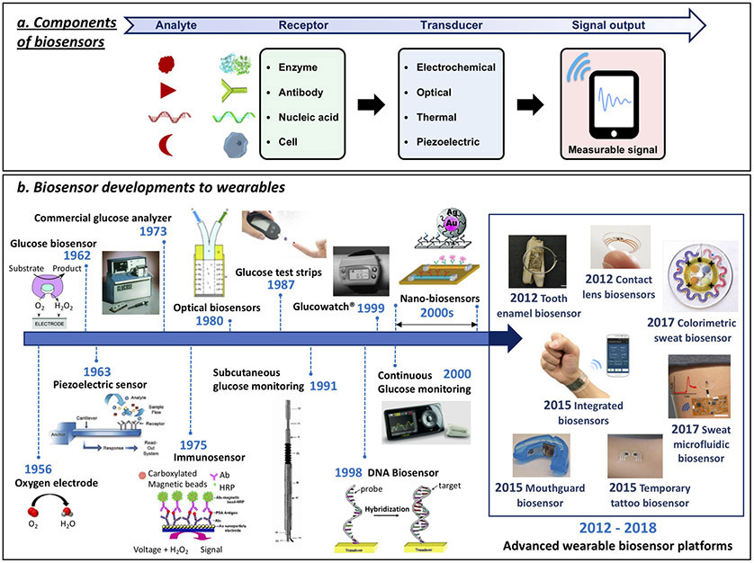
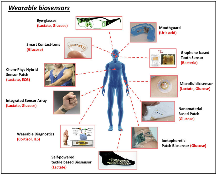
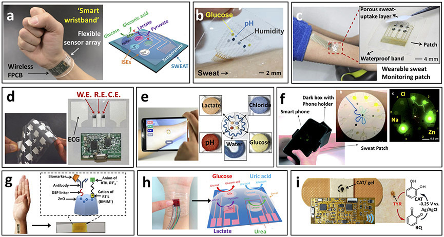
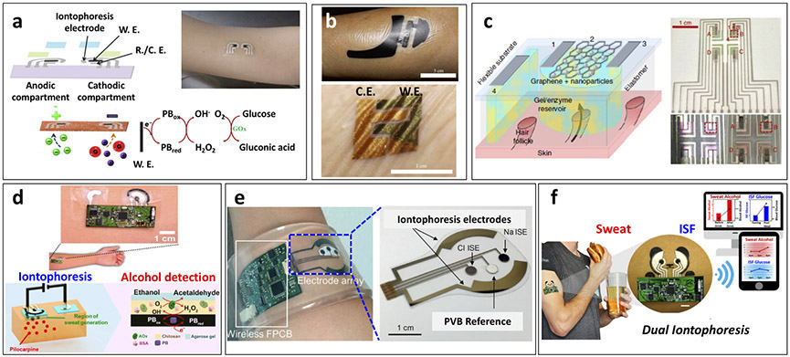
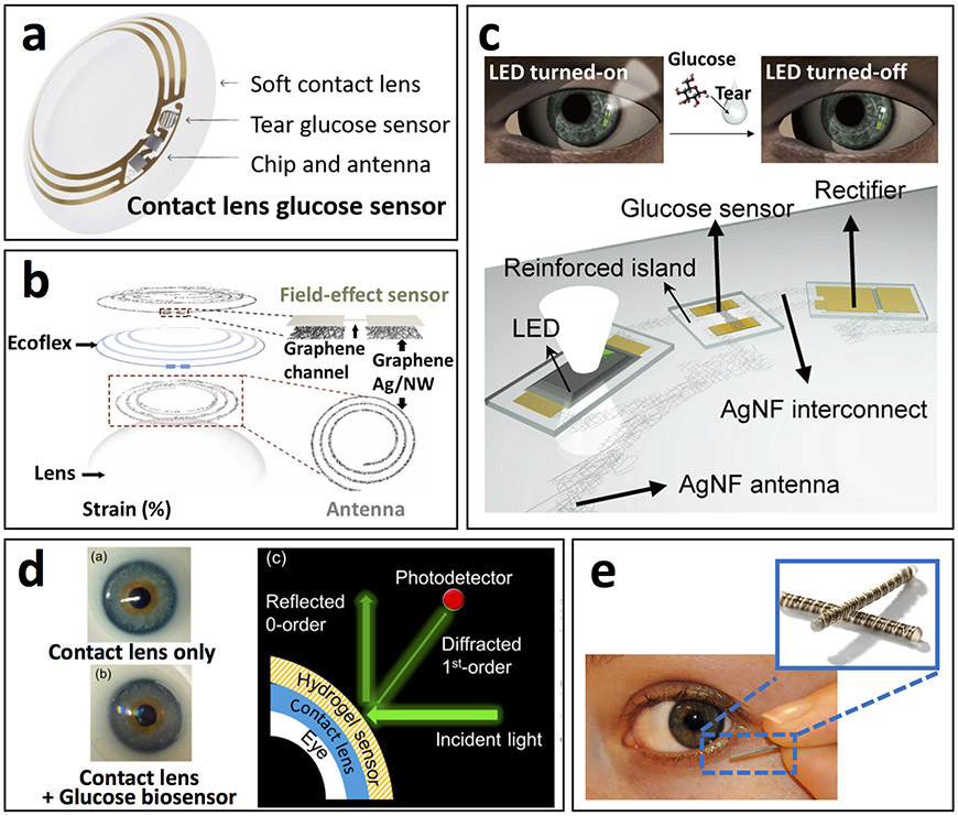
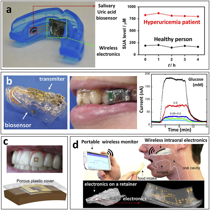
Wearable biosensors are garnering substantial interest due to their potential to provide continuous, real-time physiological information via dynamic, noninvasive measurements of biochemical markers in biofluids, such as sweat, tears, saliva and interstitial fluid. Recent developments have focused on electrochemical and optical biosensors, together with advances in the noninvasive monitoring of biomarkers including metabolites, bacteria and hormones. A combination of multiplexed biosensing, microfluidic sampling and transport systems have been integrated, miniaturized and combined with flexible materials for improved wearability and ease of operation. Although wearable biosensors hold promise, a better understanding of the correlations between analyte concentrations in the blood and noninvasive biofluids is needed to improve reliability. An expanded set of on-body bioaffinity assays and more sensing strategies are needed to make more biomarkers accessible to monitoring. Large-cohort validation studies of wearable biosensor performance will be needed to underpin clinical acceptance. Accurate and reliable real-time sensing of physiological information using wearable biosensor technologies would have a broad impact on our daily lives.


Figures

Figure 1.
Figure 1.
Biosensor components and the path of biosensor development for wearables. (a) Schematic representation of biosensor operation principles: Target analyte detection by corresponding receptor molecule followed by signal transduction method and output. (b) The concept of enzyme electrodes was proposed by Clark and Lyons in 1962. Their device relied on entrapment of the enzyme glucose oxidase (GOx) over an amperometric oxygen electrode that monitored the oxygen consumed by the biocatalytic reaction. Clark’s electrochemical biosensor technology was transferred to the Yellow Spring Instrument (YSI) Company, which launched the first dedicated blood glucose analyzer (YSI Model 23 Analyzer) in 1975. Biosensors became a ‘hot’ topic during the 1980s, reflecting the growing emphasis on biotech. New biosensor transduction principles were introduced during this decade, including fiber-optic and mass-sensitive (piezoelectric) devices-. Considerable efforts during the 1980s led also to the introduction of commercial self-testing blood glucose strips that used mediator-based enzyme electrodes,. Subsequent activity during the 1990s resulted in subcutaneously implantable needle-type electrodes for real-time in vivo glucose monitoring. These subcutaneously implantable glucose sensors moved in the early 2000 to commercial continuous glucose monitors (CGMs) that track in real-time the glucose level in the ISF, along with diabetes relevant trends and patterns,. The emergence of nanotechnology in the late 1990s has led to variety of nanomaterial-based biosensors exploiting the attractive properties of different nanomaterials, such as silicon nanowires and gold nanoparticles, for label-free or amplified biosensing, respectively,. The specific base-pair recognition of DNA sequences led to the development of different DNA biosensors in the late 1990s-. Such nucleic acid sensors are currently playing a growing role in genomic sequence analysis. These advances in biosensor technology over the past five decades paved the way to modern wearable biosensors, discussed in this article. (Glucose biosensor adapted from J.W. et al.). Piezoelectric sensor adapted from ref. . Commercial Glucose Analyzer adapted from ref. . Immunosensor adapted from ref. . Optical Biosensor adapted from ref. . Glucose test strips adapted from ref. . Subcutaneous glucose monitoring adapted from ref. . GlucoWatch adapted from ref. . DNA Biosensor adapted from ref. . Continuous glucose monitoring adapted from ref. . Top nanobiosensors adapted from ref. . Bottom nanobiosensors adapted from ref. . Tooth enamel biosensor adapted from ref. . Contact lens sensors adapted from ref.. Colorimetric sweat biosensor adapted from ref. . Integrated biosensors adapted from ref. . Mouthguard biosensor adapted from (J.K., J.W et al.). Temporary tattoo biosensor adapted from J.W. and colleagues. Sweat microfluidic sensor adapted from (J.K., A.S.C., J.W. et al.).
Figure 2.
Figure 2.
Representative examples of wearable biosensors. (clockwise from top): Eyeglasses-based wireless electrolyte and metabolite sweat sensor (adapted from J.W. et al.). Wearable salivary uric acid mouthguard-based biosensor (adapted from J.K., J.W. et al.). Graphene-based wireless bacteria sensor applied on tooth enamel (adapted from ref. 32). Wearable microfluidic sweat sampling device for colorimetric sensing of sweat (adapted from ref. 34). Graphene-based sweat sensor with thermoresponsive microneedles for diabetes monitoring and therapy (adapted from ref. 40). Integrated wearable sensor arrays for multiplexed sweat extraction and analysis (adapted from ref. 41). Stretchable self-powered sweat biosensors on textile platform (adapted from J.W. et al.). Sweat-based wearable diagnostics biosensors using room-temperature ionic liquids (adapted from ref.43). Integrated multiplexed wearable sensor arrays for in situ perspiration analysis (adapted from ref. 35). Wearable chemical-electrophysiological (lactate/ECG) hybrid biosensor for real-time health and fitness monitoring (adapted from J.W. et al.. Smart contact-lens biosensing platform for glucose monitoring in tears (adapted from ref. 33).
Figure 3.
Figure 3.
Epidermal biosensors for real-time monitoring of sweat chemistry. (a) Depiction of integrated wearable sensor arrays for multiplexed perspiration analysis applied to wrist with schematic representation of sensing array configuration. Fully integrated multianalyte sensor array for sweat-based monitoring of glucose, lactate, sodium, potassium and temperature during exercise with wearable platform containing sensing array as well as signal transduction, conditioning, processing and transmission components (adapted from ref. 35). (b) Depiction of graphene-based sweat sensor array for diabetes monitoring applied to human forearm. Multiplexed patch-type sensor array used for glucose monitoring during exercise with simultaneous measurement of pH, temperature and humidity for glucose signal correction (adapted from ref. 40). (c) Depiction of wearable sweat monitoring patch for sweat-based glucose monitoring and therapy applied to human forearm during exercise. Inset: sweat-based glucose monitoring sensor array configuration with porous sweat-uptake layer. Multiplexed glucose monitoring patch capable of operating in low sweat volumes (adapted from ref. 78). (d) Depiction of wearable chemical-electrophysiological hybrid biosensor configuration for real-time health and fitness monitoring with example of screen-printed electrodes. Simultaneous monitoring of sweat lactate levels and heart-rate for athletic performance evaluation (adapted from J.W. et al.). (e) Depiction of colorimetric microfluidic sweat sampling device configuration for chemical analysis of sweat with representation of sweat-filled device and smartphone-based signal analysis. Device exhibited enhanced microfluidic sampling of sweat during exercise with wireless quantitative measure of target pH, lactate, glucose and chloride (adapted from ref. 34). (f) Depiction of fluorometric skin-interfaced microfluidic platform for the measurement of chloride, sodium and zinc in exercise induced sweat. Fluorescent probes selectively react with target biomarkers upon sweat flow through the microfluidic system with fluorescent intensity analyzed via smartphone-based imaging module, which obviates the need for electrochemical or colorimetric analyses (adapted from ref. 84). (g) Schematic representation of wearable diagnostic antibody-based biosensor targeting detection of IL-6 and cortisol in human sweat using room temperature ionic liquids for enhanced antibody operational stability. Biosensor configuration with antibody immobilization is shown with depiction of device application onto a human forearm. This device exhibited prolonged stability in pooled human sweat with continuous combinatorial analyte detection within the physiologically relevant concentration range (adapted from ref. 43). (h) Schematic representation of self-powered multifunctional electronic skin used for continuous monitoring of lactate, glucose, uric acid, and urea in exercise-induced sweat using piezoelectric-linked enzymatic biosensors. During exercise, this device was capable of monitoring these biomarkers related to personal health status without an additional power supply through piezoelectric-enzymatic-reaction coupling (adapted from ref. 56). (i) Depiction of wearable tyrosinase sensing bandage for non-invasive melanoma screening. Inset: schematic representation of tyrosinase detection paradigm. Bandage-type wearable sensor for portable cancer biomarker detection (adapted from (J.W. et al.).
Figure 4.
Figure 4.
Epidermal iontophoretic biosensors. (a) Schematic representation of epidermal reverse iontophoretic tattoo-based glucose sensor configuration and operation paradigm with on-body depiction of device applied to human subject. Proof-of-concept demonstration of reverse iontophoretic tattoo-based ISF glucose sensor (adapted from J.W. et al.). (b) On-body depiction of iontophoretic paper battery and skin-like biosensor for non-invasive blood glucose monitoring applied to human subject. Inclusion of hyaluronic acid facilitated enhanced ISF extraction for increased ISF glucose sampling reliability (adapted from ref. 87). (c) Schematic representation of transdermal, path-selective iontophoretic ISF sampling approach using miniaturized graphene-based pixel arrays for non-invasive glucose monitoring. Configuration of pixel-type biosensor array with four individual ISF extraction and detection locations. This proof-of-concept device exhibited the capability to sample ISF through individual follicular pathways for enhanced glucose detection reliability over 6 hours by focusing on device architecture design rather than specific sensor implementation (adapted from ref. 88). (d) Depiction of epidermal iontophoretic alcohol sensing tattoo applied to human subject with schematic representation of iontophoretic drug delivery and sensing paradigms. Localized, drug-induced sweat generation for on-demand sampling of sweat alcohol at a patch-type sensor platform (adapted from J.K., J.W. et al.). (e) On-body depiction of integrated wearable sensor array band for multiplexed sweat extraction and analysis applied to human wrist with schematic representation of sensor array configuration. Simultaneous detection of chloride, sodium and glucose in iontophoretic induced sweat (adapted from ref. 41). (f) Device configuration and on-body application of simultaneous dual iontophoretic ISF and sweat sampling platform for the sampling and analysis of these two bio-fluids on a single platform without cross-contamination. This device demonstrated the capability to monitor sweat alcohol and ISF glucose simultaneously through the iontophoretic delivery of sweat inducing pilocarpine and iontophoretic extraction of ISF, which were shown to correlate to concurrent trends in blood concentrations (adapted from J.K., A.S.C., J.W. et al.).
Figure 5.
Figure 5.
Tear-based biosensors. (a) Pictorial depiction of contact lens sensor previously under co-development by Google and Novartis to measure tears glucose concentrations in a miniaturized glucose sensor. Prototype platform contained integrated electronics for sensor response processing and wireless transmission (adapted from https://sites.google.com/site/smartcontactlens/). (b) Schematic illustration of multifunctional wearable smart sensor system incorporated onto contact lenses for monitoring of glucose in tears as well as intraocular pressure using enzyme-functionalized graphene-silver nanowire hybrid nanostructures. The device proved capable of wirelessly detecting fluctuating glucose concentrations and pressure in a rabbit model in vivo and in a bovine eyeball in vitro (adapted from ref. 117). (c) Schematic representation of wireless glucose sensor incorporated into a contact lens platform with wireless power transfer circuitry and display pixels for a fully integrated and transparent platform that does not hinder vision. This device detected fluctuating tear glucose concentrations through a resistance-based enzymatic mechanism, which was demonstrate in a rabbit model (adapted from ref. 118). (d) Pictorial depiction of wearable contact lens tear glucose biosensor platform applied to an artificial eye with schematic representation of smartphone-based quantification of glucose levels through reflection of incident light by the photonic microstructure within the lens. The smart contact lens system integrated with a glucose sensitive hydrogel monitored changing glucose concentrations in vitro without complicated fabrication procedures that allowed rapid response time for continuous measurements (adapted from ref. 112). (e) Depiction of Noviosense electrochemical tear glucose sensor. A small spring-like sensing device designed to be placed within the conjunctive fornix for continuous access to tears glucose (adapted from http://noviosense.com).
Figure 6.
Figure 6.
Saliva-based biosensors. (a) Depiction of mouthguard-based wearable salivary uric acid biosensing platform with integrated wireless electronics and analysis of salivary uric acid concentrations in a healthy volunteer and a hyperuricemia patient. This platform exhibited selective uric acid detection in undiluted human saliva to monitor the response or uric acid levels of a hyperuricemia patient during treatment (adapted from J.K., J.W. et al.). (b) Depiction of mouthguard-based sensor for glucose monitoring in saliva with on-body application and analysis of increasing glucose concentrations. Fully integrated saliva glucose sensor toward continuous in-mouth glucose monitoring (adapted from refs 97,148). (c) On-body depiction and cross-sectional configuration of radiofrequency trilayer tooth-mounted sensor for wireless monitoring of food consumption. This dielectric sensor fabricated with biocompatible materials was capable of being mounted onto tooth enamel to detect foods and fluids during ingestion when functionalized with analyte sensitive layers. Projected uses were for detection of sugars, alcohol, salinity, pH and temperature (adapted from ref. 155). (d) Depiction of operational principles and electronics configuration of wireless, user-comfortable sensing platform for long-range oral monitoring of sodium intake toward hypertension management. Electrochemical sodium sensing was demonstrated in vitro as well as in vivo with the orally-mounted biocompatible sensing platform (adapted from ref. 156).

References

    1. Bandodkar AJ, Jeerapan I & Wang J Wearable chemical sensors: Present challenges and future prospects. ACS Sens. 1, 464–482 (2016).
    1. Heikenfeld J et al. Wearable sensors: Modalities, challenges, and prospects. Lab Chip 18, 217–248 (2018). - PMC - PubMed
    1. Matzeu G, Florea L & Diamond D Advances in wearable chemical sensor design for monitoring biological fluids. Sens. Actuators B 211, 403–418 (2015).
    1. Liu Y, Pharr M & Salvatore GA Lab-on-skin: A review of flexible and stretchable electronics for wearable health monitoring. ACS Nano 11, 9614–9635 (2017). - PubMed
    1. Amjadi M, Kyung KU, Park I & Sitti M Stretchable, skin-mountable, and wearable strain sensors and their potential applications: A review. Adv. Funct. Mater 26, 1678–1698 (2016).

Publication types