. 2019 May;4(5):789-799.
doi: 10.1038/s41564-019-0371-3. Epub 2019 Feb 25.

Dampened NLRP3-mediated inflammation in bats and implications for a special viral reservoir host

Affiliations

Dampened NLRP3-mediated inflammation in bats and implications for a special viral reservoir host

Matae Ahn et al. Nat Microbiol. 2019 May.

Abstract

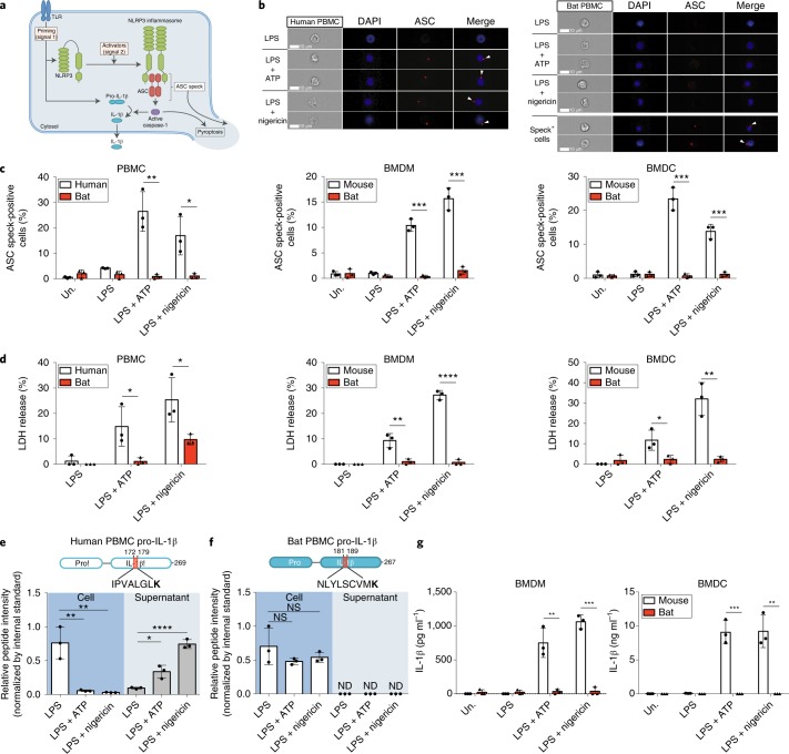
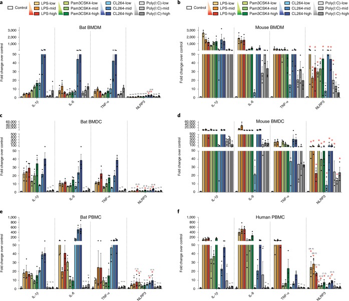
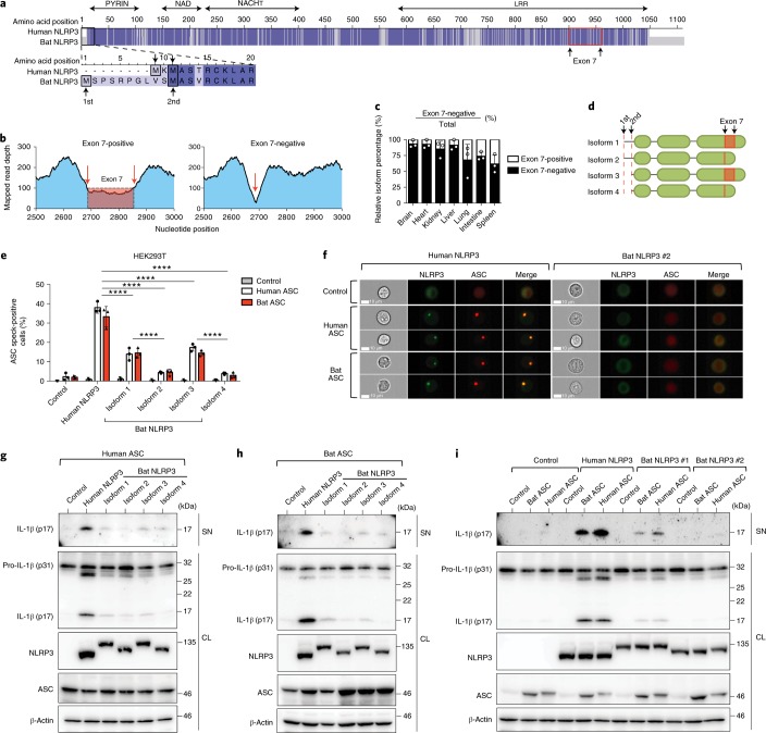
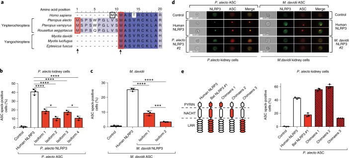
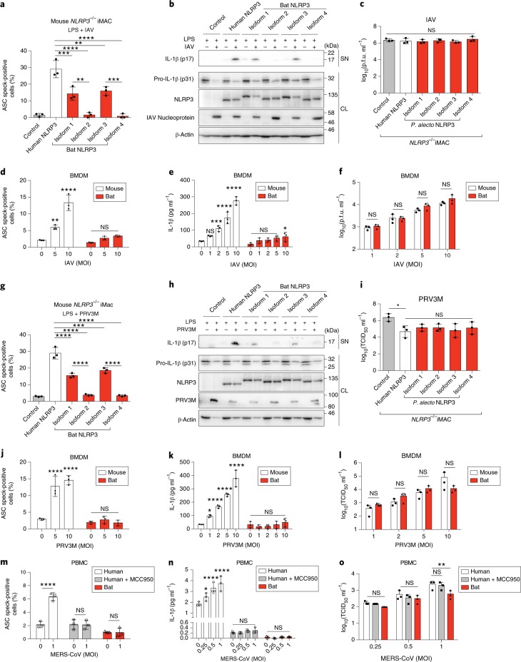
Bats are special in their ability to host emerging viruses. As the only flying mammal, bats endure high metabolic rates yet exhibit elongated lifespans. It is currently unclear whether these unique features are interlinked. The important inflammasome sensor, NLR family pyrin domain containing 3 (NLRP3), has been linked to both viral-induced and age-related inflammation. Here, we report significantly dampened activation of the NLRP3 inflammasome in bat primary immune cells compared to human or mouse counterparts. Lower induction of apoptosis-associated speck-like protein containing a CARD (ASC) speck formation and secretion of interleukin-1β in response to both 'sterile' stimuli and infection with multiple zoonotic viruses including influenza A virus (-single-stranded (ss) RNA), Melaka virus (PRV3M, double-stranded RNA) and Middle East respiratory syndrome coronavirus (+ssRNA) was observed. Importantly, this reduction of inflammation had no impact on the overall viral loads. We identified dampened transcriptional priming, a novel splice variant and an altered leucine-rich repeat domain of bat NLRP3 as the cause. Our results elucidate an important mechanism through which bats dampen inflammation with implications for longevity and unique viral reservoir status.


Conflict of interest statement

The authors declare no competing interests.

Figures

Fig. 1
Fig. 1. Activation of the NLRP3 inflammasome is dampened in bat PBMCs, BMDMs and BMDCs.
a, Schematic model for NLRP3 inflammasome activation. b, Representative single-cell images of gated monocytes of human (left) and bat (right) PBMCs, primed with LPS for 3 h, with or without stimulation by ATP or nigericin for 30 min. Cells were acquired using ImageStream. DAPI, blue; ASC, red; arrowheads, ASC specks. Scale bars, 10 μm. c, Quantification of ASC specks in gated monocytes of PBMCs (left), BMDMs (middle) or BMDCs (right) by ImageStream, unprimed (Un.) or primed with LPS for 3 h, with or without stimulation with ATP or nigericin for 30 min. d, LDH release in cell-free supernatants of PBMCs (left), BMDMs (middle) or BMDCs (right), primed with LPS for 3 h, with or without stimulation by ATP or nigericin for 1 h. e,f, Detection of human (e) and bat (f) C-terminal pro-IL-1β peptides in cell lysates (left) or supernatants (right) of PBMCs as in d, using PRM-based targeted mass spectrometry with heavy isotope-labelled internal standards. K indicates heavy labelled lysine (13C6, 15N2); ND, not detected; NS, not significant. g, Secretion of IL-1β by ELISA in the supernatant of BMDMs (left) or BMDCs (right) as stimulated in d. *P < 0.05, **P < 0.01, ***P < 0.001, ****P < 0.0001; two-tailed unpaired t-test. Exact P values are provided in Supplementary Table 5. Data are representative of three biological replicates (n = 3) in b, or mean + s.d. of three biological replicates (n = 3) in cg.
Fig. 2
Fig. 2. Transcriptional priming of bat NLRP3 is dampened independent of TLRs.
af, Fold changes of IL-1β, IL-6, TNF-α and NLRP3 measured by qPCR in BMDMs (a,b), BMDCs (c,d) or PBMCs (e,f) of bat (a,c,e) and mouse (b,d) or human (f) after stimulating with different concentrations of TLR ligands for 3 h. LPS = 10, 100, 1,000 ng ml−1; Pam3CSK4 = 10, 100, 1,000 ng ml−1; CL264 = 0.1, 1, 10 μg ml−1; poly(I:C) = 0.1, 1, 10 μg ml−1. Fold induction of cytokines is relative to mock-treated samples (control) after normalizing with GAPDH. *P < 0.05 (only labelled for NLRP3), NS, not significant (labelled for IL-1β, IL-6, TNF-α and NLRP3), by one-way analysis of variance (ANOVA) with Bonferroni’s multiple comparisons test for log fold changes. Exact P values are provided in Supplementary Table 5. Data are presented as mean + s.e.m. of three biological replicates (n = 3) in af.
Fig. 3
Fig. 3. The function of all four bat NLRP3 isoforms, but not ASC, is reduced.
a, Amino acid sequence alignment of human and bat (P. alecto) NLRP3, with identical residues highlighted in dark blue and mismatches in light blue. Black boxes, alternative translation start sites. Red box, boundaries of exon 7. b, Read-mapping analysis of two splice variants (exon 7-positive/negative) by RNA–seq read counts of the P. alecto splenocytes subsets. Arrows, exon–exon junctions. c, Percentage of exon 7-positive or exon 7-negative isoform in P. alecto tissues measured by qPCR. d, Schematic representation of four bat NRLP3 isoforms. e, Quantification of ASC specks by ImageStream in HEK293T cells stably expressing mPlum (control), human or bat ASC–mPlum, transfected with mCitrine (control), human or bat NLRP3–mCitrine. f, Representative images from e. NLRP3–mCitrine (green); ASC–mPlum (red). Scale bars, 10 μm. g–i, Immunoblot analysis of IL-1β cleavage in cell lysates and supernatant in HEK293T cells stably expressing mPlum (control), human or bat ASC–mPlum, transfected with mCitrine (control), human or bat NLRP3–mCitrine, plus human pro-caspase-1 and mouse pro-IL-1β. NLRP3, NLRP3–mCitrine stained with anti-GFP; ASC, ASC–mPlum stained with anti-mPlum; SN, supernatant; CL, cell lysate.   ****P < 0.0001 by two-way ANOVA with Bonferroni’s multiple comparisons test (e). Exact P values are provided in Supplementary Table 5. Data are presented as mean + s.d. of three biological replicates (c) or three independent experiments (e), or representative of three independent experiments (fi).
Fig. 4
Fig. 4. NLRP3 isoform activity in bat cells from both major bat lineages is dampened.
a, Amino acid sequence alignment of N-terminal NLRP3 from human and six bats species from Yinpterochiroptera or Yangochiroptera suborders, with identical residues (dark blue) and mismatches (light blue). Arrows indicate alternative translation start sites. b, Quantification of ASC specks by ImageStream in P. alecto kidney (PaKi) cells stably expressing bat ASC, transfected with mCitrine (control), human or bat NLRP3–mCitrine. c, Quantification of ASC specks by ImageStream in M. davidii kidney cells stably expressing M. davidii ASC, transfected with mCitrine (control), human or M. davidii NLRP3–mCitrine. d, Representative ImageStream images of P. alecto or M. davidii cells stably expressing P. alecto or M. davidii ASC, respectively, transfected with empty vector, human or bat NLRP3–mCitrine. NLRP3–mCitrine, green; ASC–mPlum, red. Scale bars, 10 μm. e, Left, Schematic representation for chimaeras between human NLRP3 and P. alecto NLRP3 isoform 1. Right, Quantification of ASC specks by ImageStream in PaKi cells stably expression P. alecto ASC, transfected with mCitrine (control), human or bat NLRP3 or chimaera–mCitrine. *P < 0.05, ***P < 0.001, ****P < 0.0001 by one-way ANOVA with Bonferroni’s multiple comparisons test (b,c). Exact P values are provided in Supplementary Table 5. Data are presented as mean + s.d. of three independent experiments (b,c,e) or representative of three independent experiments (d).
Fig. 5
Fig. 5. Bat NLRP3-mediated inflammation in immune cells is dampened in response to IAV, PRV3M and MERS-CoV infection.
a–c,g–i, Quantification of ASC specks by ImageStream (a,g), immunoblot analysis (b,h) and virus titration (c,i) of mouse NLRP3/− iMACs reconstituted with mCitrine alone (control), human or bat (P. alecto) NLRP3–mCitrine, primed with LPS for 3 h, followed by infection with IAV (MOI = 0.5, ac) or PRV3M (MOI = 2, g–i) for 24 h. NLRP3, NLRP3–mCitrine stained with anti-GFP; PRV3M, PRV3M viral protein stained with polyclonal macaque serum; SN, supernatant; CL, cell lysates. Titres in the supernatant are expressed as p.f.u. ml−1. df,j–l Quantification of ASC specks by ImageStream (d,j), IL-1β secretion by ELISA (e,k) and virus titration (f,l) of mouse or bat BMDMs, primed with LPS for 3 h, followed by infection with IAV (MOI = 1–10, df) or PRV3M (MOI = 1–10, jl) for 24 h. Titres in the supernatant are expressed as median tissue culture infectious dose (TCID50) ml−1 m–o, Quantification of ASC specks by ImageStream (m), IL-1β secretion by ELISA (n) and virus titration (o) of human or bat PBMCs, primed with LPS for 3 h, followed by infection with MERS-CoV (MOI = 0.25, 0.5, 1) for 24 h. MCC950 inhibitor was added 1 h before infection for human PBMCs. Titres in the supernatant are expressed as TCID50 ml−1. *P < 0.05, **P < 0.01, ***P < 0.001, ****P < 0.0001, NS, not significant, by one-way ANOVA (a,c,g,i) or two-way ANOVA (df and jo) with Bonferroni’s multiple comparisons test. Exact P values are provided in Supplementary Table 5. Data are presented as mean + s.d. of three biological replicates (df and jo) or three independent experiments (a,c,g,i), or representative of three independent experiments (b,h).

Comment in

  • "On the bat's back I do fly".
    York A. York A. Nat Rev Microbiol. 2019 May;17(5):265. doi: 10.1038/s41579-019-0177-6. Nat Rev Microbiol. 2019. PMID: 30842614 Free PMC article.
  • Bat tolerance to viral infections.
    Hayman DTS. Hayman DTS. Nat Microbiol. 2019 May;4(5):728-729. doi: 10.1038/s41564-019-0430-9. Nat Microbiol. 2019. PMID: 31015739 Free PMC article.

Similar articles

Cited by

References

    1. Leroy EM, et al. Fruit bats as reservoirs of Ebola virus. Nature. 2005;438:575–576. doi: 10.1038/438575a. - DOI - PubMed
    1. Clayton BA, Wang LF, Marsh GA. Henipaviruses: an updated review focusing on the pteropid reservoir and features of transmission. Zoonoses Public Health. 2013;60:69–83. doi: 10.1111/j.1863-2378.2012.01501.x. - DOI - PubMed
    1. Li W, et al. Bats are natural reservoirs of SARS-like coronaviruses. Science. 2005;310:676–679. doi: 10.1126/science.1118391. - DOI - PubMed
    1. Mohd HA, Al-Tawfiq JA, Memish ZA. Middle East respiratory syndrome coronavirus (MERS-CoV) origin and animal reservoir. Virol. J. 2016;13:87. doi: 10.1186/s12985-016-0544-0. - DOI - PMC - PubMed
    1. Cameron MJ, Bermejo-Martin JF, Danesh A, Muller MP, Kelvin DJ. Human immunopathogenesis of severe acute respiratory syndrome (SARS) Virus Res. 2008;133:13–19. doi: 10.1016/j.virusres.2007.02.014. - DOI - PMC - PubMed

Publication types

MeSH terms

Substances