

Wide-angled endoillumination vs traditional scleral buckling surgery for retinal detachment - a comparative study
- PMID: 30804661
- PMCID: PMC6371939
- DOI: 10.2147/OPTH.S182751
Wide-angled endoillumination vs traditional scleral buckling surgery for retinal detachment - a comparative study
Abstract
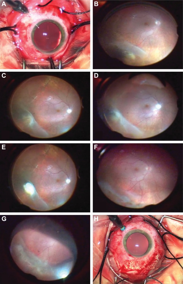
Purpose: To evaluate the surgical outcomes of traditional scleral buckling (TSB) compared to chandelier-assisted scleral buckling (CSB) for rhegmatogenous retinal detachment repair.
Patients and methods: A retrospective interventional comparative case series of 49 eyes that underwent SB procedure. Medical records of 27 and 22 eyes that underwent TSB and CSB surgery, respectively, were evaluated. Outcome measures included primary anatomical success, visual acuity (VA), and perioperative complications.
Results: Primary reattachment rate was similar with 85.2% in the TCB group and 81.8% in the CSB group (P=1.00); eight patients needed one additional operation or gas injection with a final reattachment rate of 100% at 6 months. Mean VA in the CSB group improved from 20/60 at presentation to 20/35, 6 months postoperatively. In the TSB group, VA improved from 20/80 to 20/45 (P=0.90). Among the eyes that were successfully reattached with either SB approach, two eyes in each group had cataract progression and none of them required surgery during follow-up. No cases of endophthalmitis were observed.
Conclusion: CSB is a modified technique with an advantage of superior visualization compared with the traditional surgery, which simplifies the operation, enhance competency, and could be used as a valuable educational tool. It can provide similar anatomical and functional outcomes with no additional perioperative complications.
Keywords: chandelier; endoillumination; retinal detachment; scleral buckle; wide-angled viewing system.
Conflict of interest statement
Disclosure The authors report no conflicts of interest in this work.
Figures
Similar articles
-
SCLERAL BUCKLING WITH WIDE-ANGLED ENDOILLUMINATION AS A SURGICAL EDUCATIONAL TOOL.Retina. 2016 Apr;36(4):830-3. doi: 10.1097/IAE.0000000000000792. Retina. 2016. PMID: 26447399
-
Non-contact wide-angled visualization with chandelier-assisted scleral buckling for primary uncomplicated rhegmatogenous retinal detachment.Graefes Arch Clin Exp Ophthalmol. 2020 Sep;258(9):1857-1861. doi: 10.1007/s00417-020-04737-1. Epub 2020 May 14. Graefes Arch Clin Exp Ophthalmol. 2020. PMID: 32409979
-
ENDOILLUMINATION-ASSISTED MODIFIED SCLERAL BUCKLING.Retina. 2018 Feb;38(2):320-324. doi: 10.1097/IAE.0000000000001568. Retina. 2018. PMID: 28221258
-
Comparison of Chandelier-Assisted versus Standard Scleral Buckling for the Treatment of Primary Rhegmatogenous Retinal Detachment: A Systematic Review and Meta-Analysis.Ophthalmologica. 2024;247(5-6):345-354. doi: 10.1159/000540820. Epub 2024 Aug 19. Ophthalmologica. 2024. PMID: 39159609 Free PMC article.
-
Scleral Buckling with Chandelier Illumination.J Ophthalmic Vis Res. 2016 Jul-Sep;11(3):304-9. doi: 10.4103/2008-322X.188402. J Ophthalmic Vis Res. 2016. PMID: 27621789 Free PMC article. Review.
Cited by
-
Six-Year Outcomes of 25-Gauge Chandelier Illumination-Assisted Scleral Buckling.Biomed Res Int. 2021 Oct 4;2021:4628160. doi: 10.1155/2021/4628160. eCollection 2021. Biomed Res Int. 2021. PMID: 35402605 Free PMC article.
-
Chandelier-Assisted Scleral Buckling: A Literature Review.Vision (Basel). 2023 Jun 28;7(3):47. doi: 10.3390/vision7030047. Vision (Basel). 2023. PMID: 37489326 Free PMC article. Review.
-
Updated chandelier illumination-assisted scleral buckling using 3D visualization system.Clin Ophthalmol. 2019 Sep 6;13:1743-1748. doi: 10.2147/OPTH.S218975. eCollection 2019. Clin Ophthalmol. 2019. PMID: 31564825 Free PMC article.
-
Scleral Buckling: A Look at the Past, Present and Future in View of Recent Findings on the Importance of Photoreceptor Re-Alignment Following Retinal Re-Attachment.Clin Ophthalmol. 2022 Jun 16;16:1971-1984. doi: 10.2147/OPTH.S359309. eCollection 2022. Clin Ophthalmol. 2022. PMID: 35733617 Free PMC article. Review.
-
A Comparative Study of Traditional Scleral Buckling to a New Technique: Guarded Light Pipe with Heads-Up Three-Dimensional Visualization.Clin Ophthalmol. 2022 Sep 19;16:3079-3088. doi: 10.2147/OPTH.S378179. eCollection 2022. Clin Ophthalmol. 2022. PMID: 36160731 Free PMC article.
References
-
- Custodis E. Treatment of retinal detachment by circumscribed diathermal coagulation and by scleral depression in the area of tear caused by imbed-ding of a plastic implant. Klin Monbl Augenheilkd Augenarztl Fortbild. 1956;129(4):476–495. German. - PubMed
-
- Arruga H. Le cerclage equatorial pour traiter le décollement rétinien [An equatorial cerclage to treat retinal detachment] Bull Mem Soc Fr Ophthalmol. 1958;71:571.
-
- Schepens CL, Okamura ID, Brockhurst RJ. The scleral buckling procedures. 1. Surgical techniques and management. Arch Ophthalmol. 1957;58(6):797–811. - PubMed
LinkOut - more resources
Full Text Sources
