Role of microRNA-92a in metastasis of osteosarcoma cells in vivo and in vitro by inhibiting expression of TCF21 with the transmission of bone marrow derived mesenchymal stem cells
- PMID: 30804710
- PMCID: PMC6373113
- DOI: 10.1186/s12935-019-0741-1
Role of microRNA-92a in metastasis of osteosarcoma cells in vivo and in vitro by inhibiting expression of TCF21 with the transmission of bone marrow derived mesenchymal stem cells
Abstract
Background: This study aims to investigate the role of microRNA-92a (miR-92a) in metastasis of osteosarcoma (OS) cells in vivo and in vitro by regulatingTCF21 with the transmission of bone marrow derived mesenchymal stem cells (BMSCs).
Methods: BMSCs were isolated, purified and cultured from healthy adult bone marrow tissues. The successfully identified BMSCs were co-cultured with OS cells, and the effects of BMSCs on the growth metastasis of OS cells in vitro and in vivo were determined. qRT-PCR and western blot analysis was used to detect the expression of miR-92a and TCF21 in OS cells and OS cells co-cultured with BMSCs. Proliferation, invasion and migration of OS cells transfected with miR-92a mimics and miR-92a inhibitors was determined, and the tumorigenesis and metastasis of OS cells in nude mice were observed. Expression of miR-92a and TCF21 mRNA in tissue specimens as well as the relationship between the expression of miR-92a and the clinicopathological features of OS was analyzed.
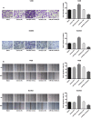
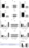
Results: BMSCs promoted proliferation, invasion and migration of OS cells in vitro together with promoted the growth and metastasis of OS cells in vivo. Besides, high expression of miR-92a was found in OS cells co-cultured with BMSCs. Meanwhile, overexpression of miR-92a promoted proliferation, invasion and migration of OS cells in vitro as well as promoted growth and metastasis of OS cells in vivo. The expression of miR-92a increased significantly, and the expression of TCF21 mRNA and protein decreased significantly in OS tissues. Expression of miR-92a was related to Ennecking staging and distant metastasis in OS patients.
Conclusion: Collectively, this study demonstrates that the expression of miR-92a is high in OS and BMSCs transfers miR-92a to inhibit TCF21 and promotes growth and metastasis of OS in vitro and in vivo.
Keywords: Invasion; MicroRNA-92a; Migration; Osteosarcoma; Proliferation; TCF21.
Figures


Similar articles
-
LncRNA XIST from the bone marrow mesenchymal stem cell derived exosome promotes osteosarcoma growth and metastasis through miR-655/ACLY signal.Cancer Cell Int. 2022 Oct 29;22(1):330. doi: 10.1186/s12935-022-02746-0. Cancer Cell Int. 2022. PMID: 36309693 Free PMC article.
-
MiR-92a modulates proliferation, apoptosis, migration, and invasion of osteosarcoma cell lines by targeting Dickkopf-related protein 3.Biosci Rep. 2019 Apr 26;39(4):BSR20190410. doi: 10.1042/BSR20190410. Print 2019 Apr 30. Biosci Rep. 2019. PMID: 30926679 Free PMC article.
-
MicroRNA-92a promotes epithelial-mesenchymal transition through activation of PTEN/PI3K/AKT signaling pathway in non-small cell lung cancer metastasis.Int J Oncol. 2017 Jul;51(1):235-244. doi: 10.3892/ijo.2017.3999. Epub 2017 May 16. Int J Oncol. 2017. PMID: 28534966
-
Overexpression of miR-92a promotes the tumor growth of osteosarcoma by suppressing F-box and WD repeat-containing protein 7.Gene. 2017 Mar 30;606:10-16. doi: 10.1016/j.gene.2017.01.002. Epub 2017 Jan 6. Gene. 2017. PMID: 28069547
-
BMSC-EV-derived lncRNA NORAD Facilitates Migration, Invasion, and Angiogenesis in Osteosarcoma Cells by Regulating CREBBP via Delivery of miR-877-3p.Oxid Med Cell Longev. 2022 Mar 1;2022:8825784. doi: 10.1155/2022/8825784. eCollection 2022. Oxid Med Cell Longev. 2022. PMID: 35281474 Free PMC article.
Cited by
-
Osteoclasts differential-related prognostic biomarker for osteosarcoma based on single cell, bulk cell and gene expression datasets.BMC Cancer. 2022 Mar 17;22(1):288. doi: 10.1186/s12885-022-09380-z. BMC Cancer. 2022. PMID: 35300639 Free PMC article.
-
MicroRNA-92a-3p Inhibits Cell Proliferation and Invasion by Regulating the Transcription Factor 21/Steroidogenic Factor 1 Axis in Endometriosis.Reprod Sci. 2023 Jul;30(7):2188-2197. doi: 10.1007/s43032-021-00734-9. Epub 2023 Jan 17. Reprod Sci. 2023. PMID: 36650372 Free PMC article.
-
Cancer Stem Cells as a Source of Drug Resistance in Bone Sarcomas.J Clin Med. 2021 Jun 14;10(12):2621. doi: 10.3390/jcm10122621. J Clin Med. 2021. PMID: 34198693 Free PMC article. Review.
-
TCF21: a critical transcription factor in health and cancer.J Mol Med (Berl). 2020 Aug;98(8):1055-1068. doi: 10.1007/s00109-020-01934-7. Epub 2020 Jun 15. J Mol Med (Berl). 2020. PMID: 32542449 Review.
-
Fibrinogen-Albumin Ratio Index Exhibits Predictive Value of Neoadjuvant Chemotherapy in Osteosarcoma.Cancer Manag Res. 2022 May 5;14:1671-1682. doi: 10.2147/CMAR.S358310. eCollection 2022. Cancer Manag Res. 2022. PMID: 35547600 Free PMC article.
References
-
- Kobayashi E, et al. MicroRNA expression and functional profiles of osteosarcoma. Oncology. 2014;86(2):94–103. - PubMed
-
- Gorlick R, Khanna C. Osteosarcoma. J Bone Miner Res. 2010;25(4):683–691. - PubMed
-
- Chou AJ, Gorlick R. Chemotherapy resistance in osteosarcoma: current challenges and future directions. Expert Rev Anticancer Ther. 2006;6(7):1075–1085. - PubMed
-
- Kansara M, Thomas DM. Molecular pathogenesis of osteosarcoma. DNA Cell Biol. 2007;26(1):1–18. - PubMed
LinkOut - more resources
Full Text Sources

