. 2019 Feb 11:13:87.
doi: 10.3389/fnins.2019.00087. eCollection 2019.

Vagal Nerve Stimulation Attenuates Ischemia-Reperfusion Induced Retina Dysfunction in Acute Ocular Hypertension

Affiliations

Vagal Nerve Stimulation Attenuates Ischemia-Reperfusion Induced Retina Dysfunction in Acute Ocular Hypertension

Meng-Nan Jiang et al. Front Neurosci. .

Abstract

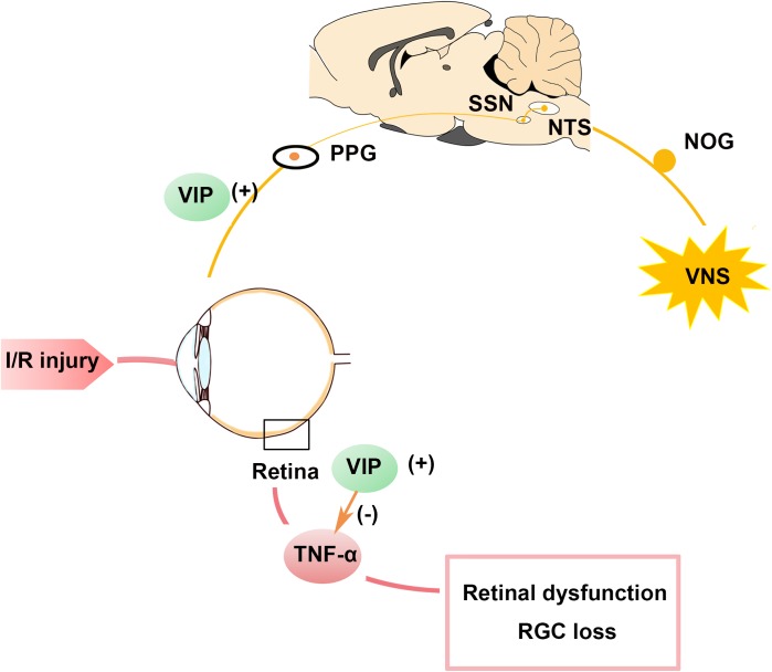
Purpose: The present study aimed to investigate whether cervical vagal nerve stimulation (VNS) could prevent retinal ganglion cell (RGC) loss and retinal dysfunction after ischemia/reperfusion (I/R) injury. Methods: First, rats were randomly divided into sham group (n = 4) and VNS group (n = 12). Activation of the nodose ganglia (NOG), nucleus of the solitary tract (NTS), superior salivatory nucleus (SSN), and pterygopalatine ganglion (PPG) neural circuit were evaluated by c-fos expression at 0 h after sham VNS and at 0 h (n = 4), 6 h (n = 4), 72 h (n = 4) after VNS. Secondly, rats were randomly assigned to I/R group (pressure-induced retinal ischemia for 1 h and reperfusion for 1 h in the right eye, n = 16) and I/R+VNS group (right cervical VNS for 2 h during the I/R period, n = 16). The left eye of each rat served as a control. Electroretinogram (ERG), RGC numbers, tumor necrosis factor-α (TNF-α) and vasoactive intestinal polypeptide (VIP) levels in retina were determined. Additionally, the level of VIP in PPG was evaluated. Results: In the first part of the study, compared with the sham group, the VNS group exhibited significantly increased expression of c-fos in NOG, NTS, SSN, and PPG tissues at 0, 6, and 72 h. In the second part of the study, compared with left eyes, retinal function in right eyes (as assessed by the a-wave, b-wave and the oscillatory potential amplitudes of ERG and RGC data) was significantly decreased by I/R. The decreased retinal function was attenuated by VNS. In addition, I/R induced an increase in inflammation, which was reflected by elevated TNF-α expression in the retina. VNS significantly attenuated the increase in I/R-induced inflammation. Moreover, VIP expression in the retina and PPG, which may contribute to the inhibition of the inflammatory response, was significantly increased after VNS. Conclusion: VNS could protect against retinal I/R injury by downregulating TNF-α. Upregulation of VIP expression due to activation of the NOG-NTS-SSN-PPG neural circuit may underlie to the protective effects of VNS.

Keywords: inflammation; ischemia/reperfusion; neuroprotection; retina; vagal nerve stimulation; vasoactive intestinal polypeptide.


Figures

FIGURE 1
FIGURE 1
Timeline of the experimental design. (A) Experiments in the first part of the study detected whether VNS can activate the NOG-NTS-SSN-PPG neuron circuit. Rats were sacrificed at 0 h after sham VNS and at 0, 6, and 72 h after VNS. (B) Experiments in the second part of the study investigated the protective effect of VNS on retinal I/R injury. All operations were performed on the rat’s right eyes. The right vagal nerve was isolated and the stimulation was applied during the I/R period. Sham procedures were performed on the left eyes, which served as control. Rats were sacrificed at 6 and 72 h after reperfusion; (n = 4/group) I/R, ischemia/reperfusion; VNS, vagal nerve stimulation; ERG, electroretinogram.
FIGURE 2
FIGURE 2
The expression of c-fos was evaluated. (A) NTS located at –13.32 mm from bregma, –4.32 mm from interaural and SSN located at –11.04 mm from bregma, –2.04 mm from interaural. C-fos positive cells in the NOG, NTS, SSN, and PPG are indicated by black arrows. The scale bar represents 50 μm. (B) Quantification of c-fos positive cell numbers in the NOG, NTS, SSN, and PPG tissues (p < 0.05); p < 0.05 vs. VNS 0 h, #p < 0.05 vs. VNS 6 h, § p < 0.05 vs. VNS 72 h; (n = 4/group); NOG, nodose ganglia; NTS, nucleus of solitary tract; SSN, superior salivatory nucleus; PPG, pterygopalatine ganglion.
FIGURE 3
FIGURE 3
Flash electroretinograms were recorded at 6 and 72 h after I/R injury. (A) Representative traces from left and right eyes in the I/R group and I/R+VNS group at 6 and 72 h after reperfusion and at different luminance intensities. (B,C) Show the hypertensive/control amplitude ratios of a- and b-waves at 6 and 72 h at luminance intensities of 0.3, 1, and 3 cd⋅s/m2. (D) Representative oscillatory potential traces from the left and right eyes of the I/R and I/R+VNS groups at 6 and 72 h after reperfusion and at luminance intensity of 3 cd⋅s/m2; p < 0.05; (n = 4/group).
FIGURE 4
FIGURE 4
Retina ganglion cells were stained with Brn3a (red), and the number of Brn3a-positive cells was determined. (A) Representative images of the middle and peripheral areas of the retina showing Brn3a-positive cells in the left and right retinas of the I/R and I/R +VNS groups, at different time points. The scale bar represents 100 μm. (B) Quantification of RGC numbers in the middle and peripheral areas of the retina; p < 0.05 vs. I/R-Left and I/R + VNS-Left groups; #p < 0.05 vs. I/R +VNS-Right group; (n = 4/group).
FIGURE 5
FIGURE 5
TNF-α expression in the retina was evaluated. (A) Representative TNF-α and β-actin bands. (B) Quantitative analysis of the protein expression level of TNF-α; ∗∗p < 0.01 vs. I/R-Left and I/R +VNS-Left groups; #p < 0.05 and ##p < 0.01 vs. I/R +VNS-Right group; (n = 4/group).
FIGURE 6
FIGURE 6
(A) Representative bands and quantitative analysis of VIP expression in the retina. (B) VIP expression in PPG as shown by immunohistochemistry. The scale bar represents 50 μm; p < 0.05 vs. the I/R +VNS-Right group; #p < 0.05 vs. the I/R +VNS-Left group; (n = 4/group).
FIGURE 7
FIGURE 7
Schematic summary of the protective role of VNS in the ocular hypertension model. Stimulation of the vagal nerve can activate the NTS-SSN-PPG neural circuit and increase VIP expression in the PPG and retina. Increased VIP expression can attenuate the increase in TNF-α expression induced by retinal I/R injury and contributes to preserving the RGC number and improving retina function. I/R, ischemia/reperfusion; NOG, nodose ganglia; NTS, nucleus of solitary tract; PPG, pterygopalatine ganglion; RGC, retinal ganglion cell; SSN, superior salivatory nucleus; TNF-α, tumor necrosis factor-α; VIP, vasoactive intestinal polypeptide; VNS, vagal nerve stimulation.

Similar articles

Cited by

References

    1. Adams C. M., Stacy R., Rangaswamy N., Bigelow C., Grosskreutz C. L., Prasanna G. (2018). Glaucoma - next generation therapeutics: impossible to possible. Pharm. Res. 36:25. 10.1007/s11095-018-2557-4 - DOI - PubMed
    1. Agassandian K., Fazan V. P., Adanina V., Talman W. T. (2002). Direct projections from the cardiovascular nucleus tractus solitarii to pontine preganglionic parasympathetic neurons: a link to cerebrovascular regulation. J. Comp. Neurol. 452 242–254. 10.1002/cne.10372 - DOI - PubMed
    1. Agassandian K., Fazan V. P. S., Margaryan N., Dragon D. N., Riley J., Talman W. T. (2003). A novel central pathway links arterial baroreceptors and pontine parasympathetic neurons in cerebrovascular control. Cell. Mol. Neurobiol. 23 463–478. 10.1023/A:1025059710382 - DOI - PMC - PubMed
    1. Atlasz T., Werling D., Song S., Szabo E., Vaczy A., Kovari P., et al. (2018). Retinoprotective effects of TAT-bound vasoactive intestinal peptide and pituitary adenylate cyclase activating polypeptide. J. Mol. Neurosci. 10.1007/s12031-018-1229-5 [Epub ahead of print]. - DOI - PMC - PubMed
    1. Cakmak A. I., Basmak H., Gursoy H., Ozkurt M., Yildirim N., Erkasap N., et al. (2017). Vasoactive intestinal peptide, a promising agent for myopia? Int. J. Ophthalmol. 10 211–216. 10.18240/ijo.2017.02.05 - DOI - PMC - PubMed

LinkOut - more resources