

Direct Stimulation of Human Hippocampus During Verbal Associative Encoding Enhances Subsequent Memory Recollection
- PMID: 30804768
- PMCID: PMC6371751
- DOI: 10.3389/fnhum.2019.00023
Direct Stimulation of Human Hippocampus During Verbal Associative Encoding Enhances Subsequent Memory Recollection
Abstract
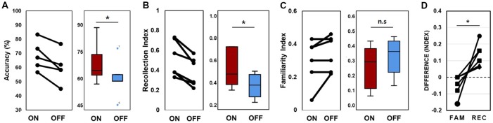
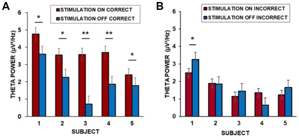
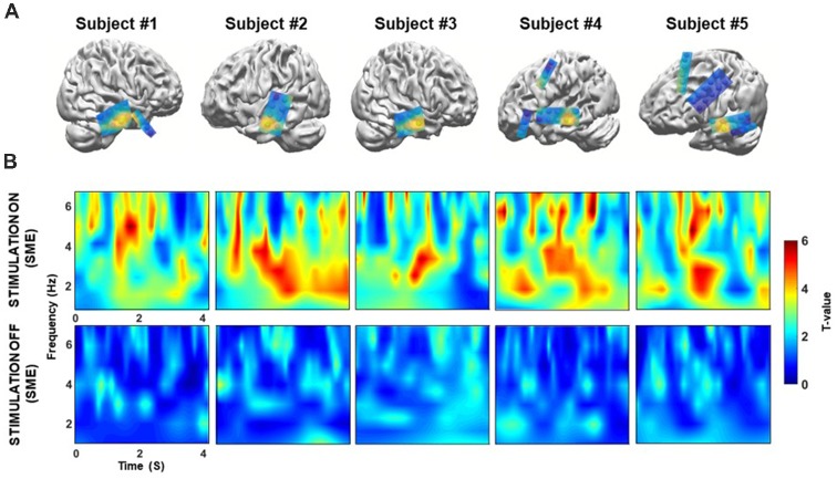
Previous studies have reported conflicting results regarding the effect of direct electrical stimulation of the human hippocampus on memory performance. A major function of the hippocampus is to form associations between individual elements of experience. However, the effect of direct hippocampal stimulation on associative memory remains largely inconclusive, with most evidence coming from studies employing non-invasive stimulation. Here, we therefore tested the hypothesis that direct electrical stimulation of the hippocampus specifically enhances hippocampal-dependent associative memory. To test this hypothesis, we recruited surgical patients with implanted subdural electrodes to perform a word pair memory task during which the hippocampus was stimulated. Our results indicate that stimulation of the hippocampus during encoding helped to build strong associative memories and enhanced recollection in subsequent trials. Moreover, stimulation significantly increased theta power in the lateral middle temporal cortex during successful memory encoding. Overall, our findings indicate that hippocampal stimulation positively impacts performance during a word pair memory task, suggesting that successful memory encoding involves the temporal cortex, which may act together with the hippocampus.
Keywords: brain stimulation; hippocampus; lateral temporal cortex; memory enhancement; recollection; theta power.
Figures
Similar articles
-
Task-dependent effects of intracranial hippocampal stimulation on human memory and hippocampal theta power.Brain Stimul. 2020 May-Jun;13(3):603-613. doi: 10.1016/j.brs.2020.01.013. Epub 2020 Jan 25. Brain Stimul. 2020. PMID: 32289685
-
Evidence for Immediate Enhancement of Hippocampal Memory Encoding by Network-Targeted Theta-Burst Stimulation during Concurrent fMRI.J Neurosci. 2020 Sep 9;40(37):7155-7168. doi: 10.1523/JNEUROSCI.0486-20.2020. Epub 2020 Aug 17. J Neurosci. 2020. PMID: 32817326 Free PMC article.
-
Dopamine Enhances Item Novelty Detection via Hippocampal and Associative Recall via Left Lateral Prefrontal Cortex Mechanisms.J Neurosci. 2019 Oct 2;39(40):7920-7933. doi: 10.1523/JNEUROSCI.0495-19.2019. Epub 2019 Aug 12. J Neurosci. 2019. PMID: 31405927 Free PMC article. Clinical Trial.
-
The role of recollection and familiarity in the functional differentiation of the medial temporal lobes.Hippocampus. 2010 Nov;20(11):1291-314. doi: 10.1002/hipo.20853. Hippocampus. 2010. PMID: 20928828 Review.
-
Principled Approaches to Direct Brain Stimulation for Cognitive Enhancement.Front Neurosci. 2017 Nov 30;11:650. doi: 10.3389/fnins.2017.00650. eCollection 2017. Front Neurosci. 2017. PMID: 29249927 Free PMC article. Review.
Cited by
-
Modulation of Human Memory by Deep Brain Stimulation of the Entorhinal-Hippocampal Circuitry.Neuron. 2020 Apr 22;106(2):218-235. doi: 10.1016/j.neuron.2020.02.024. Neuron. 2020. PMID: 32325058 Free PMC article. Review.
-
Factors affecting the efficacy of repetitive transcranial magnetic stimulation for patients with Alzheimer's disease.Zhejiang Da Xue Xue Bao Yi Xue Ban. 2021 Jun 25;50(3):383-389. doi: 10.3724/zdxbyxb-2021-0184. Zhejiang Da Xue Xue Bao Yi Xue Ban. 2021. PMID: 34402259 Free PMC article. Review. English.
-
Prediction of Successful Memory Encoding Based on Lateral Temporal Cortical Gamma Power.Front Neurosci. 2021 May 25;15:517316. doi: 10.3389/fnins.2021.517316. eCollection 2021. Front Neurosci. 2021. PMID: 34113226 Free PMC article.
-
Hippocampal Neuronal Activity Preceding Stimulus Predicts Later Memory Success.eNeuro. 2023 Feb 15;10(2):ENEURO.0252-22.2023. doi: 10.1523/ENEURO.0252-22.2023. Print 2023 Feb. eNeuro. 2023. PMID: 36720645 Free PMC article.
-
Increased T- and B-cells associated with the phenotype of autoimmune limbic encephalitis with mainly memory dysfunction.J Transl Autoimmun. 2022 Sep 28;5:100167. doi: 10.1016/j.jtauto.2022.100167. eCollection 2022. J Transl Autoimmun. 2022. PMID: 36247087 Free PMC article.
References
LinkOut - more resources
Full Text Sources