

State-Related Alterations of Spontaneous Neural Activity in Current and Remitted Depression Revealed by Resting-State fMRI
- PMID: 30804860
- PMCID: PMC6378291
- DOI: 10.3389/fpsyg.2019.00245
State-Related Alterations of Spontaneous Neural Activity in Current and Remitted Depression Revealed by Resting-State fMRI
Abstract
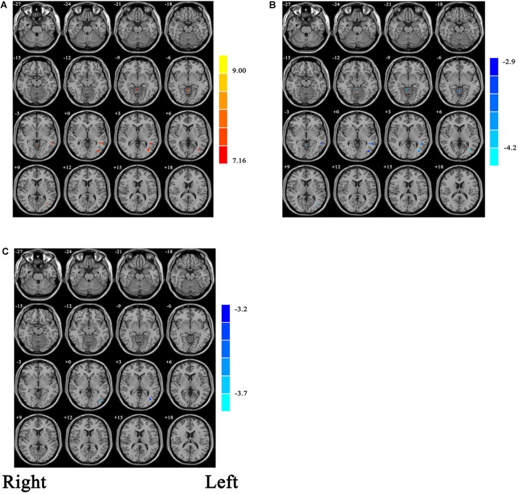
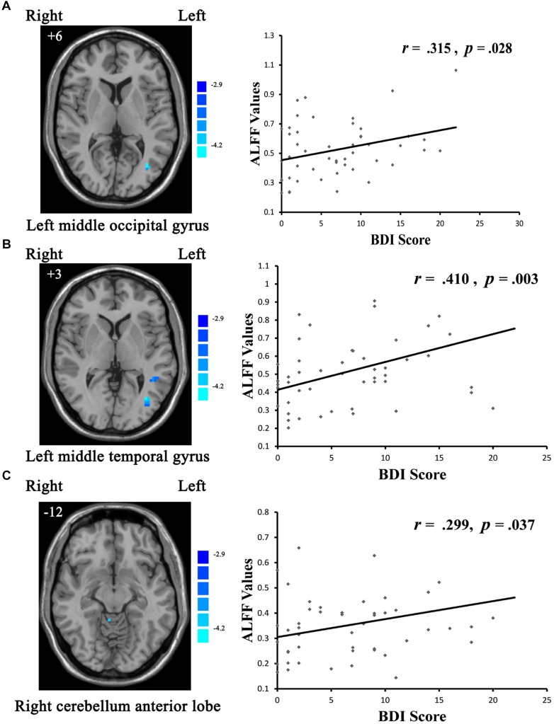
Purpose: Although efforts have been made to identify neurobiological characteristic of major depressive disorder (MDD) in recent years, trait- and state-related biological characteristics of MDD still remains unclear. Using functional magnetic resonance imaging (fMRI), the aim of this study was to explore whether altered spontaneous neural activities in MDD are trait- or state- related. Materials and Methods: Resting-state fMRI data were analyzed for 72 current MDD (cMDD) patients (first-episode, medication-naïve), 49 remitted MDD (rMDD) patients, and 78 age- and sex- matched healthy control (HC) subjects. The values of amplitude of low-frequency fluctuation (ALFF) were compared between groups. Results: Compared with the cMDD group, the rMDD group had increased ALFF values in the left middle occipital gyrus, left middle temporal gyrus and right cerebellum anterior lobe. Besides, compared with the HC group, the cMDD group had decreased ALFF values in the left middle occipital gyrus. Further analysis explored that the mean ALFF values in the left middle occipital gyrus, left middle temporal gyrus and right cerebellum anterior lobe were correlated positively with BDI scores in rMDD patients. Conclusion: Abnormal activity in the left middle occipital gyrus, left middle temporal gyrus and right cerebellum anterior lobe may be state-specific in current (first-episode, medication-naïve) and remitted (medication-naïve) depression patients. Furthermore, the state-related compensatory effect was found in these brain areas.
Keywords: amplitude of low-frequency fluctuation; major depressive disorder; remission; resting-state fMRI; state-related; trait-related.
Figures
References
-
- Beck A. T., Steer R. A., Brown G. K. (1996). Manual for the Beck Depression Inventory-II. Washington, DC: American University, 21.
LinkOut - more resources
Full Text Sources