. 2019 Jan 31:10:55.
doi: 10.3389/fneur.2019.00055. eCollection 2019.

Apraclonidine Is Better Than Cocaine for Detection of Horner Syndrome

Affiliations

Apraclonidine Is Better Than Cocaine for Detection of Horner Syndrome

Fion Bremner. Front Neurol. .

Abstract

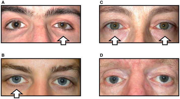
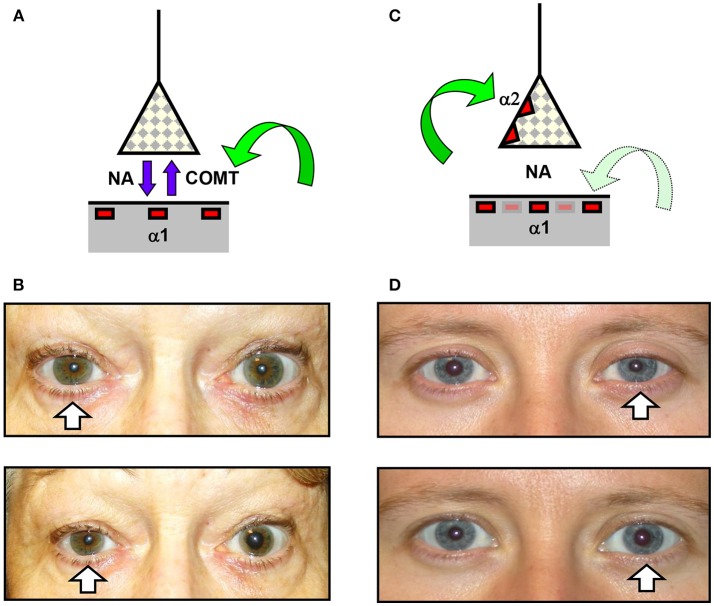
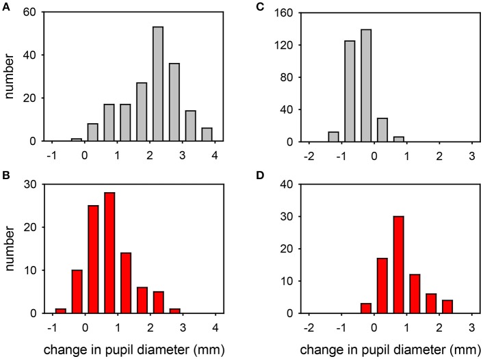
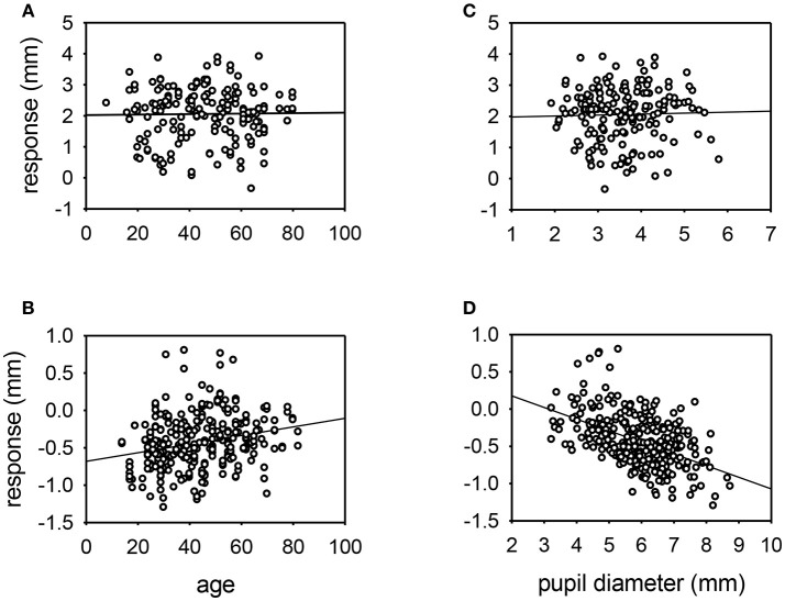
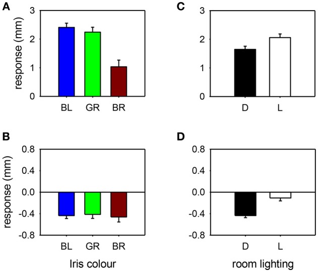
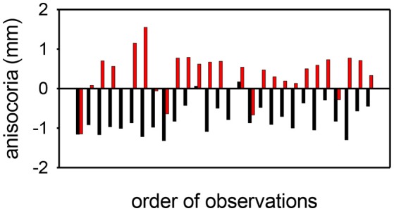
Background: In suspected cases of Horner syndrome pharmacological confirmation is often required before embarking on further investigations. There are two drugs currently used for this purpose that are commercially available for topical administration: cocaine (2-10%) and apraclonidine (0.5-1.0%). Aims: To evaluate and compare the effects of both drugs in normal eyes and eyes with Horner syndrome Methods: This is a retrospective study looking at the outcome of 660 consecutive pharmacological tests with these two drugs in one tertiary referral center over 14 years. Eyes were categorized as "normal" or "Horner syndrome" based on non-pharmacological criteria (pupillometric and clinical evidence). Pupil diameters in the dark and in bright light were measured by pupillometry before and 40 min after administration of the test drug (either 4% cocaine or 0.5% apraclonidine). Results: Cocaine dilated the normal pupil (measured in bright light: mean +2.1 mm, range -0.4 to +3.9 mm; 95% lower limit +0.5 mm); the extent of this response was not significantly affected by patient age or pupil size, but was 50% less in brown eyes compared with blue or green eyes, and 20% less if the measurements were made in the dark. In eyes with Horner syndrome cocaine had significantly less mydriatic effect (mean +0.7 mm, range -0.7 to +2.9 mm). Apraclonidine constricted the normal pupil (measured in the dark: mean -0.4 mm, range -1.3 to +0.8 mm; 95% upper limit +0.1 mm); eye color made no difference but the response was significantly greater in younger patients and larger pupils and significantly less if measured in bright lighting conditions. In eyes with Horner syndrome apraclonidine dilated the pupil (mean +0.6, range -0.4 to +2.3 mm). Applying the 95% limits identified from my normative data, I estimate the sensitivity of each drug test for detection of Horner syndrome at 40% for cocaine (criterion for abnormal: mydriasis ≤0.5 mm when measured in the dark) compared with 93% for apraclonidine (criterion for abnormal: mydriasis ≥0.1 mm when measured in the dark). Conclusions: Apraclonidine is a more sensitive test than cocaine for detection of Horner syndrome, and should be adopted as the new gold standard in routine clinical practice. However, caution is needed when using this drug within hours of a suspected sympathetic lesion, or in infants under 1 year of age.

Keywords: Horner syndrome; apraclonidine; cocaine; pharmacological testing; pupillometry; test sensitivity.


Figures

Figure 1
Figure 1
Variations in the clinical signs associated with Horner syndrome (arrows indicate side of the oculosympathetic paresis). (A) “Complete” Horner syndrome, with relative ptosis of the upper lid, elevation of the lower lid, miosis of the pupil, and injection of the conjunctiva. (B) “Incomplete” Horner syndrome, with relative miosis but no ptosis. (C) “Bilateral” Horner syndrome, with no lid or pupil asymmetry. (D) “Pseudo-Horner syndrome”: lid asymmetry is associated with right-sided enophthalmos and hypoglobus following an old orbital floor fracture; the anisocoria is physiological.
Figure 2
Figure 2
Effects of cocaine and apraclonidine on the pupil. (A,C) Schematic diagrams of the neuro-effector junction in the iris dilator muscle, showing the sympathetic nerve ending releasing noradrenaline (NA) to bind with alpha-1 adrenoceptors on the muscle membrane. (A) Cocaine blocks the enzyme cyclo-oxygenase methyl transferase (COMT) which provides the active re-uptake mechanism terminating the action of noradrenaline, and so the concentration of noradrenaline rises and the normal pupil dilates. (C) Apraclonidine is an adrenergic agonist with greater affinity for the presynaptic alpha-2 receptors (which inhibit noradrenaline release) than the post-synaptic alpha-1 receptors, so in a normal pupil the alpha-2 effect predominates and the pupil mioses. (B,D) Photographs of the pupils before (upper) and after (lower) cocaine (B) or apraclonidine (D) eye drops in two patients with unilateral Horner syndrome (arrows). Cocaine is seen to increase the degree of anisocoria, whereas apraclonidine causes the anisocoria to reverse.
Figure 3
Figure 3
Frequency distribution plots of the change in pupil diameter induced by cocaine (A,B) or apraclonidine (C,D) in normal eyes (A,C) and eyes with Horner syndrome (B,D). Cocaine measurements were made in bright light, apraclonidine measurements were made in the dark.
Figure 4
Figure 4
Scatter plots of the response of the pupil to cocaine (A,C) or apraclonidine (B,D) with respect to age (A,B) or resting pupil size (C,D). Linear regression lines are shown. No significant relationships were demonstrated for cocaine responses, but apraclonidine had significantly greater miotic effect in younger patients and in eyes with larger pupils (P < 0.001).
Figure 5
Figure 5
Pupil response to cocaine (A,C) or apraclonidine (B,D) according to iris color [(A,B): BL, blue; GR, green; BR, brown] or room lighting conditions [(C,D): D, measured in the dark; L, measured in bright light]. Cocaine had significantly greater effect on blue and green eyes compared with brown eyes, and greater effect when measurements were made in bright light compared with darkness. Apraclonidine had similar effects regardless of iris color, but significantly more effect when measurements were made in the dark compared with bright light.
Figure 6
Figure 6
Anisocoria measurements (affected eye—unaffected eye) before (black) and after (red) apraclonidine in 28 consecutive cases of unilateral Horner syndrome. In most cases (21/28) the anisocoria reverses, but in a small number of cases the anisocoria is in the same direction after as before apraclonidine, giving a sensitivity for the test using this criterion of 75%. In all but 2 cases, apraclonidine produced an abnormal degree of mydriasis (≥0.1 mm) in the pupil of the affected eye, giving a sensitivity for the test using this criterion of 93%.

Similar articles

Cited by

References

    1. Horner JF. On a form of ptosis. Klin Monatsbl Augenheilk. (1869) 7:193–8.
    1. Fulton JF. Horner and the syndrome of paralysis of the cervical sympathetic. Arch Surg. (1929) 18:2025–39. 10.1001/archsurg.1929.01140131129078 - DOI - PMC - PubMed
    1. Maloney WF, Younge BR, Moyer NJ. Evaluation of the causes and accuracy of pharmacologic localization in Horner's syndrome. Am J Ophthalmol. (1980) 90:394–402. 10.1016/S0002-9394(14)74924-4 - DOI - PubMed
    1. Mahoney NR, Liu GT, Menacker SJ, Wilson MC, Hogarty MD, Maris JM. Pediatric Horner syndrome: etiologies and roles of imaging and urine studies to detect neuroblastoma and other responsible mass lesions. Am J Ophthalmol. (2006) 142:651–9. 10.1016/j.ajo.2006.05.047 - DOI - PubMed
    1. Almog Y, Gepstein R, Kesler A. Diagnostic value of imaging in Horner syndrome in adults. J Neuro Ophthalmol. (2010) 30:7–11. 10.1097/WNO.0b013e3181ce1a12 - DOI - PubMed

LinkOut - more resources